
Abstract
The antibacterial activity of an herbal combination composed of Mume Fructus, Coptidis Rhizoma, and Schizandrae Fructus extracts on enterohemorrhagic Escherichia coli (EHEC) was evaluated in the present study. The combination demonstrated antibacterial activity against all EHEC strains tested in this study, including those resistant to multiple antibiotics; minimum inhibitory concentration values ranged from 0.49 to 31.25 mg/mL. In in vivo antibacterial activity assay, the herbal combination was administered to mice after initial E. coli O157 infection and had significant effects on mouse mortality. The effects of the herbal combination on Shiga toxin release from EHEC O26, EHEC O111, and EHEC O157 strains containing the stx1 and stx2 genes were assessed by the reversed passive latex agglutination method, and there was no increased Shiga toxin release in the strain cultures containing the herbal combination. These results suggested that the herbal combination may be a safe and effective remedy for EHEC inhibition.
Introduction
Safe and effective EHEC therapeutic protocols were evaluated in the present study using the herbal combination. The preparation was orally administered to E. coli O157–infected mice to determine its antimicrobial activity and potential in vivo applications. In addition, we also assessed the effects of the herbal combination on ST release from EHEC O26, EHEC O111, and EHEC O157 strains in the present study.
Materials and Methods
Combination preparation
The medicinal herbs Coptidis Rhizoma, Mume Fructus, and Schizandrae Fructus were used in the present study. Herbs were air-dried and ground into a powdered substance. The powder (100 g) was added to methanol (500 mL) for 20 h under mantle-reflux. Methanol was removed from extracts; extracts were dried with a rotary evaporator. The extract combination has the following compositions: 50% Mume Fructus, 30% Coptidis Rhizoma, and 20% Schizandrae Fructus.
Bacterial strains
In this study, we used 14 strains of EHEC O26, EHEC O111, and EHEC O157, including both human (n = 6) and cattle (n = 8) strains. Human strains used for antimicrobial resistance testing and ST release included EHEC O26:H28 (ATCC 25826), EHEC O111:H8 (ATCC 700840), and EHEC O157:H7 (ATCC 43889, ATCC 43894, ATCC 35150, and ATCC 43890). In addition, the cattle isolates EHEC O26 (n = 2), EHEC O111 (n = 2), and EHEC O157 (n = 4) were also used to establish the respective minimum inhibitory concentration (MIC) values. Genetic profiling for EHEC virulence markers (stx1, stx2, eae, and hly) was performed using PCR (Jo et al., 2004; Jeon et al., 2006). All strains were positive for hly and eae genes and contained either the stx1 or stx2 genes.
Antimicrobial resistance testing
E. coli resistance to various antimicrobial agents was determined with the disk-agar method, which was standardized using the Clinical and Laboratory Standards Institute (CLSI, 2009a). Quality control strains included Enterococcus faecalis ATCC29212 and E. coli ATCC25922.
Determination of MICs
A slightly modified (CLSI, 2009b) agar dilution method was used to determine MIC values. Tetracycline, amikacin, and trimethoprim/sulfamethoxazole were used as reference standards (CLSI, 2009b). Equal volumes of each bacterial strain culture (∼1 × 10⁵ CFU/mL) were applied onto Mueller Hinton broth (Difco, Detroit, MI) supplemented with the herbal combination at concentrations which ranged from 0.12 to 61 mg/mL. These serially diluted cultures were incubated at 37°C for 24 h. Cultures (100 μL) were plated on Mueller Hinton agar and were further incubated at 37°C for 24 h. Experiments were performed in duplicate, and MIC values were defined as the lowest concentration that completely suppressed colony growth.
Assessment of therapeutic effects using mice
Animal experiments were conducted with approval from the Chonbuk National University Animal Ethics Committee in accordance with the guidelines of the Korean Council on Animal Care. Mice were obtained from Laboratory Animal Center of Korea Research Institute of Bioscience and Biotechnology (Deajeon, Korea). Four- to 5-week-old female BALB/c mice (n = 90) were used in all in vivo experiments (Mohawk et al., 2010). Mice were divided into control, tetracycline, and test groups (herbal combination); these animal experiments were performed in two separate settings; each group contained 20 mice for the first set and 10 mice for the second set, respectively. Mice were given drinking water supplemented with streptomycin (5 mg/mL) during experimentation to reduce the levels of facultative anaerobic bacteria that typically colonize the mouse intestine under normal conditions (Myhal et al., 1982). EHEC O157 (ATCC43894) suspensions with 1 × 10⁹ CFU bacteria diluted in 20% sucrose were administered to all mice. Animals in the test and tetracycline groups were fed a diet containing the herbal combination (1%) or tetracycline (0.1%), respectively, 1 h postinfection. Control animals were fed the diet without supplementation. Live and dead animal numbers were quantified daily for 7 days.
Assay for release of STs
Human E. coli EHEC O26 (ATCC25826), EHEC O111 (ATCC700840), and EHEC O157 (ATCC43894) strains in the log-phase growth were prepared (1 × 10⁴ cfu/mL) in a brain heart infusion (Difco Laboratories). Antimicrobial agents and the herbal combination were added to each tube to a final concentration of one-quarter the MIC value and the finished tubes were incubated at 37°C for 18 h. The control group was prepared by incubation of each E. coli isolate without additional agents. Bacterial cultures for three strains were collected at 6, 12, and 18 h and centrifuged at 1100 g for 15 min at 4°C. Supernatants were sterilized through a 0.22 μm pore-size filter (Millipore, Bedford, MA) and stored at −20°C until needed. STs 1 and 2 activity were measured with the reversed passive latex agglutination method (RPLA) using anti-ST1 and anti-ST2 polyclonal antibodies (VTEC-RPLA kit; Denka Seiken, Tokyo, Japan).
Statistical analysis
Significant differences in ST2 release between the groups of antibiotics and herbal combination to the control group were determined by the one-way analysis of variance using the SPSS 16.0 program (SPSS, Chicago, IL). Differences in the mortalities between the groups of herbal combination and control were analyzed by a chi-square test using the SPSS 16.0 program.
Results and Discussion
MIC values were assessed for the herbal combination to identify any antimicrobial properties against various EHEC O26, EHEC O111, and EHEC O157 strains. All strains tested in the present study were affected by the combination. MIC values ranged from 0.49 to 31.25 mg/mL. MIC data also demonstrated that the combination was effective against antibiotic-resistant EHEC strains including. Animals were infected with E. coli O157 (1 × 10⁹ CFU), and in vivo therapeutic activity of the herbal combination was examined with a mouse EHEC infection model. Results are summarized in Table 1. Administration of this combination resulted in significant effects on mortality. Control group mice deaths were identified on day 2 postinfection, whereas deaths in the herbal combination and tetracycline groups were identified on day 3 postinfection. Deaths were not observed in the herbal combination group after day 4, whereas tetracycline and control group animals died until day 6. Total deaths in the control (20/30, 67%) and tetracycline (17/30, 57%) groups were greater than deaths in the herbal combination (3/30, 10%).
Escherichia coli O157:H7 ATCC43894.
Experiments were performed in two separate settings; each group contained 20 mice for the first set and 10 mice for the second set, respectively.
Number of dead animals.
Indicates significant differences in mortality between the combination (p < 0.05) and control groups.
The herbal combination was composed of Coptidis Rhizoma (the root of Coptis chinensis), Mume Fructus (the fruit of Prunus mume), and Schizandrae Fructus (the fruit of Schizandra chinensis). These medicinal herbs have been traditionally used for the treatment of infectious diseases in countries, including Korea, China, and Japan. This herbal combination has resulted in effective antibacterial activity against Salmonella (Kwon et al., 2008). Results from the present study revealed that MIC values of the herbal combination exhibited antimicrobial activity against all tested EHEC strains. In addition, the combination demonstrated high activity against the EHEC strains, which were all somewhat resistant to many common antibiotics, suggesting that the herbal combination antimicrobial activity was not affected by antibiotic resistance. The combination might also be effective for antibiotic-resistant EHEC strains. Several metabolites from herbal species (including alkaloids, tannins, saponins, and sterols) have previously been associated with antimicrobial activity (Leven et al., 1979). However, further studies to accurately characterize the antibacterial effects of such medicinal herbs are required. The herbal combination significantly reduced mouse mortality in an in vivo assay (Table 1). Our data suggested that the herbal combination could serve as a novel therapy in EHEC infections. Biological effects of medicinal herbs derived from phytochemicals may reduce the risk of infectious diseases (Sinclair, 1998; Nakajima et al., 2006). It is possible that the herbal combination functioned in bacterial inhibition rather than enhancement of host immunity since MIC results suggested direct antibacterial effects.
We also examined the effects of the herbal combination and of antibiotics (tetracycline, amikacin, and trimethoprim/sulfamethoxazole) on the release of STs. Bacterial cultures of E. coli EHEC O26, EHEC O111, and EHEC O157 strains (containing both stx1 and stx2 genes) grown with or without antimicrobial agents collected at 6, 12, and 18 h were tested for STs 1 and 2 activity through RPLA measurement with anti-ST1 and anti-ST2 antibodies. ST1 levels in all groups were not significantly different (data not shown). In contrast, ST2 levels in the supernatants of cultures treated with tetracycline, amikacin, and trimethoprim/sulfamethoxazole were higher than those of the controls, particularly at 12 and 18 h incubations (Fig. 1). In contrast, ST2 levels from cultures treated with the herbal combination were similar to those from control levels (Fig. 1).
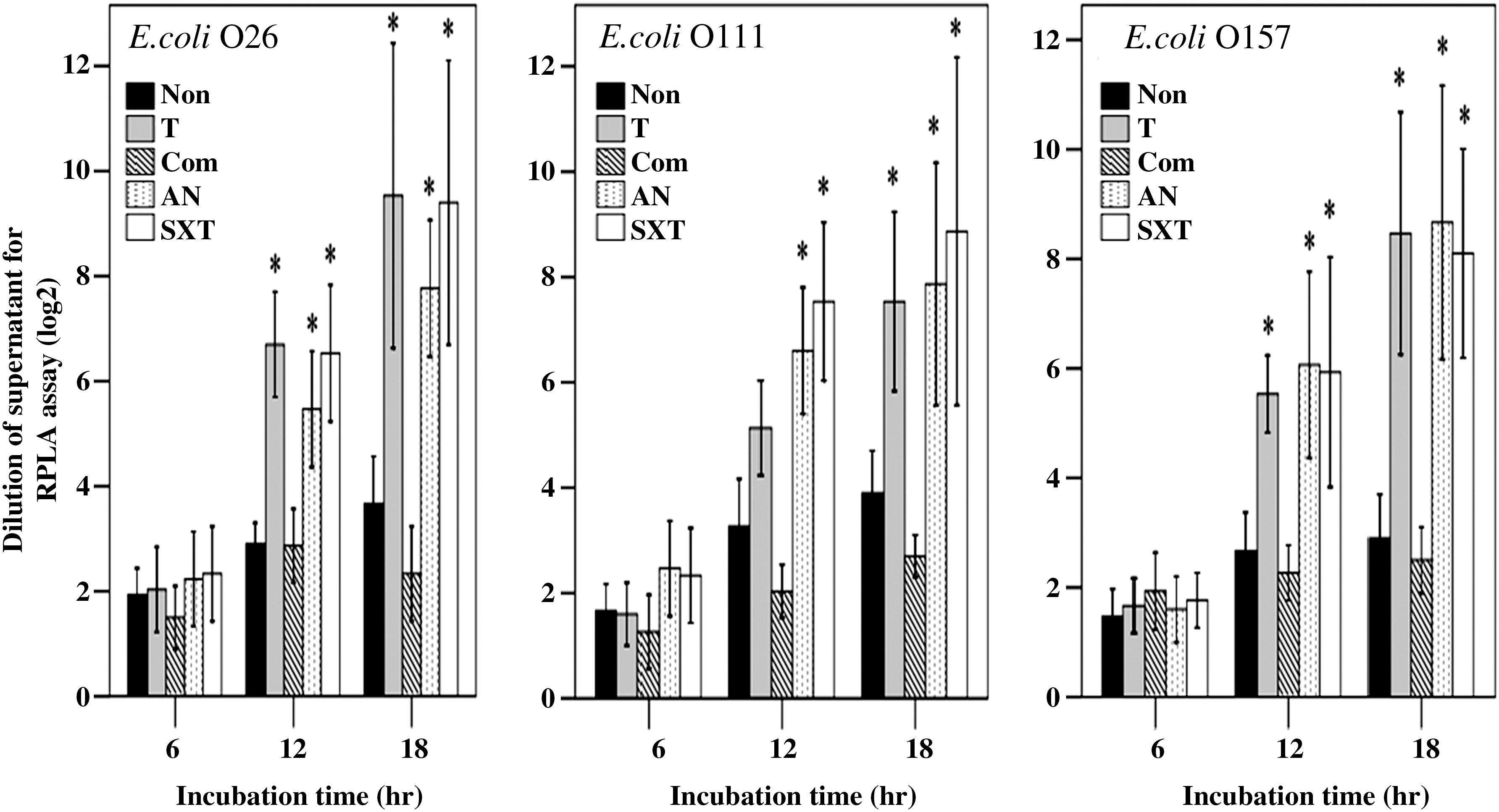
Effects of antibiotics and the herbal combination on Shiga toxin 2 release from enterohemorrhagic Escherichia coli strains as measured by reversed-passive latex agglutination. Mean values of three determinations are represented: E. coli O26, E. coli O111, and E. coli O157. Data are the mean values on Shiga toxin 2, and error bars demonstrated standard deviation. Asterisks indicate significant differences between group T, Com, AN, and SXT values (*p < 0.01) and those of control (Non). Non, nontreated control; T, tetracycline; Com, herb combination; AN, amikacin; SXT, trimethoprim/sulfamethoxazole.
The use of antimicrobial agents in EHEC infection is controversial since their use may increase the risk of more severe clinical conditions (Ostroff et al., 1989; Pavia et al., 1992). Certain antibiotics induce ST-encoding bacteriophages that result in increased expression of ST genes. In addition, some antibiotics may also cause bacterial lysis, which can lead in increased levels of free ST in the intestinal tract (Wong et al., 2000; Zhang et al., 2000). Many antibiotics, including tetracycline, amikacin, and trimethoprim-sulfamethoxazole, increased the yield of STs in previous studies; such toxins are associated with complications such as HUS (Herold et al., 2005). ST2 levels in antibiotic-treated cultures were higher in control levels in the present study. In contrast, ST2 release in cultures treated with the herbal combination were similar or suppressed compared to controls, suggesting that this combination did not increase ST yield. This suggested that the herbal combination could serve as a safe component in the treatment of EHEC infection. Further studies should clarify the use of the herbal combination as an associated treatment for EHEC infections.
Footnotes
Acknowledgments
This work was supported by a Korea Research Foundation Grant funded by the Korean Government (MOEHRD)" (KRF-2007-313-E00535), grant No. RTI05-03-02 from the Regional Technology Innovation Program of the Ministry of Commerce, Industry and Energy (MOCIE), and the international collaborative research funds of Chonbuk National University, 2010.
Disclosure Statement
No competing financial interests exist.