
Abstract
Pulsed electric field (PEF) treatments, a nonthermal process, have been reported to injure and inactivate bacteria in liquid foods. However, the effect of this treatment on bacterial cell surface charge and hydrophobicity has not been investigated. Apple juice (pH 3.8) purchased from a wholesale distributor was inoculated with cocktail of Escherichia coli O157:H7 at 7.4 log CFU/mL, processed with a PEF at a field strength of 18.4 kV/cm and 32.2 kV/cm at 25°C, 35°C, and 45°C with a treatment time of 160 μs and a flow rate of 120 mL/min. Bacterial cell surface charge and hydrophobicity of untreated and PEF-treated E. coli O157:H7 were determined immediately and after storage at 5°C and 23°C using hydrophobic and electrostatic interaction chromatography. Similarly, the populations surviving the PEF treatments including injured cells were determined by plating 0.1 mL of the sample on sorbitol MacConkey agar and tryptic soy agar (TSA) plates. The surviving populations of E. coli cells after PEF treatment varied depending on field strength and treatment temperature used. Percent injury in the surviving populations was high immediately after PEF treatment and varied among treatment temperatures. Cell surface charge of E. coli bacteria before PEF treatment averaged 32.10±8.12. PEF treatments at 25°C, 35°C, and 45°C reduced the above surface charge to 26.34±1.24, 14.24±3.30, and 6.72±2.82, respectively. Similarly, the surface hydrophobicity of untreated E. coli cells at 0.194±0.034 was increased to an average of 0.268±0.022, 0.320±0.124, and 0.586±0.123 after PEF treatments at 25°C, 35°C, and 45°C, respectively. The results of this study indicate that PEF treatment affects the outer cell envelope of E. coli bacteria as evidenced by the changes in surface hydrophobicity and cell surface charge leading to injury and subsequent inactivation of the cells.
Introduction
C
Several nonthermal processing technologies are emerging (FDA, 2002; Geveke and Brunkhorst, 2003, 2004a, 2004b), including pulsed electric fields (PEF) (Qin et al., 1995; Barbosa-Canovas et al., 1998; Selma et al., 2006). The microbial safety of PEF-treated juices such as apple juice, orange juices, tomato watermelon, and melon juices has been investigated during refrigerated storage (Evrendilek et al., 2000; Min et al., 2003; Mosqueda-Melgar et al., 2008). However, the current knowledge of PEF processes and its mechanisms for inactivation of bacterial pathogen is limited. It was hypothesized that PEF inactivation is caused by rupture of bacteria membrane structure through application of high-voltage electric fields to the bacteria (Zimmerman et al., 1976; Zimmerman, 1986). Several studies have reported that inactivation of bacteria by PEF is through cell membrane permeabilization and injury after a critical value is achieved (Barbosa-Canovas et al., 1997; Lebovka and Vorobiev, 2004; Aronsson et al., 2005; Garcia et al., 2007). More information on the exact mechanism of bacteria inactivation by PEF is needed. The reason is that liquid food matrix, such as vegetable juices, fruit juices, liquid egg, sauces, beer, wine, and soups, may require a particular technology that works best for it. Bacterial cell surfaces have net negative charge due to the presence of ionized phosphoryl and carboxylate moieties on the outer envelope exposed to the extracellular environment. Therefore, if the membrane structure of the bacteria is damaged due to electrostatic charge separation in the cell membrane, the injured bacteria may lose its biological activities. In this study, we used hydrophobic interaction chromatography (HIC) and electrostatic interaction chromatography (ESIC) to estimate changes on net negative charge and hydrophobicity of E. coli cells due to the effect of sublethal and lethal PEF treatment. Also, the relationship between changes in surface charge and injury was estimated.
Materials and Methods
Bacterial strains, growth conditions, and preparation
Bacterial strains used in this study were E. coli ATCC 25922 (type strain), O157:H7 strains SEA13B88, and Oklahoma (apple juice cider-related outbreaks) from the Eastern Regional Research Center, Agricultural Research Service, U.S. Department of Agriculture, culture collection. Individual cell cultures were maintained on trypticase soy agar (TSA) at 4°C. Before use the cells were cultured by loop transfer to tryptic soy broth (TSB: Remel, Inc.) with incubation at 37°C for 16–18 h with shaking. A 0.1 mL aliquot of cell culture was transferred to 20 mL of TSB and incubated statically at 37°C for 24 h. The overnight cell suspensions were harvested by centrifugation at 3000 g for 10 min at 5°C. The cell pellets were washed with 100 mL of sterile phosphate-buffered saline (PBS; pH 7.2) solution. Finally, the washed cells were resuspended in PBS and used as the inoculum (109 CFU/mL). The first inoculum type consisted of the individual bacterial strains at 109 CFU/mL. The second inoculum type consisted of a cocktail containing strain of individual strains at 109 CFU/mL. The mixed inoculum was used to inoculate commercial pasteurized apple juice at 2.5×10⁷ CFU/mL as determined by plate count methods. Similar bacterial suspensions were also used for the chromatographic assay described below.
PEF treatment and processing variables
Commercial pasteurized apple juice (pH 3.2) obtained from a local manufacturer was inoculated with a cocktail of bacterial inoculum to a final concentration of 2.53×10⁷ CFU/mL as determined by plate count methods. The inoculated apple juice was allowed to stand at room temperature for up to 2 h before being pumped through the PEF generator (OSU-4H). The PEF system consisted of pulse generator (Model 9410; Quantum Composer, Inc.) that produced bipolar square-waves of 2.6 μs width, and three pairs of treatment chambers each with a diameter of 0.23 cm, a gap distance of 0.29 cm connected in series. The treated sample was cooled by a cooling coil submerged in a water bath (Multitemp Water Bath III; Pharmacia Biotech) after passing each pair of treatment chambers to reach the final outlet temperature. The PEF conditions used were 25°C, 35°C, and 45°C at a field strength of 18.6 kV/cm with a treatment time of 160 μs. In another study, the treatment temperatures and time were left as is; however, the field strength was increased to 32.2 kV/cm. The inlet and outlet temperatures were monitored by type K thermocouples attached to the dual input digital thermometer (Omega HH509; Omega Engineering Inc.). A two-channel digital real-time oscilloscope (model TDS 210; Tektronix Inc.) was used to monitor voltage and current, and a digital pump (Digital Pump75211-30; Cole-Parmer, Vernon Hills, IL) was used to pump the samples through the PEF system at a set rate of 120 mL/min. Approximately, 20 mL of each sample at each outlet temperatures was individually collected in 50 mL polystyrene snap cap test tubes and placed in an ice bath to stop thermal effects. The bacterial cells in the control and PEF-treated apple juice including the populations in PBS buffer were analyzed for cell surface hydrophobicity and charge, survivors, injured cells, and viability loss.
Chromatography
HIC and ESIC columns were prepared according the procedure described by Mafu et al. (1991) and Pedersen (1980) with slight modification. For the HIC, pasteur capillary pipettes (14.59 cm long; Macalaster Bicknell Co.) were plugged with glass wool, and washed sequentially with 5 mL of 75% ethanol and 10 mL of 0.02 M sodium phosphate (NaPO4), pH 6.8 buffer. Columns for HIC were packed with 8 mL of octyl-sepharose CL-4B gel (Sigma) equilibrated overnight at 4°C in 12 mL of 1 M ammonium sulfate (NH4SO4), pH 6.8 buffer. Approximately 4 mL of the equilibrated gel was added to the column to obtain a 0.7 mL bed volume. The gel bed was washed with 12 bed volumes of 1 M (NH4SO4), pH 6.8 buffers to remove traces of ethanol added to the gel as preservatives. The ESIC columns were packed with 2 mL distilled water (1:2 wt/vol) suspension of the ion-exchange resin (Dowex chloride form; Sigma) for the anionic resin and Dowex hydrogen form (capacity, 1.7 meq/mL, 50 by 8) for the cation resin (Bio-Rad Laboratories). The mesh size was 100 to 200 μm for both resins. Chromatography was done according to Dickson and Koohmariae (1989).
Surface hydrophobicity and the bacterial surface charge
For bacterial surface hydrophobicity, a sample (0.1 mL) of washed bacterial cell suspension for each strain stated above was loaded onto the surface of the column followed by elution with 10 mL of 1 M NH4SO4 buffer at a flow rate of 2.6 mL/min. The eluted fractions were collected at 2 mL interval and the bacterial populations in each milliliter fraction were determined by plating on TSA. From this experiment, it was established that ∼5 mL of the buffer is needed to desorb most of the bacteria from the gel. For the rest of the study, 0.1 mL of the PEF-treated cells was placed on the gel, 5 mL of 2 M NH4SO4 buffered with sodium phosphate (NaPO4, pH 6.8) was passed continuously through the gel and the eluted fraction was collected and named eluate (e). Similarly, the gel also was collected and named g. Finally, 0.1 mL of washed or PEF-treated cells was mixed with 4 mL of 2 M NH4SO4 buffer solutions and were individually named X (Mozes and Rouxhet, 1987). The same procedure was used for the ESIC study and the relative ion values for PEF-treated and untreated E. coli cells were determined and expressed as r/e. r represent the number of bacteria retained by the gel in the columns while e is the numbers eluted from the gel. The bacterial cell populations in all the suspensions including each eluted sample and the populations remaining in the gel were determined using TSA (BBL/Difco). The relative hydrophobicity of PEF-treated and untreated E. coli cells were expressed as the g/e ratio. According to Dahlback et al. (1981), a log g/e ratio of<0 is considered to indicate hydrophilicity.
Microbial analysis
Aliquot (0.1 mL) of PEF-treated and untreated apple juice described above was plated on TSA and sorbitol MacConkey agar plates and incubated at 36°C for 48 h. When necessary, depending on the treatment temperature, samples were diluted in 0.1% peptone water (PW) before plating onto the agar plates. Bacterial inactivation was calculated using the following formula:
where No=concentration of bacteria before PEF processing, and N=concentration of bacteria after PEF processing.
Percent injury for bacteria cells in PEF-treated apple juice was calculated using this formula:
Untreated PEF inoculated apple juice was used as controls for each experiment. All experiments were performed at least three times with duplicate determinations.
Data analysis
All experiments were done in triplicate with duplicate samples being analyzed at each sampling time. Data were subjected to analysis of variance using the Statistical Analysis System Program (SAS Institute, Cary, Version 9.12). Relationship between percent injury and changes in surface charge at optimum treatment temperatures were analyzed. Similarly, the significant differences (p<0.05) between mean values of number of cells and treatment type were determined by the Bonferroni LSD method (Miller, 1981).
Results and Discussion
Desorption of bacteria
To determine the approximate volume of buffer needed to desorb each strain of E. coli ATCC 25922 and E. coli O157:H7 (SEA13B88 and Oklahoma) deposited on octyl-sepharose gel, 15 mL volume of 1 M ammonium sulfate buffer was used. During the desorption of E. coli bacteria, 1 mL of the eluted fraction of each 15 mL was collected and 0.1 mL plated on TSA to determine CFU/mL of each strain of E. coli cells (Fig. 1). Bacterial populations determined from each eluted fraction varied especially in the first 4 mL. Highest bacterial populations were detected in the first 2 to 3 mL fraction than any other subsequent fraction. The total plate counts for all E. coli strains determined after the first initial 4 mL fraction averaged 5 log CFU/mL and the populations slightly declined but remained approximately the same in subsequent mL fractions after that. For the rest of the study, we used 5 mL volume of the buffer to desorb the E. coli cells and to estimate the surface hydrophobicity and cell surface charge of each strain.
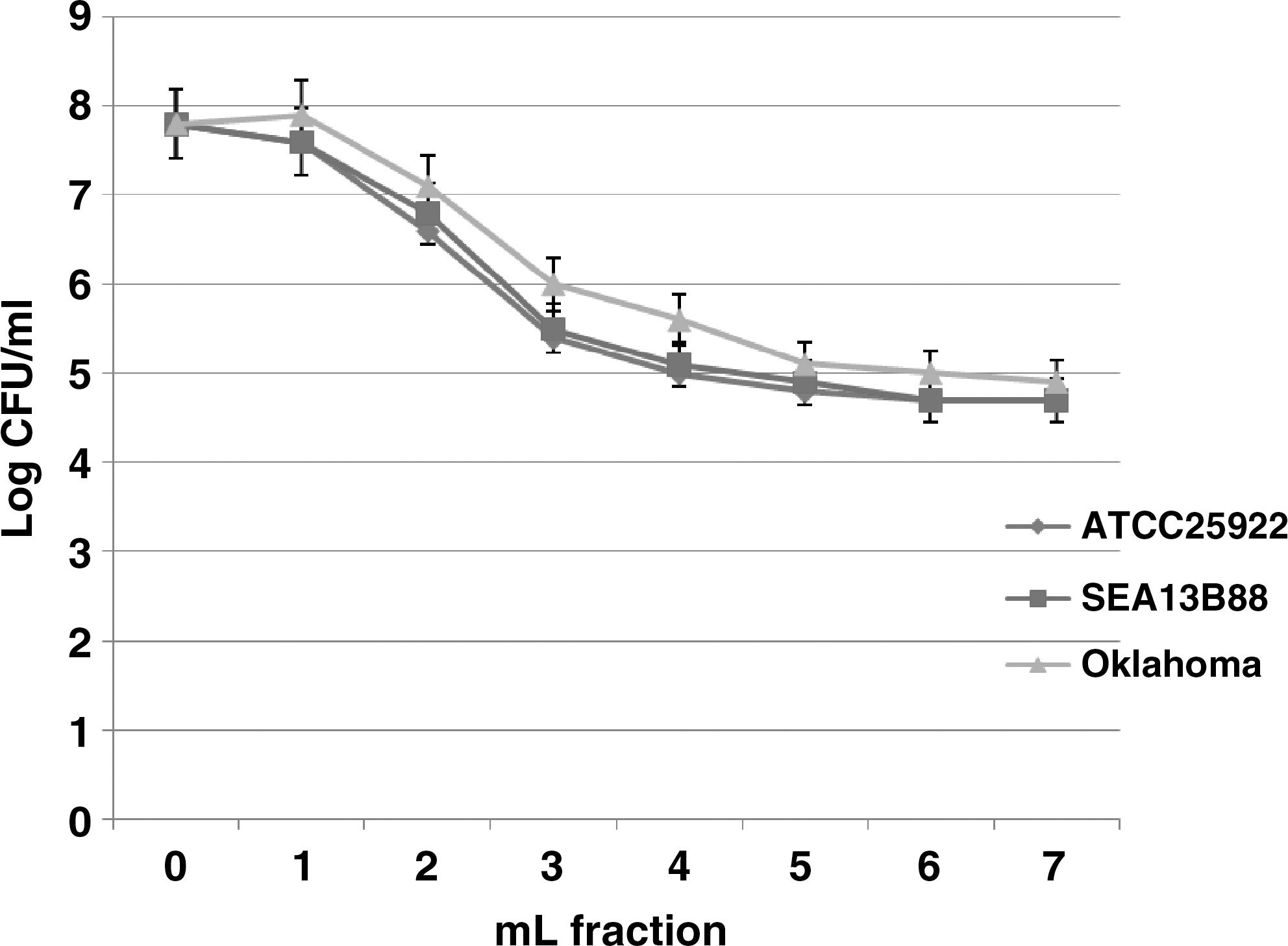
Desorption of Escherichia coli cells deposited on octyl-sepharose gel using 1 M (NH4)2SO4. Values are means±standard deviation of three experiments with duplicate determinations. Determination (log CFU/mL) was performed by plaiting 0.1 mL of the each eluted fraction on tryptic soy agar with incubation at 36°C for 24 h.
Injury and inactivation of bacteria by PEF
A higher population of bacteria was inactivated at 32.2 kV/cm field strength irrespective of treatment temperatures than at 18.6 kV/cm (Fig. 2). PEF surviving E. coli cells at 18.6 kV/cm averaged approximately less than 6 log but higher than 5 log CFU/mL and these populations were used to monitor the impact of PEF on bacterial cell surface charge and hydrophobicity (Table 1), injury (Fig. 3), and the behavior of the surviving cells during storage at different temperatures (Fig. 4). Populations of injured E. coli cells in PEF-treated samples were determined immediately after PEF treatments (Fig. 3). The injured populations of E. coli cells in apple juice treated with at 18.6 kV/cm at 35 and 45°C averaged 58 and 84%, respectively. These populations increased to 96% and 98% at 35°C and 45°C, respectively, when the field strength was increased to 32.2 kV/cm. Further inactivation of E. coli cells occurred in samples left at room temperature (∼23°C) and refrigerated (5°C) storage for 24 h. This observation can be attributed to the dying off of injured populations in samples stored at 5°C due to cold shock (Fig. 4). The population reduction in samples treated at 32.2 kV/cm and stored at 5°C was significantly (p<0.05) different from 18.6 kV/cm treatment.
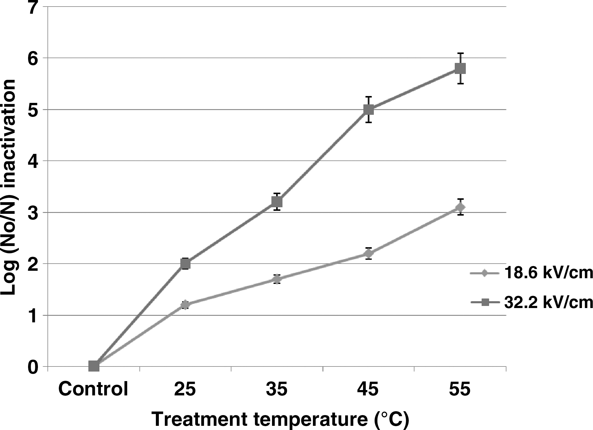
Effect of pulse electric field treatment at different temperatures on inactivation of E. coli cells in apple juice. Values are means±standard deviation of three experiments with duplicate determinations.
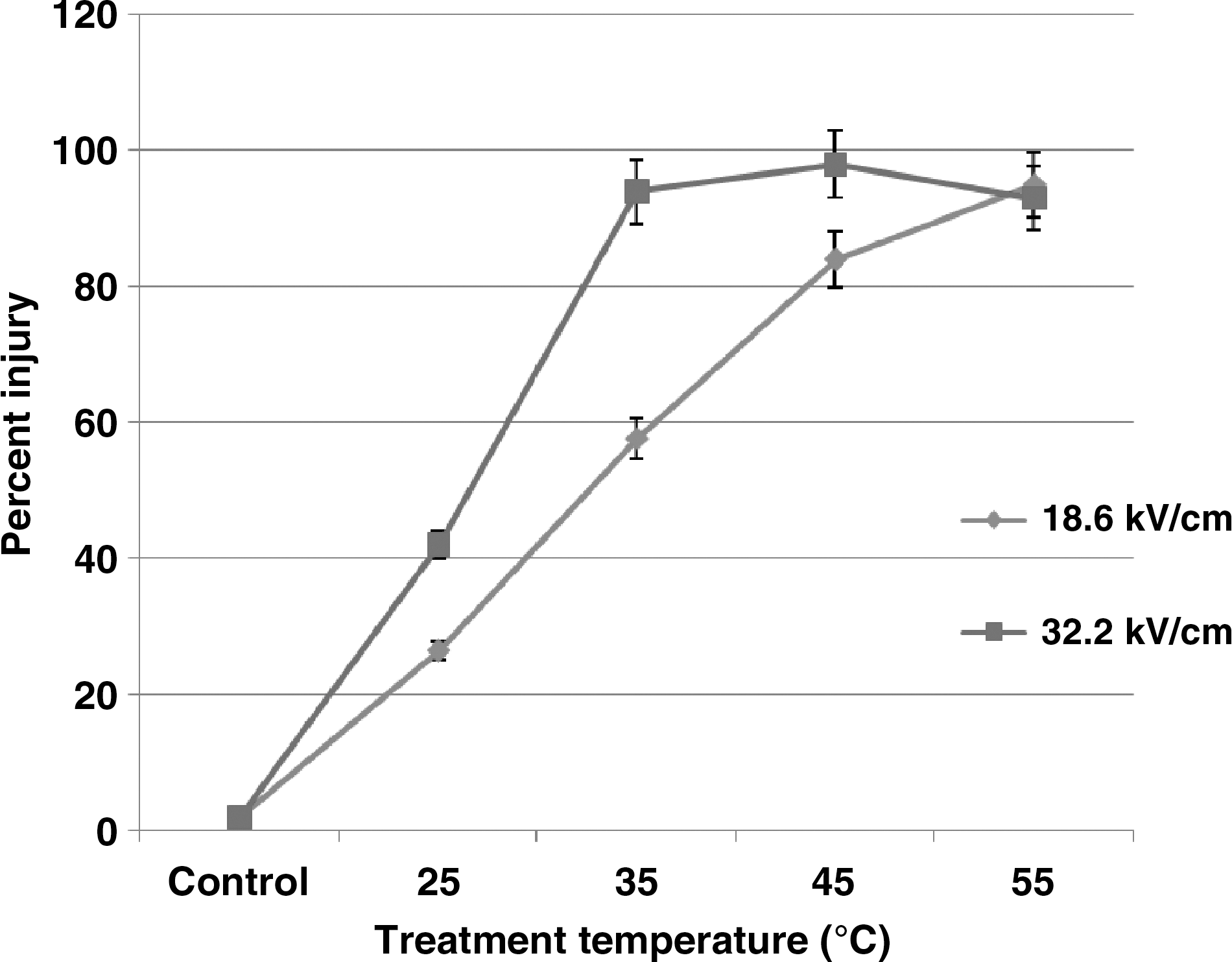
Effect of pulsed electric field (PEF) and treatment temperature on injured populations of E. coli cells in apple juice. Values are means±standard deviation of three experiments with duplicate determinations.
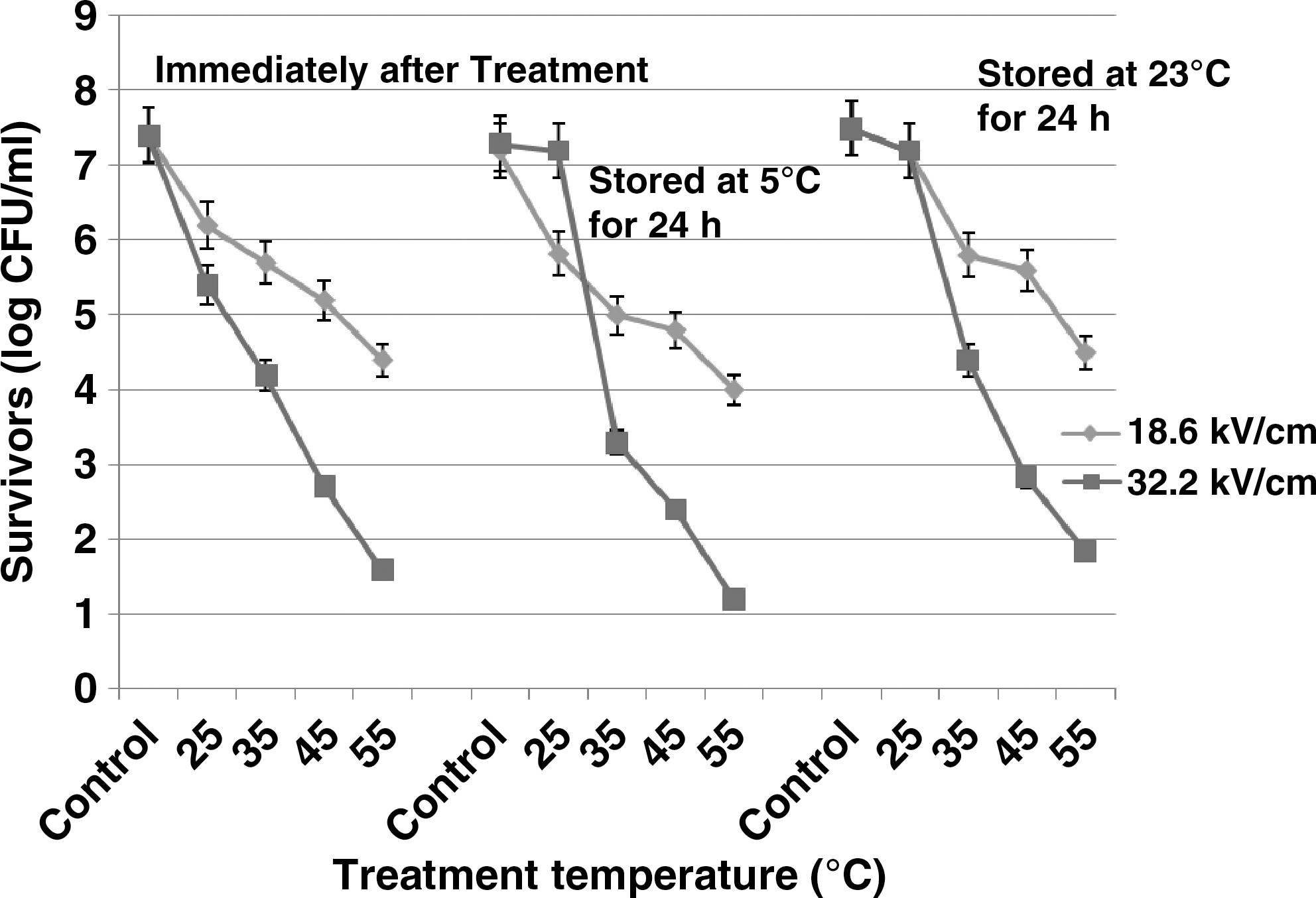
Effect of storage temperature on PEF-injured populations of E. coli cells in apple juice. Values are means±standard deviation of three experiments with duplicate determinations.
Values are averages of three separate determinations±standard deviation. About 2 M (NH4)2SO4 buffer was used to encourage surface hydrophobicity of Escherichia coli cells to the octyl-sepharose gel.
Means in each row and column not followed by the same letter are significantly (p<0.05) different.
PBS, phosphate-buffered saline; PEF, pulsed electric field.
Bacterial cell surface hydrophobicity and charge
To understand the sublethal effect of PEF on bacterial cell surfaces, we concentrated our effort in investigating the effect of lower field strengths (18.6 kV/cm) and a treatment time of 160 μs. The results on HIC of all E. coli bacteria in PBS and apple juice tested before and after the PEF treatment is shown in Table 1. Surface hydrophobicity of E. coli ATCC 25922 and E. coli O157:H7 (SEA13B88) in PBS averaged 0.201, 0.198, and 0.220, respectively. After PEF treatments, these values slightly increased and only Oklahoma cells were not significantly (p>0.05) different from untreated bacterial cells in PBS. The HIC of E. coli ATCC 25922, E. coli O157:H7 (SEA13B88), and Oklahoma in apple juice before PEF treatment averaged 0.189, 0.185, and 0.193, respectively. After PEF treatment, the surface hydrophobicity of E. coli ATCC 25922 and O157H7 (SEA13B88 and Oklahoma) increased significantly (p<0.05). The bacterial cell surface hydrophobicity measured by HIC and ESIC in this study is a relative value and is expressed as the g/e ratio.
Surface hydrophobicity and the relative negative and positive ions of mixed culture containing all bacterial strains were investigated (Table 2). The relative negative and positive ions for all bacteria in PBS averaged 1.6 and 0.14, respectively. For apple juice, these values averaged ∼34 and 0.02. The hydrophobicity of the mixed culture in PBS averaged 0.219 and this value increased after PEF treatment at all temperature tested. These values were significantly (p<0.05) increased again when PEF treatment temperature was set at 25°C, 35°C, and 45°C. However, the relative negative and positive ions for all bacteria decreased in PEF-treated PBS and apple juice. When 2 M (NH4)2SO4 buffer solutions was used, all PEF-treated E. coli cell surface hydrophobicity was affected. For example, cell surface hydrophobicity for E. coli ATCC 25922 increased from 0.127 to 0.310 log g/e ratio, cell surface hydrophobicity for E. coli O157:H7 (Oklahoma) increased from−0.335 to 0.597 log g/e, whereas cell surface hydrophobicity for E. coli (SEA13B88) decreased from 0.127 to 0.00 log g/e. These values were within the range reported for E. coli bacteria in an attachment study on cantaloupe surfaces (Ukuku and Fett, 2002, 2006).
Values are averages of three separate determinations±standard deviation
No positive (+) ion value was determined when 2 M (NH4)2SO4 buffer was used to desorb the E. coli cells from the hydrogen ion gel.
Means in each column between not followed by the same letter are significantly (p<0.05) different.
ESIC, electrostatic interaction chromatography.
HIC and ESIC are the most widely used techniques to study bacterial cell surface charge because bacterial cell surface properties can only be measured indirectly, through phenomena that reflect more or less the nature of molecular interactions with surfaces (Noda and Kanemasa, 1986; Mozes and Rouxhet, 1987; Irvin, 1990). Bacterial surfaces are heterogeneous with physicochemical properties determined primarily by teichoic acid (Gram-positive strains) or other polysaccharides (Gram-negative strains) along with proteinaceous appendages (fimbriae) (Pringle and Fletcher, 1986; Romantschuk, 1992; Fletcher, 1996). This surface heterogeneity in bacterial populations may help explain the differences in cell reaction to environments. The results of this study indicate that the overall total relative cell surface charge for the E. coli O157:H7 cells were reduced by the PEF treatment. In another study, we estimated the effect of treatment temperature alone on relative negative ions, sublethal injury, and viability loss and found a minimal effect only on the bacterial cells surfaces treated at 45°C (Data not shown). This observation seems normal since these temperatures (35°C and 45°C) are within the range for survival and growth of E. coli cells.
This is the first study in which HIC or ESIC techniques were used to investigate the relationship between bacterial cell surface charges or hydrophobicity and injury of PEF-treated E. coli cells in apple juice. E. coli cells used in this study are Gram-negative bacteria that have lipopolysaccharide and protein units on its outer layer. The lipopolysaccharide and protein forms a highly charged surface that is stabilized by cation binding (Peterson et al., 1985). It is possible that the disruption of the surface structure of E. coli in this study was due to charge–charge interactions between the bacterial negative charge and the energy charge produced by the PEF.
During PEF treatments, the outlet treatment temperatures (34.1°C, 45.9°C, and 55.3°C) were higher than the inlet temperatures (25°C, 35°C, and 45°C), respectively. A relationship between percent injuries at these outlet temperatures and changes in the surface charge of treated E. coli cells were analyzed immediately after treatments and during storage (Fig. 5). The correlation between relative negative ions on the surface of E. coli cells and PEF injured bacterial cells determined immediately after treatment was R 2=0.994. This value decreased to R 2=0.932 when the E. coli cells were stored at 23°C for 24 h. There was a significant (p<0.05) decrease in correlation (R 2=0.687) between the negative ions in relation to surface injury when the E. coli cells were stored at 5°C for 24 h. The slight decrease of R 2=0.932 in samples stored at 23°C for 24 h could be attributed to recovery of PEF injured E. coli cells as this value was significantly (p<0.05) higher than values determined in samples stored at 5°C for 24 h.

Relationship between relative negative ions of E. coli cells surfaces and injury during storage. Values are means±standard deviation of three experiments with duplicate determinations.
Conclusion
The results of this study suggest that the mechanism of bacterial inactivation by PEF treatment involves among other phenomenon the reduction of surface charge and changes in surface hydrophobicity. Also, the results of this study suggest storage of PEF-treated juices immediately after treatment at 5°C to increase the microbial safety of processed juices.
Footnotes
Acknowledgments
The authors wish to thank Ms. Donyel Jones and Lee Chau for technical support and Dr. John G. Phillips for assistance in statistical analysis of the data.
Disclaimer
Mention of trade names or commercial products in this article is solely for the purpose of providing specific information and does not imply recommendation or endorsement by the U.S. Department of Agriculture.
Disclosure Statement
No competing financial interests exist.