
Abstract
Salmonella enterica serovar Enteritidis (S. Enteritidis) is a major serovar associated with human salmonellosis. A total of 425 clinical S. Enteritidis isolates of human origin were collected between June 2009 and September 2010 from North Carolina. The isolates were further characterized for antimicrobial susceptibility, antimicrobial resistance coding determinants, virulence genes, and fingerprint profiles to determine whether they were similar or different to the S. Enteritidis strain responsible for the human outbreak due to consumption of contaminated eggs. Ten different antimicrobial resistance phenotypes were observed with the highest frequency of resistance exhibited to ampicillin (n=10; 2.35%). The isolates were predominantly pansusceptible (n=409; 96.23%); however, seven isolates were multidrug resistant (MDR; i.e., resistant to three or more antimicrobials). Extended spectrum β-lactamase (ESBL) coding genes (bla TEM and bla PSE) were detected in the ampicillin-resistant isolates, whereas a single MDR isolate tested positive for class 1 integron (1 kb). The majority of the isolates (n=422; 99.3%) carried the invA, mgtC, stn, sopB, sopE1, and sefA virulence genes. However, 37 (8.7%) and 46 (10.82%) S. Enteritidis isolates tested negative for the plasmid encoded genes spvC and rck, respectively. Pulsed-field gel electrophoresis (PFGE) typing of 118 S. Enteritidis isolates by restriction enzymes XbaI and BlnI resulted in seven clusters, each with a discriminatory index (DI) of 0.715 and 0.785, respectively. The combination of XbaI-BlnI patterns generated a dendrogram with 14 clusters and a higher DI of 0.914. The PFGE profile of 80 isolates matched 100% with the S. Enteritidis strain that has been cited for the recent outbreak in the United States due to consumption of contaminated eggs. In conclusion, we identified a genotypic similar S. Enteritidis population in our study based on antimicrobial susceptibility, virulence gene, and PFGE fingerprint profiles.
Introduction
Salmonella pathogenicity depends on a variety of virulence factors that help the pathogen in adhesion and invasion mechanisms. The Salmonella plasmid virulence (spv) operon, which consists of five genes (spvRABCD), potentiates the systemic spread of the pathogen and aids in its replication in extra-intestinal sites (Gebreyes et al., 2009; Hur et al., 2011). Other important virulence factors include the rck gene product, which encodes an outer membrane protein and induces resistance to complement-mediated killing (Fluit, 2005) and mediates adherence and invasion to cultured eukaryotic cell line (Ho et al., 2010). The Sop proteins (sopA-E) (sop) and the heat-labile Salmonella enterotoxin (stn) serve as effector proteins, which are involved in the pathogenesis of salmonellosis (Wallis and Galyov, 2000; van Asten and van Dijk, 2005). The mgtC gene encodes MgtB Mg2+ transporter and helps the pathogen to survive within macrophages (Alix and Blanc-Potard, 2007). Invasion gene (invA) exists in the majority of Salmonella strains and is related to intestinal mucosa invasion (Fluit, 2005; Chuanchuen et al., 2010), whereas the sef gene encodes sef14 fimbriae element and is reported to be involved in adhesion (van Asten and van Dijk, 2005). It is important to identify the virulence genes that are responsible for establishing the S. Enteritidis infection in humans.
Pulsed-field gel electrophoresis (PFGE) has been used by public health and surveillance agencies for strain identification and source tracking in outbreak investigations (Ribot et al., 2006; Swaminathan et al., 2001). However, the conventional PFGE method of using a single restriction enzyme does not have a good discriminatory power (DP) to differentiate between two unrelated S. Enteritidis strains and place them in separate clusters (Zheng et al., 2011). Therefore, recent studies have recommended the use of multiple restriction enzymes to increase the DP of PFGE when used for typing S. Enteritidis strains (Zheng et al., 2007, 2011).
The objective of this study was to determine the antimicrobial susceptibility, virulence gene, and genotype profile of S. Enteritidis human clinical isolates from North Carolina. A total of 425 S. Enteritidis isolates from clinical human cases were received from the North Carolina State Public Health Laboratory (NCSPHL) from June 2009 to September 2010, a time period that overlapped with the national S. Enteritidis outbreak. The isolates were characterized at the phenotypic and genotypic levels using a combination of antimicrobial susceptibility, virulence genes and PFGE profiles to determine the diversity or similarity of S. Enteritidis isolated from humans in North Carolina.
Methods
Bacterial isolates and culture conditions
A total of 425 clinical S. Enteritidis isolates were collected and received from the NCSPHL between June 2009 and September 2010. All the isolates that were submitted to the NCSPHL were forwarded to our laboratory at the end of every month. We received an average of 20 isolates every month from June 2009 to April 2010. Subsequently, we observed an increase in the total number of S. Enteritidis isolates received by our lab beginning May 2010 (n=35) and extending into June (n=59), July (n=51), August (n=43), and September (n=28), which was concurrent with the peak similar reached by the S. Enteritidis outbreak epidemic curve in the 2010 nationwide outbreak due to consumption of contaminated eggs (CDC, 2010). Stock cultures were stored on Luria Bertani (LB) agar slants (Difco, Becton Dickinson, Franklin Lakes, NJ) at room temperature; cultures were also stored on Brucella broth (Difco, Becton Dickinson) containing 20% glycerol at −80°C for future reference.
Antimicrobial susceptibility testing
The antimicrobial susceptibility of the S. Enteritidis isolates was tested for 12 antimicrobials by the Kirby-Bauer disc diffusion method on Mueller-Hinton agar using commercially available discs following the manufacturer's recommendations (Becton Dickinson, Franklin Lakes, NJ). The antimicrobials tested and the disk potency (μg/mL) used were as follows: Amikacin (AN; 30 μg), Amoxicillin/Clavulanic Acid (AMC; 20/10 μg), Ampicillin (AM; 10 μg), Ceftriaxone (CRO; 30 μg), Cephalothin (CF; 30 μg), Chloramphenicol (C; 30 μg), Ciprofloxacin (CIP; 5μg), gentamicin (G; 10 μg), Kanamycin (K; 30 μg), Streptomycin (S; 10 μg), Tetracycline (TE; 30 μg), and Trimethoprim-Sulfamethoxazole (SXT; 1.25/23.75 μg). Isolates were categorized as susceptible or resistant, according to the guidelines of the Clinical and Laboratory Standards Institute (CLSI, 2010) breakpoint established. Escherichia coli (ATCC 25922) was used as a control strain.
Characterization of antimicrobial resistance determinants
All the resistant isolates were screened for the presence of class 1 integron and corresponding resistance genes based on their resistance phenotypes. Bacterial DNA was purified using the DNeasy blood and tissue kit (Qiagen, Valencia, CA) according to the manufacturer's recommendations. The primers used in this study for detection of different AR genes include the ESBL bla TEM and bla PSE genes (Carlson et al., 1999), tetracycline resistance coding tet(A), tet(B), and tet(G) genes (Ng et al., 1999), aminoglycosides resistance coding aad A1/A2 and strA/B genes (Madsen et al., 2000), chloramphenicol resistance coding cml gene (Briggs et al., 1999), kanamycin resistance coding aphAI gene (Frana et al., 2001), trimethoprim/sulfamethoxazole resistance coding sul1 gene (Briggs et al., 1999), and sul2 (Aarestrup et al., 2003) and class 1 integrons (Ng et al., 1999). Amplification reagent concentrations and polymerase chain reaction (PCR) cycling conditions were followed as described in the above studies.
Detection of virulence genes
All the S. Enteritidis isolates were screened for a group of eight virulence genes, including invA, sefA (Oliveira et al., 2002), spvC, sopB, mgtC, sopE1 (Huehn et al., 2010), rck (Guerra et al., 2004), and stn (Murugkar et al., 2003). Multiplex PCR reactions were standardized using the above primer-pair combinations. Multiplex PCR sets included set A (invA and spvC), B (rck and mgtC), and C (stn and sopE1). Singleplex-PCR was used to amplify virulence genes sefA and sopB. Amplification reagent concentrations and PCR cycling conditions were followed as described in the above studies.
PFGE analysis
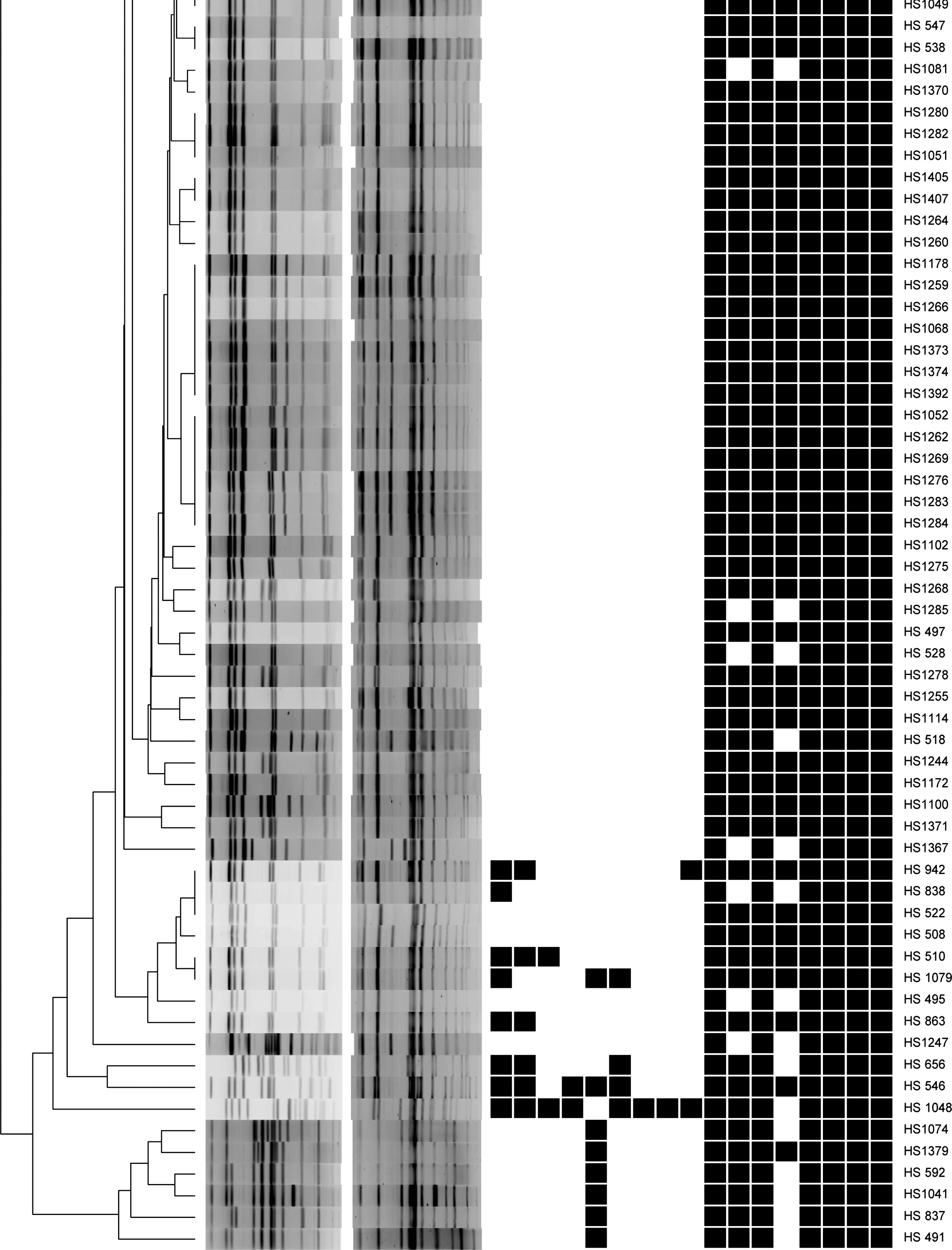
A total of 118 S. Enteritidis isolates selected for genotyping were representative of the month of isolation and different antimicrobial resistance and virulence gene profiles. Clonal relationships were determined by PFGE using the PulseNet protocol (Ribot et al., 2006). Briefly, 400 μL of overnight culture cells were lysed, and intact genomic DNA was digested in agarose-embedded plugs with XbaI and BlnI restriction enzymes (Roche, Mannheim, Germany). The restriction fragments were separated by electrophoresis in 0.5×TBE buffer, 1% Seakem Gold agarose (Lonza, ME), for 18 h at 14°C in a CHEF DR-III (Bio-Rad, Hercules, CA) using pulsed times of 2.2–63.8 s. XbaI digested S. Braenderup H9812 were used as reference DNA marker. Clonal relationships among these isolates were analyzed using Bionumerics version 6.1 (Applied Maths, Austin, TX). Simpson's index of diversity was calculated to determine the DP of PFGE using single and double enzymes in this study (Hunter and Gaston, 1988).
Results
Antimicrobial susceptibility profile
Out of 425 S. Enteritidis isolates, 96.23% (n=409) were susceptible to all of the antimicrobials tested (pansusceptible). The highest frequency of resistance was detected to ampicillin (n=10; 2.35%), followed by streptomycin (n=9; 2.11%) and cephalothin (n=6, 1.41%) (Table 1). The isolates also exhibited resistance to other antimicrobials, including amoxicillin/clavunanic acid, chloramphenicol, kanamycin, tetracycline, and trimethoprim/sulfamethoxazole. No resistance was detected to amikacin, ceftriaxone, and ciprofloxacin. We detected seven multidrug resistant (MDR) patterns, including a single isolate (ID: HS1048) that exhibited resistance to eight out of the 12 antimicrobials on the panel (AM-AMC-CF-C-GM-K-TE-SXT). The other six MDR patterns (AM-AMC-CF, AM-S-TE, AM-CF-TE, AM-CF-SXT, AM-AMC-C-S-TE, AM-CF-C-S-TE) were represented by a single isolate each.
Different antimicrobials used to test antimicrobial susceptibility.
Concentrations of antimicrobials used in disc diffusion test.
Molecular characterization of antimicrobial resistance determinants
We identified 11 different antimicrobial resistance genes conferring resistance to eight classes of antimicrobials. All the ampicillin-resistant isolates (n=10) harbored extended spectrum β lactamase (ESBL) producing bla TEM gene, with the exception of a single isolate that carried the bla PSE gene. Three out of the five tetracycline-resistant strains tested PCR positive for the tet(A) gene, while the other two isolates harbored the tet(B) and tet(G) genes separately. The cml and aphAI genes encoded chloramphenicol and kanamycin resistance in the S. Enteritidis isolates in our study, respectively. All the MDR strains exhibiting resistance to aminoglycosides harbored either the strA/B (75%) or the aad A1/A2 genes (25%) genes. A single S. Enteritidis resistant strain (AR Profile AM-AMC-C-S-TE) tested positive for the presence of class 1 integron (1 kb).
Virulence genes characterization
All the isolates (n=425) tested PCR positive for the presence of virulence genes responsible for invasion (invA), Mg2+ transportation system (mgtC), production of enterotoxocin (stn), Salmonella outer protein production (sopB), Salmonella plasmid virulence operon (spvC), and the complement killing resistance (rck) genes. The majority of the isolates carried the Salmonella outer protein production (sopE1; 99.5%) and S. Enteritidis fimbriae (sefA; 98.2%) genes with the exception of two and three isolates, respectively. However, 37 (8.7%) and 46 (10.82%) S. Enteritidis isolates were negative for the plasmid genes spvC and rck, respectively.
PFGE
A total of 118 S. Enteritidis isolates were genotyped by PFGE using XbaI and BlnI restriction enzymes. The use of XbaI and BlnI restriction enzymes gave consistent reproducible fingerprints consisting of 10–14 and 9–14 bands, respectively. Single enzyme restriction analyses by XbaI distributed the 118 isolates into seven major clusters (consisting of isolates with similar PFGE profiles) representing 102 isolates and another 16 unique PFGE patterns represented by a single isolate each. The overall DP of the method using the XbaI enzyme was 0.715. The PFGE profile of 80 isolates matched 100% with the S. Enteritidis PFGE pattern that has been identified as the outbreak strain in the recent outbreak in the United States. DNA digestion with restriction enzyme BlnI also generated seven clusters represented by 99 isolates. We also detected 19 unique PFGE patterns that were represented by a single isolate. The DP of BlnI (0.785) was higher, as it was able to further differentiate isolates that were grouped in the same cluster by the XbaI enzyme. The combined XbaI-BlnI dendrogram showed the highest number of clusters (n=14), unique patterns (n=43), and DP of 0/914 when compared to individual XbaI and BlnI dendrograms (Fig. 1). Overall, the S. Enteritidis population in this study appeared to be genotypically similar.
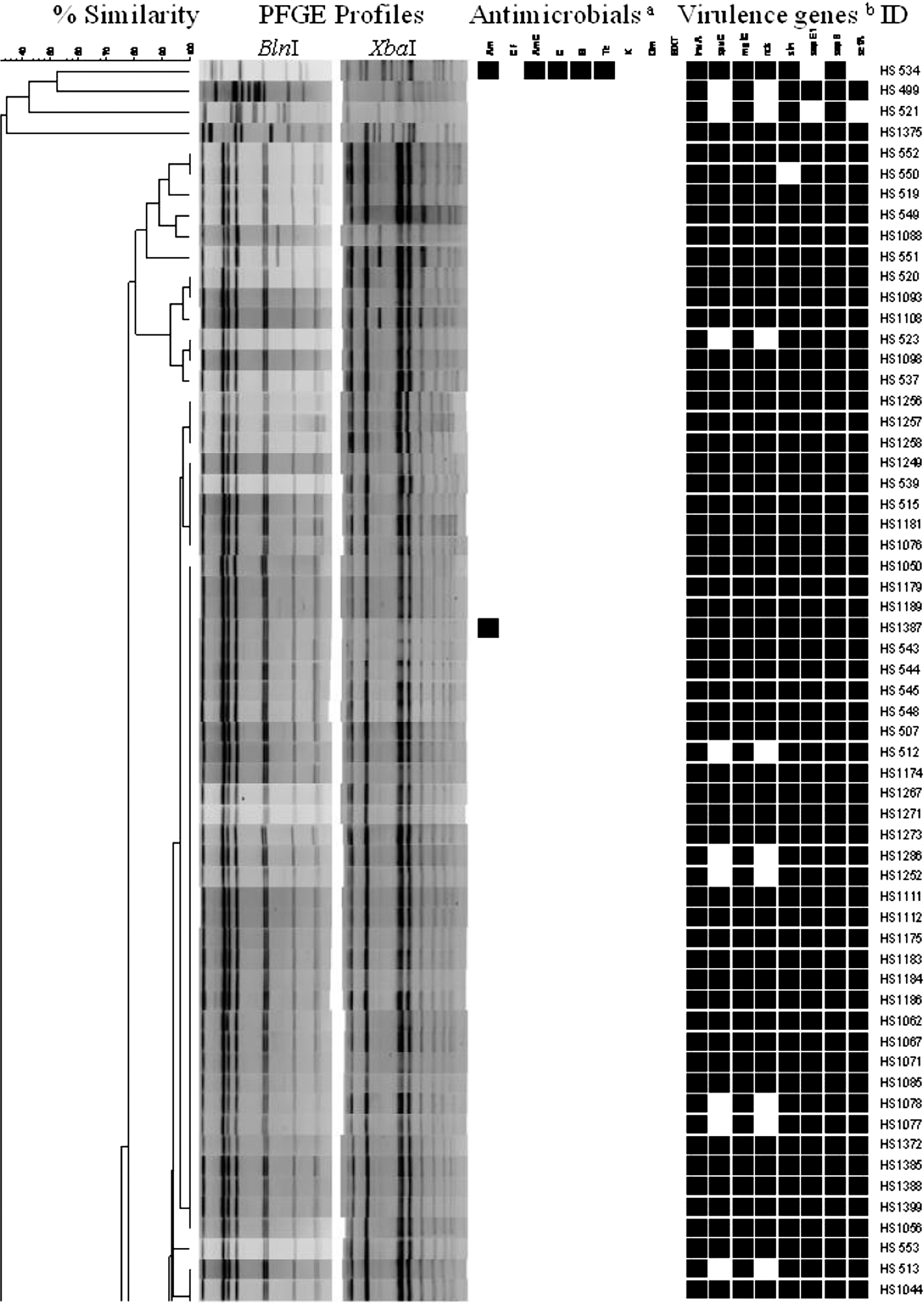
Dendrogram highlighting the pulsed-field gel electrophoresis (PFGE) profiles of XbaI and BlnI enzymes combination analysis of 118 human clinical Salmonella Enteritidis isolates.
Discussion
The recent CDC report attributes an estimated 1.0 million cases, 19,336 hospitalizations, and 378 deaths annually in United States to infections caused by nontyphoidal Salmonella serovars (Scallan et al., 2011). The predominant Salmonella serovars causing infections in humans in 2009 in the United States include S. Enteritidis (19.2%) and S. Typhimurium (16.1%) (MMWR, 2010). Contaminated egg products and foods products containing egg ingredients have been identified in both the United States and the European Union as the most common vehicles for transmission of infections caused by S. Enteritidis in humans (Frank et al., 2009; CDC, 2010). A recent multistate outbreak extending from May 2010 to November 2010 in the United States was attributed to consumption of S. Enteritidis contaminated eggs, which resulted in 1,939 reported illnesses (CDC, 2010).
Salmonellosis is usually a self-limiting disease and, as such, antimicrobial therapy is not prescribed. However, Salmonella infection caused by AR strains is a huge public health concern and a constant drain on communities. The majority of the isolates (n=409; 96.2%) in our study were susceptible to all the 12 antimicrobials tested. Only a few isolates exhibited resistance to antimicrobials, including ampicillin (2.35%), streptomycin (2.17%), and cephalothin (1.4%; Table 1). This finding is in agreement with previous reports that have described S. Enteritidis to exhibit a lower frequency of resistance to antimicrobials, including in the United States (Crump et al., 2011; NARMS, 2008), Finland (Lukinmaa et al., 2006), Senegal (Cardinale et al., 2005), and Brazil (Vaz et al., 2010). In our study, seven isolates (1.6%) were MDR isolates, which is comparable to other studies conducted in humans and animals (Nair et al., 1995; Yang et al., 2002). Although the frequency of resistance to antimicrobials in S. Enteritidis is low, continuous surveillance is important to monitor the emergence of AR strains.
Molecular characterization of AR S. Enteritidis isolates was done to determine the genes responsible for coding resistance to the different antimicrobials. Antimicrobial resistance to the aminoglycosides was predominantly coded by the strA/B genes (75%) followed by the aadA1/A2 genes (25%), while tetracycline resistance was encoded by the tet(A) (60%) followed by tet(B) (20%) and tet(G) (20%) genes as reported previously (French and Schwarz, 2000; Hur et al., 2011; Randall et al., 2004). All ampicillin-resistant strains harbored the bla TEM gene thereby concurring with published data, which describe resistance to penicillin class of antimicrobials due to production of TEM lactamase enzymes (Hur et al., 2011).
Class I integrons play an important role in the dissemination of antimicrobial resistance and reported from members of the Enterobactericeae family in humans and animals (Kang et al., 2005; Gebreyes et al., 2005). Out of seven MDR isolates, we are able to detect class I integron from a single MDR isolate with AR pattern AM-AMC-C-S-TE. Class I integrons are not widespread in S. Enteritidis (Randall et al., 2004), unlike in S. Typhimurium, which has a 1.0- and 1.2-kb class I integron integrated in its chromosome for stable transfer from one generation to the next (Vo et al., 2010; Karuland et al., 2009). Even though we did not find any resistance to third generation cephalosporins in our study isolates, presence of ESBL producing bla TEM and bla PSE genes is a growing concern towards resistance to extended spectrum cephalosporins.
The distribution of eight virulence genes that play an important role in S. Enteritidis for invasion, enterotoxin production and pathogenesis in the host were investigated. The majority of isolates harbored six virulence genes, except for the spvC and rck genes, indicating that these virulence genes are widespread in S. Enteritidis serotype. The spvC and rck genes were found in 91.3% and 89.2% of strains, respectively. These findings are in agreement with previous studies (Kwag et al., 2008; Hur et al., 2011; Huehn et al., 2010; Bolton et al., 2011), which have also reported the high prevalence of spvC and rck genes in salmonella isolates. A self-transferable virulence-resistance plasmid (pUO-StVR2) carrying the spvA, spvB, spvC, and rck gene was earlier reported in S. Typhimurium (Guerra et al., 2002). The presence of the stn gene in all the clinical isolates highlights the role of the stn gene in the production of enterotoxin, which is responsible for causing acute gastroenteritis. Although it is not possible to predict whether a particular serovar of Salmonella will cause the disease merely by the presence or absence of a few virulence genes, the high prevalence of multiple virulence genes from the clinical isolates could explain the increased potential of this serovar in causing severe infections in humans.
Due to the good discrimination, high sensitivity, and epidemiologic relevance, PFGE is recognized as a gold standard for genotyping bacterial foodborne pathogens by the CDC (Hunter et al., 2005). The most commonly used restriction enzyme for genotyping Salmonella is XbaI (Thong et al., 1995). However, due to clonality of the S. Enteritidis (Seno et al., 2004; Zheng et al., 2007), we decided to use a second restriction enzyme (BlnI) as it has been shown to attain a better DP when typing this particular serotype (Fernandez et al., 2003). The combination of these two enzymes resulted in 14 clusters and a significant increase in DP to 0.914, compared to using either XbaI (seven clusters, DP 0.715) or BlnI (seven clusters, DP 0.785). The results are in accordance with a previous study which shows that using two or more enzymes results in more discrimination of isolates (Zheng et al., 2007). Although six enzyme PFGE profile provides a better DP (Zheng et al., 2007), the combination of XbaI-BlnI PFGE profile analysis was a more powerful discriminator than either of the enzymes in this study.
Out of 118 study isolates, 80 of them matched 100% with PFGE profile of S. Enteritidis strain, which was responsible for the recent outbreak of S. Enteritidis by eggs in the United States (CDC, 2010). A restaurant in Durham, North Carolina had a recent S. Enteritidis outbreak causing gastrointestinal illness among 65 people. The Durham County Health Department (DCHD) investigated and identified the likely source of this pathogen as commercially manufactured egg white, which was used to make meringue in the restaurant. This outbreak occurred on April 23, 2010, which was concurrent with a multistate outbreak of S. Enteritidis due to contaminated shell eggs in the United States. Although we do not know the patient history in our study due to confidentiality issues, it is possible that these isolates could have originated from the above outbreak. It is important to note the effect of repeatedly isolating the same clonal strain of S. Enteritidis population (highlighted by 96.23% pansusceptible isolates and identical PFGE profiles) among human patients in North Carolina.
Conclusion
In conclusion, S. Enteritidis isolated from clinical cases in North Carolina were phenotypically and genotypically similar. Even though few isolates were resistant to some antimicrobials, none of them were resistant to ciprofloxacin and third generation cephalosporins, the two drugs of choice to treat salmonellosis. Most of the isolates were tested positive for eight virulence genes, implying that these strains have the necessary virulence gene capable of playing an important role in causing severe infection. Based on PFGE genotyping, the S. Enteritidis population isolated from humans in North Carolina is clonal. PFGE with two enzymes improved the DP of the method when compared to using a single enzyme and should be an important consideration when genotyping S. Enteritidis isolates.
Footnotes
Acknowledgments
Human Salmonella Enteritidis clinical isolates were kindly provided by the North Carolina State Laboratory of Public Health.
Disclosure Statement
No competing financial interests exist.