
Abstract
Cronobacter (formerly known as Enterobacter sakazakii) is a genus comprising seven species regarded as opportunistic pathogens that can be found in a wide variety of environments and foods, including powdered infant formula (PIF). Cronobacter sakazakii, the major species of this genus, has been epidemiologically linked to cases of bacteremia, meningitis in neonates, and necrotizing enterocolitis, and contaminated PIF has been identified as an important source of infection. Robust and reproducible subtyping methods are required to aid in the detection and investigation, of foodborne outbreaks. In this study, a pulsed-field gel electrophoresis (PFGE) protocol was developed and validated for subtyping Cronobacter species. It was derived from an existing modified PulseNet protocol, wherein XbaI and SpeI were the primary and secondary restriction enzymes used, generating an average of 14.7 and 20.3 bands, respectively. The PFGE method developed was both reproducible and discriminatory for subtyping Cronobacter species.
Introduction
C
While Cronobacter species can be regarded as ubiquitous, being cultured from a wide variety of environments and foods, powdered infant formula (PIF) has been epidemiologically linked with several cases of illness in neonates following the ingestion of contaminated product (Anonymous, 2002; Healy et al., 2010; Kim et al., 2008; Lai, 2001; van Acker et al., 2001). Since Cronobacter species were first implicated in a case of neonatal meningitis in 1958, more than 123 cases of neonatal Cronobacter spp. infection have been reported. It is likely that many cases may have been missed, because of a failure to recognize the links to food (Agostini et al., 2004; Lai, 2001; Nazarowec-White and Farber, 1997; O'Brien et al., 2009; Urmenyi and Franklin, 1961). While the incidence of reported infection is low, the mortality rates in infants who present with Cronobacter-related meningitis are high (Bowen and Braden, 2006; Drudy et al., 2006). Cronobacter infections have also been reported in adults but are generally regarded as non-life-threatening (Hawkins et al., 1991; Lai, 2001; Nazarowec-White and Farber, 1997; Ongradi, 2002; Pribyl et al., 1985).
Molecular subtyping methods are useful tools to aid our understanding of microbial epidemiology. Robust and reproducible methods can help with the detection of outbreaks through the identification of clusters, thus preventing/limiting national and international foodborne outbreaks. Pulsed-field gel electrophoresis (PFGE) is widely considered as the gold-standard method for molecular subtyping of many bacteria in outbreak investigations (Gerner-Smidt et al., 2006; Kim et al., 2008; Mullane et al., 2007b; Nazarowec-White and Farber, 1997; Proudy et al., 2008).
PulseNet International is a molecular subtyping network for the surveillance of foodborne diseases, and is currently established in five regions worldwide (Gerner-Smidt et al., 2006; Swaminathan et al., 2006). Since its establishment in 1996, this network has focused on the rapid detection, investigation, and control of local and international outbreaks. A key feature of the PulseNet surveillance program is the use of standardized PFGE protocols for inter- and intra-laboratory comparison of bacterial subtyping data (Boxrud et al., 2010; Cooper et al., 2006; Ribot et al., 2006).
The aim of this study was to develop a robust PulseNet PFGE protocol for Cronobacter species for use by all the laboratories within the network and worldwide. This method was thoroughly validated as part of a multi-center trial.
Methods
Bacterial isolates
A selection of 11 Cronobacter spp. isolates from Argentina recovered from PIF was used for the initial protocol development. For the subsequent validation process, a panel of 44 isolates was selected from the University College Dublin Cronobacter strain collection to include six species of this genus; Cronobacter condimenti was not included here, as this new species had not been described prior to the commencement of this study. The identities of these isolates were blinded to all participants. All isolates were recovered from a range of backgrounds, including clinical, environmental, and food samples (Table 1). Slants of half-strength trypticase soy agar (TSA) were inoculated with each of the isolates and distributed to participating laboratories. As required by PulseNet International protocols, Salmonella enterica serotype Braenderup H9812 was used as molecular weight standard for all PFGE gels; DNA underwent digestion reaction with the restriction enzyme XbaI (Hunter et al., 2005).
Initial protocol development: selection of restriction enzymes and electrophoretic conditions
To prepare agarose plugs for each of the Cronobacter isolates, the PulseNet One-Day (24–28 h) Standardized Laboratory Protocol for Escherichia coli O157:H7, Salmonella serotypes, and Shigella sonnei was used (Ribot et al., 2006).
An initial assessment of the suitability of existing PulseNet protocols for Shigella sonnei (Ribot et al., 2006) and Yersinia pestis (modified protocol from the Centers for Disease Control and Prevention [CDC]) were evaluated at the Instituto Nacional de Enfermedades Infecciosas-ANLIS “Dr. Carlos G. Malbrán” (INEI-ANLIS) using the XbaI restriction enzyme (Table 2). A panel of rare-cutting restriction enzymes, including NotI, SfiI, and SpeI, were tested in parallel for their suitability as a secondary enzyme, when required. The DNA profiles obtained with these different enzymes and electrophoretic conditions were evaluated and compared with particular emphasis in regard to the distribution of the digested bands and overall resolution as described in detail below.
IST, initial switch time; FST, final switch time; RT, Run time.
Descriptive steps during method validation
The first multi-center validation process aimed to assess the robustness and reproducibility of the PFGE protocol as standardized during the initial development phase. The five participating laboratories were the CDC, USA; the INEI-ANLIS, Argentina; the National Microbiology Laboratory of Health Canada, Canada; the World Health Organization (WHO) Cronobacter Reference Laboratory, University College Dublin, Ireland; and the Institute for Food Safety and Hygiene, University of Zurich, Switzerland. Participating laboratories analyzed the complete set of isolates (n=44) by PFGE (CHEF-DR III System, BioRad; CHEF Mapper XA System, BioRad) with enzymes XbaI and SpeI applying the electrophoretic conditions of the Y. pestis modified protocol with an initial switch time (IST) 1.8 s, final switch time (FST) 25 s, and running time (RT) 17–18 h. Each laboratory performed four XbaI-PFGE and four SpeI-PFGE gels, each including 11 Cronobacter strains along with four lanes containing S. Braenderup H9812 as the molecular weight standard.
Following analysis of this data set during the first validation round, it was collectively decided to further evaluate additional electrophoretic conditions, in an attempt to improve minor band resolution issues as observed in some isolates.
Four different electrophoretic conditions (Table 2) were tested at INEI-ANLIS using a subset of seven isolates, including six strains that displayed resolution issues among high molecular weight (HMW) bands and a strain that demonstrated an optimal resolution. One of these conditions was subsequently selected for further assessment in a second round of validation, as it appeared to improve band resolution.
For the final international validation using the optimized electrophoretic conditions developed in the early stage of the study, a subset of 19 strains representing different species were analyzed by all five laboratories. The resulting images were analyzed and compared at the INEI-ANLIS and at the UCD Centre for Food Safety. Data from each validation round was analyzed and compared, and optimal electrophoretic conditions used were proposed by the working group to be implemented as the subtyping protocol for Cronobacter species.
To resolve the PFGE profiles of three isolates that were recorded as non-typeable in the first round of development, four laboratories tested these strains again and added 50 μM thiourea to the electrophoresis buffer, as recommended by PulseNet and proposed by other authors (Corkill et al., 2000; Römling and Tümmler, 2000; Silbert et al., 2003).
Data analysis
PFGE profiles were subjected to computer-assisted DNA fingerprint analysis using BioNumerics version 4.6 software (Applied Maths, Sint-Martens-Latem, Belgium), which measures band position differences against DNA size standard Salmonella Braenderup H9812. Dendrograms were constructed using unweighted pair group method with averages (UPGMA), DICE coefficient, and a 1.5% band position tolerance window. The analysis of the different electrophoretic conditions was made by visual comparisons of profiles obtained for each isolate with every electrophoresis program.
To compare results obtained for each international validation round, dendrograms were constructed for each isolate comparing patterns obtained in the five laboratories. Pattern differences were identified visually and classified either as caused by resolution issues associated with closely migrating bands or as not clearly explainable differences (bands in which it was unclear whether the variation observed in different laboratories was the result of incomplete DNA restriction digestion or genetic variation between isolates, possibly related to culture or storage conditions).
Results
Initial protocol development: selection of restriction enzymes and electrophoretic conditions
The electrophoretic conditions from the Y. pestis modified protocol provided better resolution and band distribution than other methods evaluated and were thus selected for further testing using a selection of different restriction enzymes, in addition to XbaI (Fig. 1A). Enzymes SfiI and NotI generated profiles that were difficult to analyze due mainly to the concentration of the high number of bands located at the bottom portion of the gel. With the SpeI enzyme, the number and distribution of bands in the PFGE profiles appeared to be adequate for band resolution and subsequent analysis (Fig. 1B). Based on these early observations, electrophoretic conditions of the Y. pestis modified protocol, including the enzymes XbaI and SpeI, were selected for more detailed testing in the international multicenter validation trial.
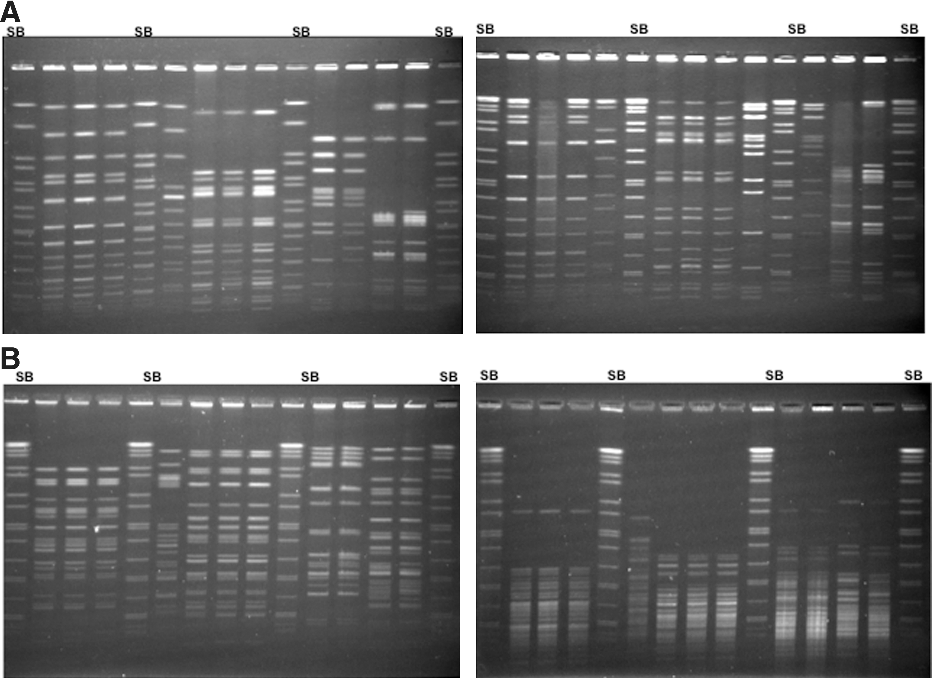
Development of pulsed-field gel electrophoresis (PFGE) protocol for Cronobacter species. Selection of electrophoretic conditions and a suitable restriction enzyme. SB, standard strain of Salmonella enterica serotype Braendeup H9812, used as a molecular weight standard.
Method validation
From the first round of the international multicenter validation trial, three of 44 Cronobacter isolates tested were non-typeable with both enzymes. Following the addition of thiourea to the running buffer, as tested in four laboratories, one of these isolates could be typed by the four participants, while the other two could only be resolved by PFGE in one of the laboratories. These isolates were not considered for further analysis, as not all laboratories were able to obtain a PFGE pattern for them. The remaining 41 isolates could be typed in the five laboratories showing between 40 and 41 unique XbaI-PFGE DNA banding profiles per laboratory, with each pattern having an average of 14.7 bands (range, 8–19). When comparing dendrograms of each isolate among laboratories, 18 showed indistinguishable patterns across all the laboratories. Among the remaining 23 isolates, minimal differences were identified when comparing DNA patterns of isolates between different laboratories, with an average of 1.1 polymorphic bands per bacterial isolate (range, 1–6). Approximately 80% of these variations were classified as being due to resolution issues mainly concentrated at the top portion of the gel. Other band differences, not clearly explainable, were found only in five of the Cronobacter isolates, and these were possibly related to incomplete restriction or genetic variation.
In the case of the SpeI enzyme, 41 PFGE patterns were resolved among the 41 isolates, in each of the five laboratories. The patterns showed an average of 20.3 bands (range, 16–26 bands). Fourteen of the isolates did not demonstrate any resolution issues and were recorded as indistinguishable by PFGE in all laboratories, with an average of 1.5 band differences per isolate (range, 1–7) being noted in the remaining 27 strains. Band resolution variations accounted for most of these differences. As with the XbaI enzyme, five isolates presented pattern differences possibly caused by incomplete restriction or genetic changes.
When the three additional electrophoretic conditions were evaluated, Programs 1 and 2 (denoted as P1 and P2), showed no relevant improvements. Conversely, Program 3 (denoted as P3) when evaluated, improved the resolution of high molecular weight bands with XbaI for the six strains that previously showed compression at these bands (Fig. 2). However, in several of these isolates, some compression in the lower sized bands was observed when using SpeI under these conditions. To address this, a fourth electrophoretic condition was tested with SpeI (denoted as P4). Data when evaluated, did not demonstrate any further improvement compared to P3 (data not shown). Thus P3 was selected for a second round of evaluation by all collaborating laboratories, and on this occasion the evaluation was carried out with a subset of 19 strains.
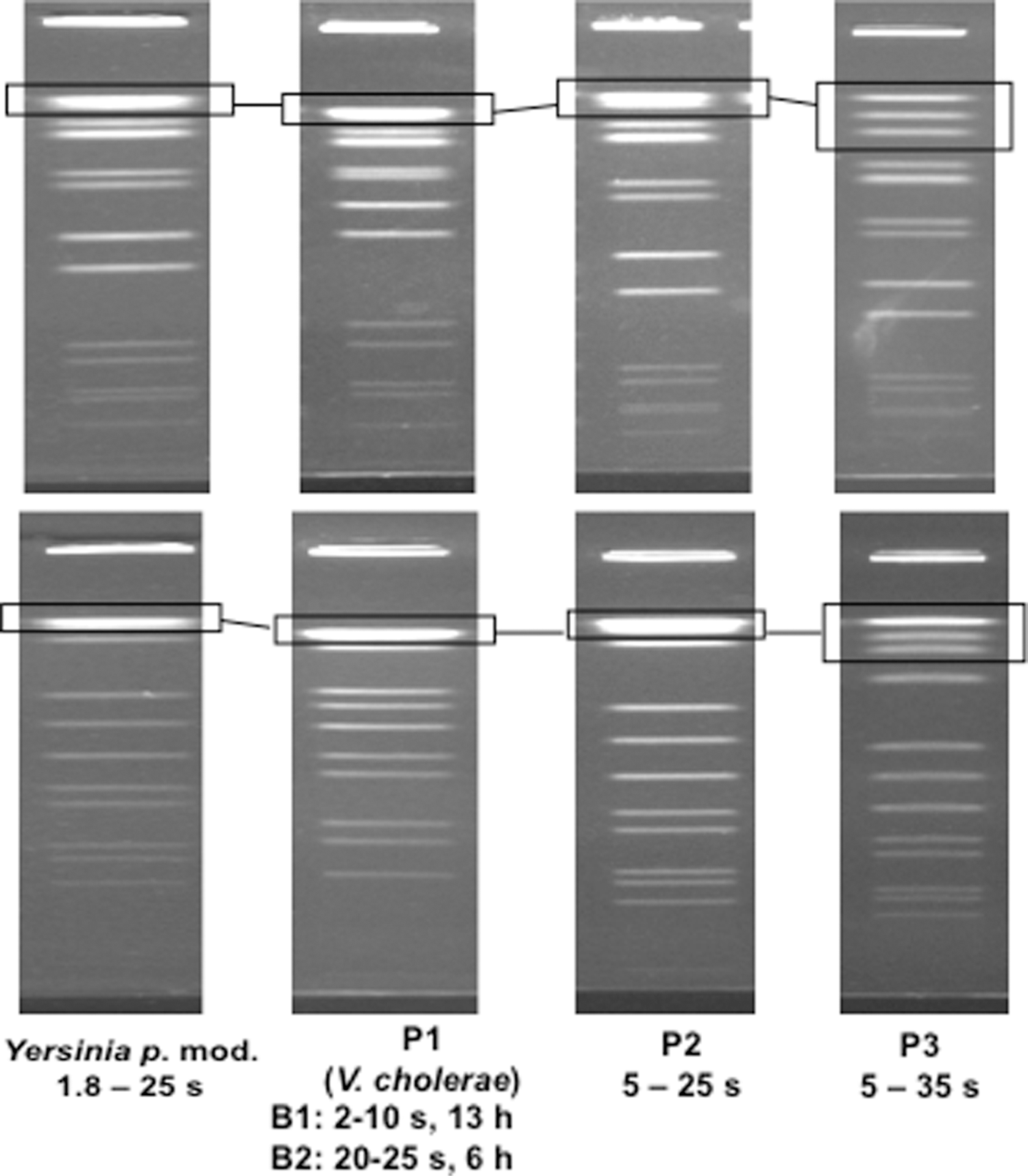
XbaI–pulsed-field gel electrophoresis (PFGE) comparison of two isolates of Cronobacter dublinensis, using the electrophoretic conditions of the modified Yersinia pestis protocol and three additional programs (denoted as P1, P2, and P3). Squares highlight the improved resolution of high molecular weight bands applying P3.
The results of the application of P3 showed that when digested with XbaI, the average number of bands per Cronobacter isolate was 15.5 (range, 10–20). Average numbers of band differences per isolate identified among laboratories was more than double compared to the previous validation round (range, 1–8), with almost 75% of these differences classified as being due to resolution issues, and affecting 15 strains. Seven isolates appeared to have different band patterns when compared among laboratories, due to other causes, not clearly explainable.
SpeI-PFGE produced profiles with an average of 18.4 bands per strain (range, 15–23). There was on average 2.10 band differences per isolate noted (range, 1–7) and which were due mainly to resolution issues.
Comparing both validation protocols (including the modified Y. pestis and the P3 electrophoretic conditions described here), the P3 protocol demonstrated a superior ability to resolve high molecular weight bands in seven of the 19 isolates tested with the XbaI enzyme and for SpeI in six of the 19 isolates. However, these improvements were not consistently reported among all the laboratories, leading to differences among the profiles obtained by these participants, while greater reproducibility was reported with the modified Y. pestis protocol (Fig. 3). In addition, the P3 conditions resulted in compression of the middle and lower sections of the DNA profiles in two isolates when digested with XbaI and three with SpeI. Table 3 summarizes the comparison of P3 and the modified Y. pestis protocol results, comparing the same set of 19 isolates analyzed during the second validation round. It can be seen that the average band difference attributable to resolution issues is higher in P3 with both enzymes, particularly with XbaI.
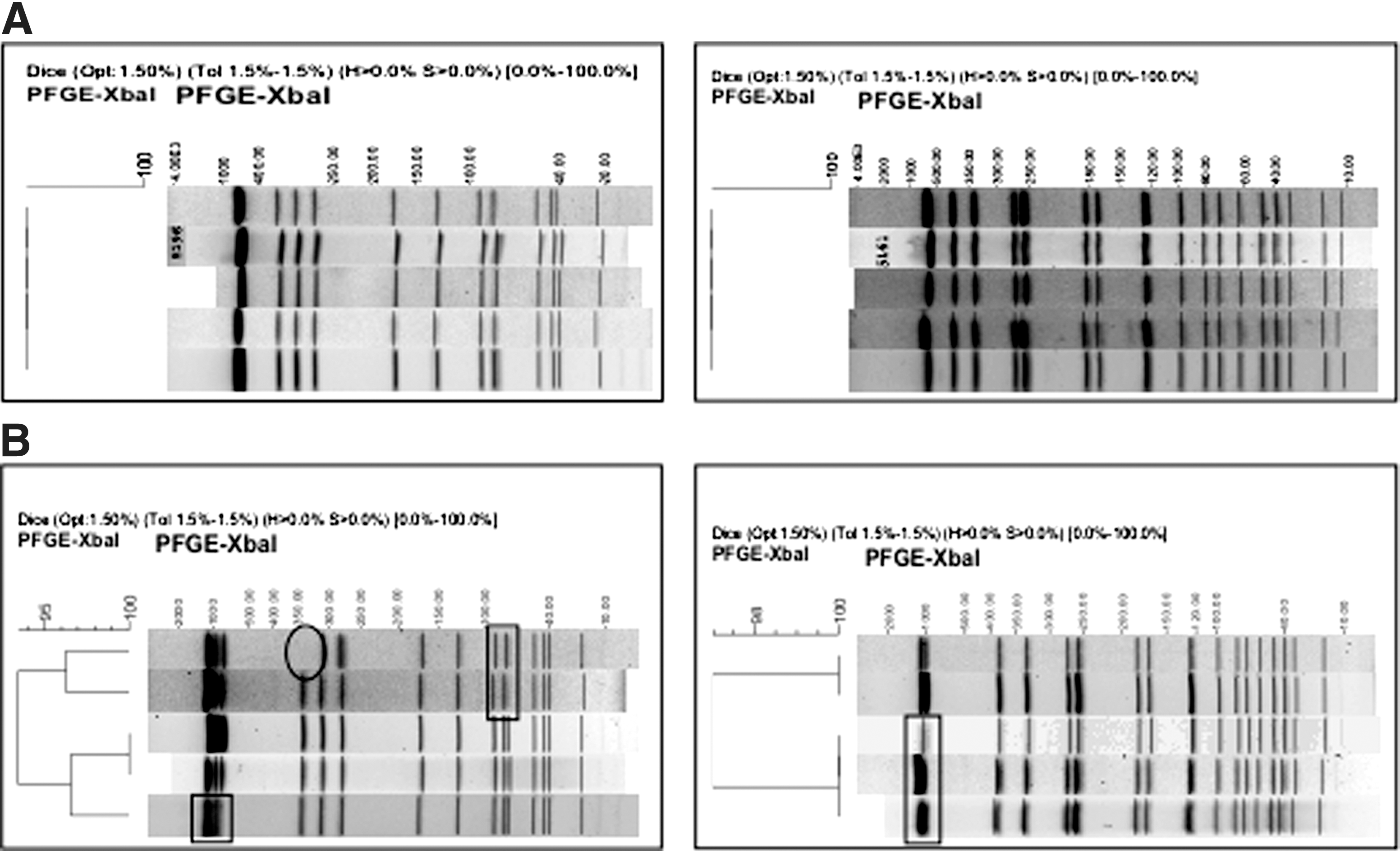
Dendograms of two strains of Cronobacter sakazakii comparing pulsed-field gel electrophoresis (PFGE) patterns among the five laboratories using the XbaI enzyme.
Classification of band differences identified among the profiles of 19 isolates analyzed in the five laboratories.
Based on these results from both validation rounds we proposed that the Y. pestis modified conditions are appropriate to use for Cronobacter species PFGE subtyping, and can be applied with XbaI and SpeI restriction enzymes (Table 4).
IST, initial switch time; FST, final switch time; RT, run time; MW, molecular weight.
Discussion
The aim of this study was to develop and evaluate a robust and reproducible PulseNet PFGE subtyping protocol for Cronobacter species for use by all the laboratories within the network and also for recommendation to other institutions for intra- and inter-laboratories comparisons.
The importance of applying discriminatory molecular subtyping methods to characterize foodborne pathogens is well known, as it facilitates the detection of outbreaks, sources of infection, and transmission pathways (Fields et al., 2011). This technique has been shown to be particularly useful in powdered infant formula production environments, where it can be used to improve surveillance and control (Mullane et al., 2007b, 2008; Proudy et al., 2008). Furthermore, this approach can also lend itself to the identification of persistent strains and to trace routes of dissemination within a PIF manufacturing plant (Mullane et al., 2007b; Terragno et al., 2009). Nevertheless, the application of multiple protocols, lacking standardization, limits the accurate comparison of results, as has been observed with other pathogens prior to the development of standardized procedures (Cooper et al., 2006; Kam et al., 2008; Ribot et al., 2006).
The Cronobacter species included in this study were recovered from different samples including clinical, food and environmental sources and represented the currently recognized species. Following the testing of a number of different electrophoretic running conditions, two were selected for a more detailed analysis, and these were included in a multi-center international validation trail. The electrophoretic condition selected as the most appropriate one was derived from a modified Y. pestis PulseNet protocol (Table 4), as this demonstrated greater reproducibility and robustness compared with P3 conditions.
Two restriction enzymes were evaluated during the processes as previous PulseNet experience has demonstrated that the use of two enzymes increases the discriminatory power of the technique (Cooper et al., 2006; Kam et al., 2008; Swaminathan et al., 2006). XbaI produced a smaller number of DNA fragments compared with SpeI. Furthermore, XbaI is more commonly available commercially and less expensive; therefore, this enzyme is to be recommended as the primary one for PFGE analysis of Cronobacter spp.
Strains from six known species of Cronobacter isolated from a range of different backgrounds were analysed in this study. Difficulties were found for the PFGE analysis of only three strains, and this arose due to degradation of DNA during the process. After the addition of thiourea to the electrophoresis buffer, this effect could be overcome (Corkill et al., 2000; Ray et al., 1995; Silbert et al., 2003).
Forty-one strains from different species could be discriminated with both XbaI and SpeI, showing highly diverse PFGE patterns at all five laboratories. Minor differences of 1–1.5 band averages for each enzyme were observed when comparing the patterns of the same strains produced by the participating laboratories. These differences were most likely attributable to differences in the types of equipment used, different batches of laboratory reagents, and user variation. Such factors have been described as inherent PFGE weaknesses and further emphasize the importance of carefully following standardized procedures for PFGE, as well as applying two enzymes when possible for a stronger validation of the relationship observed among independent isolates. Minor variations in patterns were observed in different laboratories, and this feature could reflect genetic changes that occurred during storage and/or culture passage. This issue was not evaluated further in this study; nonetheless, it has been described for other enteric bacteria (Pichel et al., 2012).
Conclusion
The method described was derived from a modified Y. pestis PulseNet protocol with modifications: XbaI and SpeI as primary and secondary restriction enzymes respectively, under electrophoresis conditions of initial switch time 1.8 s to a final switch time of 25 s, at 6 volts/cm. This validated PulseNet PFGE protocol for subtyping Cronobacter spp. isolates, was demonstrated to be robust and, reproducible and could be used for the surveillance and investigation of this pathogen.
Footnotes
Acknowledgments
We gratefully acknowledge the collaboration of Dr. Peter Gerner Smidt, Centers for Disease Control and Prevention, USA, and Dr. Enrique Pérez Gutierrez from the PanAmerican Health Organization/World Health Organization, for their support and advice throughout the development of this PulseNet protocol.
Disclosure Statement
No competing financial interests exist.