
Abstract
A suspension array assay was developed for molecular serotyping of the seven most prevalent Shiga toxin–producing Escherichia coli (STEC) serogroups (O26, O45, O103, O111, O121, O145, and O157). Fluorescence values of 59 STEC were 30 to >270 times greater than the signals of negative controls, demonstrating the method's effectiveness for the molecular serotyping of STEC.
Introduction
S
Molecular methods have been developed for serotyping Salmonella and E. coli as an alternative for traditional serotyping. These methodologies target serogroup genes (wzx and/or wzy) that encode serogroup-specific proteins forming the O antigen of Gram-negative bacteria (Samuel and Reeves, 2003). When specific sequences are detected, a serotype is attributed to the tested isolate. However, most assays are not suitable for the identification of multiple targets in a single reaction, or for the application to a large number of samples. We explored a bead-based suspension array (Bio-Plex™) that allows discrimination of the seven STEC serogroups in a single reaction. In this assay, nucleic acids are linked to beads and hybridized to the target of interest (Dunbar, 2006), followed by a detection using a flow cytometry–like device that identifies the beads and quantifies the interaction with the target.
Materials and Methods
Sequences of wzx and wzy genes from GenBank were used for designing serogroup-specific primers and capture probes. Since STEC identification requires the identification of stx 1 /stx 2 genes, primers and capture probes for stx 1and stx 2 were also incorporated in the assay (Table 1). Simulations for multiplex PCR and hybridizations were performed using Visual OMP software (DNA Software Inc., Ann Arbor, MI). Biotinylated primers and probes were from Biosearch Technologies (Novato, CA). The best panel (Table 1) was selected based on probe performance.
Primers and probes were purchased from Biosearch Technologies.
stx1 FWD and REV described by Paton and Paton (1998).
stx2 REV described by Cebula et al. (1995).
FWD, forward primer; REV, reverse primer.
A total of 103 bacterial strains were used to evaluate the panel specificity, including 59 STEC (4–11 isolates for each of the seven STEC serogroups), 23 of other E. coli serogroups, and 21 of non-E. coli species including four Shigella spp., four Salmonella serotypes, Proteus vulgaris, Pseudomonas aeruginosa, Enterobacter aerogenes, Citrobacter freundii, Hafnia alvei, Enterobacter cloacae, Acinetobacter baumannii, three Listeria spp., Streptococcus faecalis, and Bacillus subtilis. DNA was extracted from pure culture, and standardized at 80 ng/μL. For multiplex polymerase chain reaction (PCR), bacterial DNA (1 μL), HotStarTaq Plus Master mix (12.5 μL) (Qiagen, Valencia, CA), 40 nM of forward primers and 80 nM biotinylated reverse primers, and water were mixed to reach a final reaction volume of 25 μL. The PCR parameters included initial denaturation at 95°C for 5 min, 30 cycles of 94°C for 30 s, 56°C for 90 s, 72°C for 90 s, and a final elongation at 72°C for 10 min.
A previously described method was applied for probe conjugation (Fulton et al., 1997) where polystyrene microbeads (Bio-Rad, Hercules, CA) were linked to probes with an amino C12 spacer on the 5′ end modification. For probe hybridization, individual coupled beads were diluted 1:100 in 1.5x TMAC buffer (1.5 M tetramethylammonium chloride, 75 mM Tris, 6 mM EDTA, and 0.15% sarkosyl at pH 8.0) to create a bead pool, and 33 μL of the bead mixture were added to 5 μL of PCR product and 12 μL of TE buffer followed by a 15-min incubation at 55°C. Immediately, fluorescence detection was performed using Bio-Plex 200 ™ (Bio-Rad) at 55°C (Fitzgerald et al., 2007). Median fluorescence intensity (MFI) from 100 beads per region per sample was recorded, and P/N ratios (MFI/negative control MFI) were calculated using Bio-Plex software. Data were analyzed using analysis of variance with SAS software (9.2v).
Results and Discussion
The MFI values of positive samples were significantly greater (p<0.05) than those of negative samples in each serogroup. MFI values for positive samples ranged from 6500 to 19,000, whereas the average for negative samples was 140 (Fig. 1). The results were similar to those for a Salmonella serotyping panel (Fitzgerald et al., 2007). Average P/N ratios for positive samples/targets ranged depending on target: 32.8±2.49 for O26 (n=11), 127.6±9.91 for O45 (n=5), 53.0±2.83 for O103 (n=11), 144.6±16.91 for O111 (n=11), 207.0±6.53 for O121 (n=4), 153.7±3.89 for O145 (n=6), 270.9±11.03 for O157 (n=11), 121.5±19.49 for stx 1 (n=48), and 30.7±1.83 for stx 2 (n=18). The average P/N ratio for nonspecific interactions was 1.08. Since nonspecific interactions never gave P/N values over 13, a cutoff ratio of 15 was used for serogroup identification.
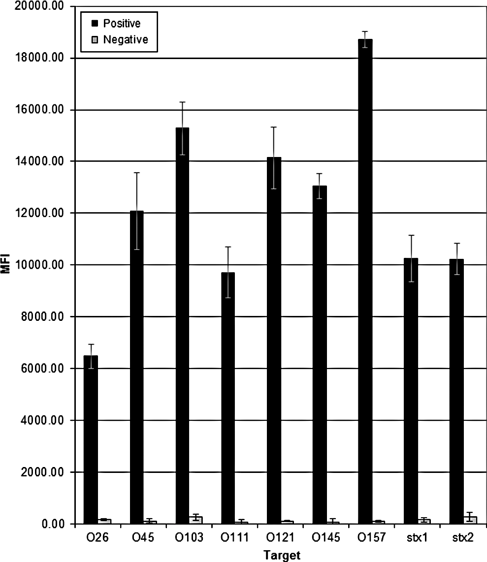
Average Median Fluorescence Intensity (MFI) by target in suspension array assay for Shiga toxin–producing E. coli. Bars represent average of Median Fluorescence Intensity (MFI) for strains analyzed in a serogroup. Lines represent standard deviation.
None of the 23 non-STEC E. coli and none of the 21 non-E. coli strains gave a positive reaction (P/N ratio ≥15) for any of the probes tested, except for Shigella dysenteriae, which was tested positive for stx 1 as it carried the gene. An O26 strain showed a P/N ratio of 13 for O45 probe, and an O145 strain gave a ratio of 11 for O111 probe. However, these values did not interfere with serogroups identification because they were much lower than those of typical positive samples. Therefore, sensitivity and specificity values were 100% for all the targets.
Molecular methods for serotyping of E. coli have been developed for important serotypes of STEC (Fratamico et al., 2009). A recent study using a suspension array was able to detect 10 STEC serogroups but did not incorporate stx 1 and stx 2 (Lin et al., 2011). Since these virulence markers define an E. coli strain as STEC, they were included in the present study. Our test panel provided a fast, reliable, and improved alternative for the identification of important STEC, and can be useful to better understand the epidemiology of STEC infections and enhance outbreak investigations.
Footnotes
Acknowledgments
We thank Drs. Pina Fratamico and Peter Feng for providing some STEC strains for this study, and to Dr. Lydia Rump for her thoughtful comments. We are grateful to Dr. Frank Siewerdt for advising in the statistical analysis. The study was supported in part by funding from the Joint Institute for Food Safety & Applied Nutrition (JIFSAN) at the University of Maryland.
Disclosure Statement
No competing financial interests exist.