
Abstract
This article presents the major differences in the exoproteomes of Listeria monocytogenes strains grown at 11°C and 20°C, and their comparison to 37°C, the optimal temperature of growth of this foodborne pathogenic bacteria. A set of four strains previously characterized and representing the genetic diversity of the species was used. Two were virulent, of which one was persistent, and two were low virulent strains. The proteins secreted by the strains grown in minimal medium were separated by sodium dodecyl sulfate–polyacrylamide gel electrophoresis and identified by liquid chromatography tandem mass spectrometry. The heterogeneity among the four strains concerning the 15 major proteins detected was noticed. No clear association of exoproteome with virulence or genotype was found. Cluster analysis of the protein patterns of the strains suggests an increasing differentiation of strain response with low temperatures, highlighting the importance of the study of the exoproteomes. The main finding was the lack of some proteins in the exoproteome of the persistent strain, namely, flagellin (FlaA) and of OppA/oligopeptide ABC transporter, when compared to the other strains. In fact, these two proteins differ in abundance between strains grown at low temperature. Moreover, FlaA was the only glycoprotein identified in the exoproteomes. An attempt is made here to assess the relevance of the major exoproteins differentially detected. The investigation of the exoproteomes of other persistent and sporadic strains will allow identification of proteins involved in adaptation of particular L. monocytogenes strains to low temperatures in use throughout the food chain.
Introduction
L
Several investigators have demonstrated that certain strains of L. monocytogenes may become established in particular food-processing facilities, integrating the resident microbiota for months or years in a way that suggests the presence of niche-adapted bacteria (Waak et al., 2002; Leite et al., 2006; Wulff et al., 2006; Keto-Timonen et al., 2007; D'Amico and Donnelly, 2008). Refrigeration is a widespread process used in the food chain to extend product shelf-life as it is the most common hurdle to microbial growth, but L. monocytogenes is able to grow at low temperatures (Tasara and Stephan, 2006).
The bacterium extracellular proteome or exoproteome, an important subset of the total proteome, is marked by its functional nature, undergoing variations and adaptations according to the bacterial surrounding conditions in each moment (Conte et al., 2000; Lee and Schneewind, 2001; Midelet-Bourdin et al., 2006). Altered expression of multiple cytoplasmic proteins was detected in a strain of L. monocytogenes grown at low temperature (Cacace et al., 2010). At the host temperature (37°C), some studies reported differences in extracellular proteins among several strains of this species (Dumas et al., 2008; Dumas et al., 2009; Cabrita et al., 2010). However, to our knowledge, no studies performed at low temperatures have yet examined the exoproteomes of different L. monocytogenes strains.
In the present work, four strains representing three of the four evolutionary lineages (I, II, III, and IV) of L. monocytogenes (Ward et al., 2008) were used to search for differences in the corresponding low temperatures (11°C and 20°C) that induced exoproteomes. The methodology used in this study constitute a relatively rapid and an accurate approach to better characterize the behavior of L. monocytogenes strains under low-temperature conditions.
Materials and Methods
Bacterial strains
The four L. monocytogenes strains used were the following: strain 3006 (CIP 78.39=ATCC 19116; serovar 4c), lineage III; strain 3049 (serovar 1/2a), lineage II; strains 3077 (serovar 4b) and 3993 (serovar 4d/4e), lineage I. The virulence potential of these strains was previously evaluated (Cabrita et al., 2004; Neves et al., 2008; Cabrita et al., 2010) as virulent (3049 and 3077) and low virulent (3006 and 3993). Moreover, clones of strain 3077 were persistently collected from three different dairies over a 1-year period (Leite et al., 2006). Cultures were maintained on Tryptic Soy Broth (TSB, Difco, Detroit, MI) containing 15% (vol/vol) glycerol (Merck, Darmstadt, Germany) and stored at −80°C until use.
Bacterial culture supernatants and protein precipitation
Bacterial culture supernatants were obtained as previously described (Cabrita et al., 2010). Briefly: strains were cultured in minimal medium (Modified Welshimer Broth [MWB]) (Premaratne et al., 1991) at 11°C or 20°C, with shaking (120 rpm), until reaching exponential growth phase, according to the growth conditions used (A600≈0.5), and were then removed by centrifugation (3000×g, 10 min, 4°C). After filtering the supernatant through Millex-GP filters, with 0.22-μm Millipore Express Membranes (Millipore, Carrigtwohill, Ireland), 0.2 mM phenylmethanesulfonyl fluoride (PMSF) (AppliChem, Darmstadt, Germany) was added.
L. monocytogenes proteins present in supernatants were precipitated, solubilized, and protein concentration was determined as previously described (Cabrita et al., 2010).
Sodium dodecyl sulfate–polyacrylamide gel electrophoresis (SDS-PAGE) and glycoprotein detection
For each tested growth temperature and strain, three biological replicates were performed. For each biological assay, samples were subjected to three independent SDS-PAGE experiments (technical replicates). Samples containing 50 μg total exoprotein were dissolved in sample buffer and submitted to SDS-PAGE as described previously (Cabrita et al., 2010).
For visualization of glycoproteins, SDS-PAGE gels were stained following a modification of the periodic acid–Schiff method, using the GLYCOPRO glycoprotein detection kit (Sigma, St. Louis, MO) according to the manufacturer instructions.
Protein sample preparation for liquid chromatography tandem mass spectrometry
Sixty three gel bands, corresponding to 21 proteins detected in supernatants of the cultures at 11°C and consistently present among triplicates, were manually excised from the gels, extracted and digested with trypsin according to Perrot et al. (2007). Ten μL of digest was used for mass spectrometric analysis. Liquid chromatography tandem mass spectrometry was performed using a setup of a trapping 300SB C-18 column (0.3×50 mm) (Agilent Technologies, Basel, CH) and a separating column (0.1 mm×10 cm) packed with Magic 300 Å C18 reverse-phase material (5-μm particle size, Michrom Bioresources, Inc., Auburn, CA). The column efluent was continuously directed into an Orbitrap FT hybrid instrument (Thermo Finnigan, San José, CA). The eluting peptides were analyzed in a data-dependent fashion. The precursor scan was done in the Orbitrap set to 60,000 resolutions, while the fragment ions were mass analyzed in the LTQ instrument. The five most intense precursors ions were selected for fragmentation. The MS/MS spectra were searched with Mascot (Perkins et al., 1999) with the following parameters: (1) National Center for Biotechnology Information (NCBI) nonredundant 2009 databank; (2) two maximum missed cleavages; (3) fixed modifications, carbamidomethylation (C); (4) variable modifications, oxidation (M); (5) peptide mass tolerance up to 10 ppm; (6) fragment mass tolerance up to 0.5 Da; (7) ions score cutoff, 25; and (8) maximum p-value, 0.05. The results were obtained with Mascot version 02.02.2004 by setting the taxonomy to “Other Firmicutes.”
Regarding the 75 gel bands, corresponding to 25 polypeptides detected in supernatants of the cultures at 20°C, the protocol used was according to Santos et al. (2010). Protein identification experiments were carried out on a hybrid quadrupole/linear ion-trap mass spectrometer (4000 QTrap, ABSiex) using a nano-electrospray source and a dual-gradient pump (Ultimate 3000, Dionex). Peptides were eluted into the mass spectrometer using an Onyx Monolithic C18 column (0.1×150 mm, 130 Å, Phenomenex) and a binary gradient (1 μL/min 2% ACN, 0.1% FA to 98% ACN, 0.1% FA in a multiple-step gradient for 42 min) (Ultimate 3000, Dionex). Peptide identification was performed with Mascot and Protein Pilot software (v2.0.1, ABSciex) against the Swiss-Prot and NCBInr databases. Search parameters included β-mercaptoethanol modification of cysteine and oxidation of methionine as fixed and variable modifications, respectively. Mascot search parameters also included peptide and fragment tolerance of 0.8 Da, and up to one miss-cleavage was allowed. Positive identifications were considered in Protein Pilot when the protein score was above 1.3 (95%) and in Mascot when at least one peptide had a score above identity threshold. Protein identification based on a single peptide hit had a minimum individual score of 95% and a minimum sequence tag of three amino acids (four consecutive peaks in the MS/MS spectrum). Protein identification was only considered when the first significant match results were equal in at least two out of three independent biological experiments.
Transmission electron microscopy (TEM)
For TEM, 5 μL of bacterial cultures, grown at 11°C as described before (A600≈0.5), were adsorbed onto carbon-coated collodion film supported on 200-mesh copper grids and observed in a Jeol JEM 1400 120 kV transmission electron microscope (Tokyo, Japan).
Image scanning, protein quantification, and data analysis
Electronic images of the SDS-PAGE gels were acquired with a calibrated densitometer ImageScanner (Amersham Biosciences, Buckinghamshire, UK) operating with the software LabScan (Amersham Biosciences, Buckinghamshire, UK).
Exoproteome patterns were analyzed using program GelCompar II version 5.1 (Applied Maths, Kortrijk, Belgium). For cluster analysis, the unweighted-pair group matching algorithm was used with an optimization value of 0.0% determined by the program. The levels of similarity were based on the Pearson product–moment correlation coefficient (Pearson correlation).
Protein relative quantification was performed by peak area calculation, according to Silva et al. (2006) with the software Proteome Discoverer (Thermo Scientific, San José, CA), calculating the area of the protein as the average of the three most abundant distinct peptides identified for the protein. Only peptides with different sequences were considered distinct. The area of the protein divided by the total area of all correspondent proteins was used for quantification by calculating means±standard deviation (n=3). The normal distribution and the homogeneity of variance of the data were confirmed, and the significant differences between the amounts of each selected protein were determined using one-way analysis of variance with Tukey's honest significant difference multiple comparison test (α=0.05) by running the program STATISTICA for Windows, version 7 (StatSoft, Inc., Tulsa, OK).
Results
SDS-PAGE and clustering analysis of L. monocytogenes exoproteins
Figure 1 shows the SDS-PAGE profiles of the polypeptides detected in the supernatants of the four strains grown at 11°C and 20°C and compared to the ones from 37°C, respectively. Corresponding biological replicates at 11°C and 20°C are shown in Supplementary Figs. S1 and S2, respectively (Supplementary Data are available online at

Sodium dodecyl sulfate–polyacrylamide gel electrophoresis profiles of the polypeptides detected in the supernatants of the four Listeria monocytogenes strains grown at different temperatures. Three biological replicates were performed for each strain and temperature (see Supplementary Fig. S1). Results from 37°C are from our previous publication (Cabrita et al., 2010) and are shown here only for comparison. Lanes: M, molecular mass marker (14.2–66 kDa; Sigma, St. Louis, MO); 1–4 corresponds to polypeptide profiles from strains 3006, 3049, 3077, and 3993 grown at each indicated temperature. The 46 polypeptide bands (25 at 20°C and 21 at 11°C) marked were identified as 15 proteins as follows:
Clustering analysis of the corresponding polypeptide patterns is presented in Figure 2. At about 56.5% similarity, these patterns were clustered in two groups: the exoproteomes of the four strains at 37°C (90.1%) and at 20°C (75.7%), respectively. Protein profiles from strains grown at 11°C were also categorized into these two groups: proteins from strains 3006 and 3049 clustering with the 37°C subgroup; proteins from strains 3077 and 3993 clustering with the 20°C subgroup (Fig. 2).
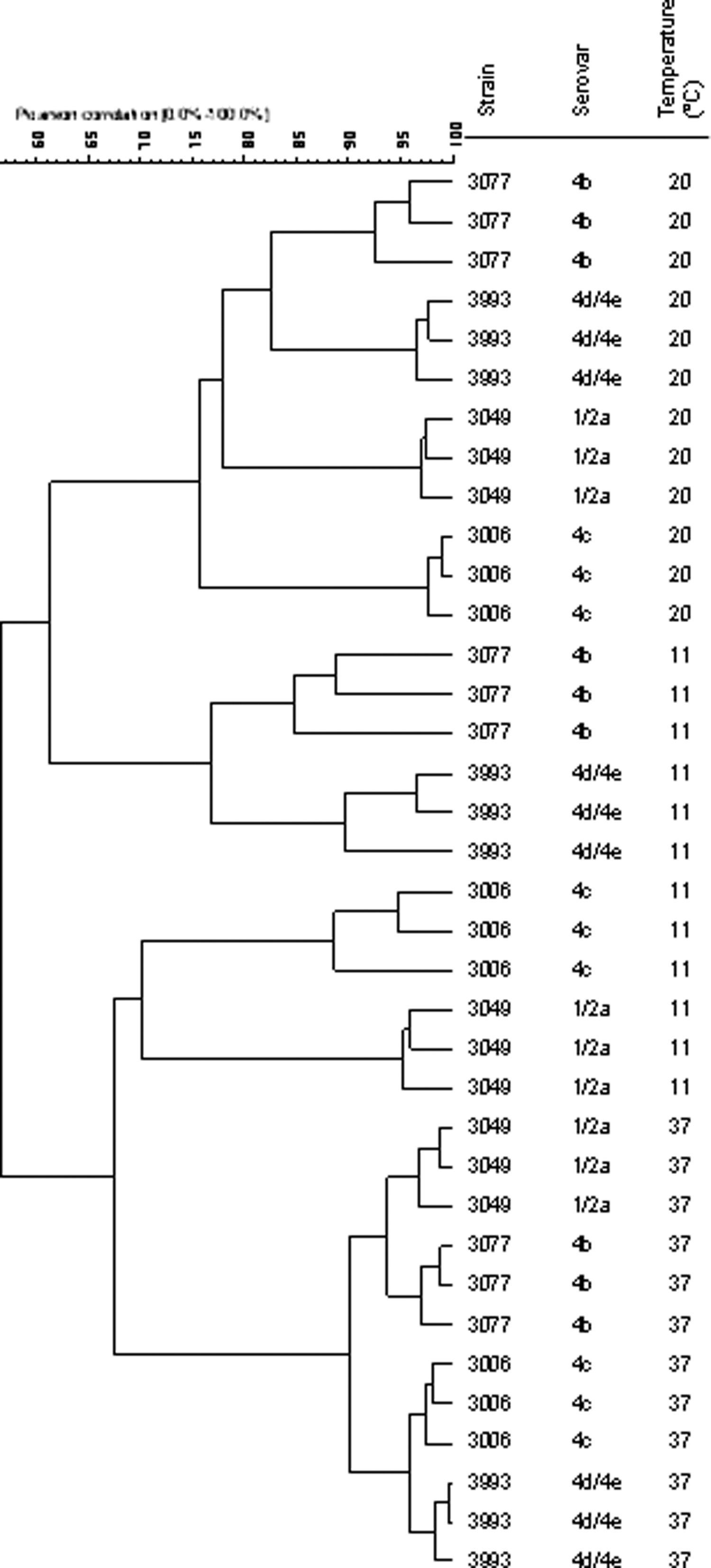
Dendrogram (unweighted-pair group matching algorithm clustering based on Pearson product–moment correlation) of the sodium dodecyl sulfate–polyacrylamide gel electrophoresis profiles of the polypeptides detected in the supernatants of the four Listeria monocytogenes strains grown at 11°C, 20°C, and 37°C. Three biological replicates were performed for each strain and temperature. GelCompar version 5.1 was used with optimization values of 0% for densitometric curve comparisons. The references of the isolates as well as their serovars are indicated.
L. monocytogenes exoproteome protein identification and TEM analysis
At 11°C and 20°C, 46 major polypeptides were selected among the four exoproteomes (Fig. 1) and subsequently identified as corresponding to 15 different L. monocytogenes proteins: 12 at 11°C (Table 1) and 14 at 20°C (Supplementary Table S1). Proteins with secretion signal and without secretion signal were detected (Table 1). Regarding the first ones, two main functional categories were recognized: specific virulent factors (listeriolysin O, at 20°C, Supplementary Table S1) and cell envelope and cellular processes (nine proteins at 11°C and one at 20°C). Concerning proteins without secretion signal, two main categories were detected: glycolysis (enolase and Gap protein) and detoxification and adaptation to atypical conditions (Lmo0927) (Table 1). At 20°C, one predictive protein (LMFG_00543) was detected in the supernatant of strain 3006 (Supplementary Table S1).
Band identification (Fig. 1): Bands “06” and “93” and “49” and “77” belong to the low virulent strains 3006 and 3993, and to the virulent strains 3049 and 3077, respectively.
Functional category (bold) and subcategory (underlined) of the proteins according to
“Accession no.” refers to the UniProtKB accession number.
The theoretical Mr is given.
Average e-value: each peptide match has an expectation value (e-value) which is the number of times we would expect to obtain an equal or higher score, purely by chance. The lower this value, the more significant the result. The “Average e-value” is the average of the matched peptides e-values.
The sequence coverage is expressed as a percentage of the complete protein sequence.
Results (20°C): + means that an equivalent protein was identified when the strain was grown at 20°C. Data concerning to parameters and results from database searches are listed in Supplementary Table S1.
The most represented functional category, cell envelope and cellular processes, may be further divided into three subcategories: cell wall processes (p60, P45, a NLP/P60 family protein and a LysM domain protein) (Table 1); transport/binding proteins and lipoproteins (OppA, oligopeptide ABC transporter, CD4+ T-cell stimulating antigen) (Table 1), and Lmo0135 (Supplementary Table S1); and mobility and chemotaxis (FlaA and cap protein [FliD]) (Table 1).
Protein OppA was detected in the supernatants of strains 3006 and 3049, at 11°C and 20°C. The very closely related oligopeptide ABC transporter (difference of only one amino acid) was detected in the supernatants of strain 3993 at 11°C and 20°C. Nevertheless, bands with similar size were not detected in the supernatants of strain 3077 grown at the three tested temperatures (Table 1 and Fig. 1). At 11°C, the relative concentration of OppA protein from strain 3049 was significantly higher (p<0.05) than the corresponding protein from strains 3006 and 3993 (data not shown).
Flagelin (FlaA) was not detected in strain 3077 supernatant at any tested temperature. At 11°C, the relative concentration of FlaA from strain 3049 was significantly higher (p<0.05) than the concentration of the FlaA from strains 3006 and 3993 (data not shown). A glycoprotein assay confirmed that FlaA is glycosylated (Renier et al., 2010), and it was the only glycoprotein detected in the exoproteome of L. monocytogenes cultured at 11°C (Supplementary Fig. S3).
Additionally, TEM results clearly showed that, at 11°C, strain 3049 is heavily flagellated (Supplementary Fig. S4II), compared with the poorly flagelatted 3077 strain (Supplementary Fig. S4III), while the other strains (3006 and 3993) exhibited intermediate levels of flagellation, at the same temperature (Supplementary Fig. S4I and IV, respectively).
Discussion
Tolerance to low temperatures is an important feature of L. monocytogenes, strongly contributing to the dissemination of the pathogen throughout the food chain. The importance of extracellular proteins in environmental adaptation has been reported in L. monocytogenes (Midelet-Bourdin et al., 2006; Soni et al., 2011). The temperature of 11°C was chosen as one at which growth is not excessively slow, but also because it is an abusive refrigeration temperature occurring more often than would be expected in the food chain (including in the consumer's home). The temperature of 20°C is a room temperature in most food industries also corresponding to the saprotrophic existence of the bacteria. In opposition, 37°C is the optimal temperature for growth and the temperature of the host (parasitic way of life).
The visible exoproteome heterogeneity detected among the four strains (Fig. 1) is particularly noticeable in the range of 29–66 kDa, where the absence or presence of specific polypeptides is not consistent among strains and/or growth temperatures. However, the profiles obtained at 11°C and 20°C are clearly distinct from those previously observed in our group, with the same strains growing at 37°C (Cabrita et al., 2010) (Fig. 1). In fact, unlike growth at 37°C, growth at 20°C resulted in clustering together virulent and low virulent strains (3049 and 3006 in one group, 3077 and 3993 in another) (Fig. 2). Moreover, the strains 3049 and 3006, representative of genetic lineages II and III, respectively, clustered together at 11°C and 20°C, supporting our previous suggestion (Cabrita et al., 2010) on a lack of correlation between exoproteome patterns and phylogenetic lineage. Moreover, the levels of similarity of the polypeptide profiles of the strains, decreased with decreasing growth temperature, suggesting an increasing differentiation of strain response with low temperatures.
The most represented functional subcategory of proteins detected in the exoproteome of L. monocytogenes grown at 11°C and 20°C are those involved in cell wall processes, with the four proteins identified (Table 1) presumably exhibiting peptidoglycan hydrolase activity. The transport/binding proteins and lipoproteins functional subcategory of L. monocytogenes exoproteome was represented by four proteins, with three of them (OppA/ABC transporter [Table 1] and Lmo0135 [Supplementary Table S1]) possibly involved in oligopeptide transport. ABC transporter, an oligopeptide-binding protein found in strain 3993 exoproteome (Table 1), exhibits 99% similarity to the OppA protein of strains 3006 and 3049. The peptide-binding protein OppA mediates oligopeptide transport to the inside of the bacterium and is essential for L. monocytogenes growth at 5°C, as oppA deletion mutant failed to grow at this temperature (Borezee et al., 2000). In fact, at 37°C, no OppA/ABC transporter protein was detected (Cabrita et al., 2010), but at lower temperatures (11°C and 20°C) OppA was detected in the supernatants of all strains except strain 3077. Nevertheless, strain 3077 was able to grow at 5°C in MWB, and the presence of the oppA gene was confirmed in the four strains (data not shown). In Salmonella Typhimurium and Escherichia coli, the Opp systems, including OppA and MppA, respectively, are involved not only in nutrient uptake but also in recycling cell wall peptides for the synthesis of new peptidoglycan (Goodell and Higgins, 1987). Since MWB is a minimal medium, under these conditions, the peptides released from peptidoglycan turnover could be the major substrates of OppA. The cold induction of OppA is likely to counteract the low diffusion rate of solutes at low temperature, thus enabling the efficient uptake of peptides. Since the strains presented no significant differences in specific growth rates at the tested temperatures, our results suggest that some strains of L. monocytogenes do not need to increase their OppA production for growth at this temperature.
Enolase and glyceraldehyde 3-phosphate dehydrogenase (Gap), encoded by the eno and gap genes, respectively, were detected in strains 3006, 3049, and 3993 at low temperatures. In fact, according to Cacace et al. (2010) they present increased expression levels at low temperature (4°C). These proteins, although lacking an N-terminal signal peptide, are reported to be exposed on the bacterial cell surface or present in the extracellular milieu, and could thus be secreted by uncharacterized secretion pathway(s) in L. monocytogenes (Glaser et al., 2001; Schaumburg et al., 2004; Trost et al., 2005; Desvaux and Hebraud, 2006).
L. monocytogenes strains are motile and flagellated below 30°C. In fact, previous analysis of the proteins secreted at 37°C by the same strains failed to demonstrate the presence of flagellin (Cabrita et al., 2010). When the strains were grown at 11°C and at 20°C, with the single exception of strain 3077, flagellin was detected in all the exoproteomes, although the amounts differed widely among strains. The TEM results (Supplementary Fig. S4I–IV) suggest a clear association between flagella number and amount of flagellin in the exoproteome, as strain 3049 was heavilly flagellated (Fig. 1 and Supplementary Fig. S4).
Conclusions
The results obtained in this work indicate a differentiation of the exoproteome of L. monocytogenes strains in response to temperature. The main finding was the nonappearance of some proteins in the supernatant of the persistent strain grown at low temperatures. It would be important to investigate low-temperature exoproteomes of other persistent and sporadic strains, in order to identify key secreted proteins involved in adaptation of particular strains.
These strains will end up more easily in refrigerated ready-to-eat foods, consequently threatening public health.
Footnotes
Acknowledgments
The authors thank Rui Fernandes (Institute for Molecular and Cell Biology, University of Porto, Portugal) for the TEM images, and INIAV, IP for allowing Paula Cabrita's stay at Laboratório de Microbiologia (CBAA/DRAT), ISA. The authors also thank Fundação para a Ciência e a Tecnologia (Project REEQ/348/AGR/2005) and Fundação Marquês de Pombal for financial support.
Disclosure Statement
No competing financial interest exist.
References
Supplementary Material
Please find the following supplemental material available below.
For Open Access articles published under a Creative Commons License, all supplemental material carries the same license as the article it is associated with.
For non-Open Access articles published, all supplemental material carries a non-exclusive license, and permission requests for re-use of supplemental material or any part of supplemental material shall be sent directly to the copyright owner as specified in the copyright notice associated with the article.