
Abstract
Listeria monocytogenes is difficult to control in food and processing environments due to its widespread nature and ability to survive in a range of adverse conditions, including low temperatures, pH, and high salt concentrations. The objective of this study was to evaluate the efficacy of Photohydroionization™ (PHI; RGF Environmental Group, Inc., Riviera, Beach, FL), a novel advanced oxidation technology, as a surface treatment to control L. monocytogenes on food-contact surfaces, sliced American cheese, and ready-to-eat (RTE) turkey. A five-strain cocktail of L. monocytogenes was used to inoculate sample surfaces. Food-contact surfaces were exposed to ultraviolet and other oxidative gases produced by the PHI system for 10, 20, 30, 45, 60, and 120 s and 5, 10, and 15 min; cheese and turkey samples were treated for 30, 60, and 120 s and 5 min. For each matrix at each time point, seven samples were treated and enumerated by plating appropriate dilutions onto modified oxford medium and thin-agar-layer modified oxford medium. Results showed reductions (p<0.05) in L. monocytogenes: 4.37 log colony-forming units (CFU)/coupon on stainless steel after 15-min treatment. A 1.39 and 1.63 log CFU/sample after 120 s and 2.16 and 2.52 log CFU/sample after 5 min were seen on American cheese and ready-to-eat turkey, respectively. Lipid oxidation analyses performed on cheese and turkey samples indicated that PHI treatment did not affect (p>0.05) thiobarbituric acid–reactive substances values. This study demonstrates the efficacy of PHI treatment to reduce L. monocytogenes on stainless steel and RTE foods and may serve as a processing intervention to ensure safe production of food.
Introduction
A
Listeria monocytogenes is an important foodborne pathogen that has a low infective dose (Schuchat et al., 1991) and exists widely in food-processing environments (Bell and Kyriakides, 2005; Ryser and Marth, 2007). Foods can become contaminated by contact with a surface on which Listeria spp. is present. Stainless steel is a food-contact surface commonly found in food-processing environments (Wilmcow, 2012) that may harbor Listeria if not properly cleaned or sanitized. Furthermore, L. monocytogenes can survive and grow at refrigerated temperatures (FDA and USDA/FSIS, 2003; Swaminathan and Smidt, 2007). Packaged ready-to-eat (RTE) foods become prime carriers of L. monocytogenes if they become contaminated postprocessing or during packaging (Zhu et al., 2005) because these foods are consumed without additional cooking (Saulo, 2005). Sliced American cheese and RTE deli-style turkey are common packaged RTE food products that are consumed without any additional cooking at the consumer level.
The objective of this study was to determine the efficacy and applicability of advanced oxidation technology with PHI as a surface treatment for control of L. monocytogenes on stainless steel coupon surfaces, sliced American cheese, and RTE turkey.
Materials and Methods
Bacterial cultures and inoculum preparation
The bacterial cultures used in this study were obtained from the American Type Culture Collection (Rockville, MD) and included five strains of L. monocytogenes (ATCC 13932, 19112, 19115, 19113, and SLR-2249). To prepare the inoculum, cultures were grown individually in 9 mL of tryptic soy broth (TSB, BD/Difco; Franklin Lakes, NJ) for 24 h at 35°C. For inoculation purposes, each strain was combined into a single mixed culture suspension to obtain a five-strain bacterial cocktail for inoculating food and food-contact surfaces. The cell density of the suspension was determined by plating appropriate dilutions onto modified oxford medium agar (MOX, Oxoid, UK) for enumerating L. monocytogenes. Plates were incubated at 35°C for 24 h. Cultures were then confirmed for their purity by biochemical analysts using BBL Crystal Gram Positive rapid test kits (BD/Difco). The target cell density of the bacterial cocktail used for inoculation purposes was 8 log colony-forming units (CFU)/mL.
Sample preparation
Three matrices were studied: stainless steel coupons (food-contact surface), American cheese, and deli-style turkey (Table 1). Polished stainless steel coupons of #316 finish, 6.4×1.9×0.7 cm, were cleaned and autoclaved for use. Individually wrapped sliced American cheese and low-sodium oven-roasted turkey were obtained from a local grocery store. RTE turkey was sliced at 40 barr, 0.5-cm thickness, and cored to obtain 15.9-cm2 surface area.
Sample inoculation
Stainless steel coupons were inoculated by dipping coupons in the five-strain cocktail of L. monocytogenes for 1 min and allowing them to drip dry on racks for 30 min. For inoculating sliced American cheese, 0.1 mL of the five-strain bacterial cocktail was pipetted onto the top surface of the cheese slice and spread across the entire top surface area using a sterile loop. Slices were left to dry for 10 min in a biosafety cabinet. Cored turkey slices were inoculated similarly to sliced cheese, except 0.05 mL of inoculum was pipetted onto the surface of the turkey and spread across the entire top surface area using a sterile loop. These samples were also left to dry for 10 min in a biosafety cabinet. The target inoculum level on the surface of stainless steel, sliced American cheese, and RTE turkey after allowing initial attachment was 7–8 log CFU.
Sample treatment
Samples were treated in an enclosed chamber equipped with the PHI unit manufactured by RGF Environmental Group Inc. (West Palm Beach, FL). According to the manufacturer, the 6-lamp hood (35.3×10.2×13.6 in), at a distance of 15.24 cm or 6 in, delivers an average 16.65 mJ/cm2 germicidal 254 nm UV energy, and requires lamp replacement after 8000 h or annually. During the process, 254-nm wavelength breaks down to create advanced oxidation processes on the surface, while 185-nm wavelength splits the oxygen molecule to create ozone.
Stainless steel coupons were treated for 10, 20, 30, 45, 60, and 120 s and 5, 10, and 15 min. Sliced American cheese and turkey samples were treated inside the chamber at 30, 60, and 120 s and 5 min. The treatment times were chosen based on the nature of product to be treated. The treatment time was based on maximum exposure time of the product to oxidative species to keep the product properties intact and be practically feasible in a food production setting. At each time point, 7 samples were treated and analyzed, making a total of n=21 for 3 independent replications per time point.
Ozone and hydrogen peroxide measurements
The ozone and hydrogen peroxide measurements were taken using the Draeger Tube System (Draeger Safety, Inc., Pittsburgh, PA).
Sampling and enumeration
Stainless steel coupons
After treatment, each stainless steel coupon was individually placed in a 50-mL conical tube with 15 mL of 0.1% sterile peptone water. Bacteria attached to the coupon were dislodged by vortexing for 1 min. Serial dilutions were prepared from the coupon wash suspension spread-plated (0.1 mL) onto MOX agar (MOX, Oxoid, UK) and thin agar layer MOX agar (TALMOX) (Kang and Fung, 1999).
Sliced American cheese and RTE oven-roasted turkey
Each slice of cheese and turkey sample was individually placed in a stomacher bag after treatment, and 50 mL of 0.1% peptone water was added and stomached (Seward Stomacher 400, UK) for 1 min. Serial dilutions were prepared and spread-plated (0.1 mL) onto MOX and TALMOX.
All plates were incubated at 35°C for 48 h. Recovered populations of L. monocytogenes were calculated (detection limit [DL]≤50 CFU/individual sample unit) and reported as log CFU/individual sample units.
Thiobarbituric acid–reactive substances (TBAR) analysis
One sample per time point (120 s and 5 min) of each cheese and turkey was analyzed from 3 replications (n=3). TBA (0.375% thiobarbituric acid, 15% trichloroacetic acid, and 0.25N HCl) stock solution was added to treated cheese and turkey (0.5 g, finely chopped). Samples were heated in a boiling water bath for 10 min, then cooled and centrifuged (Model no. J2-21, Beckman Coulter, Brea, CA) at 5000×g for 10 min to obtain a clear supernatant. Absorption of the supernatant was measured at 532 nm in a spectrophotometer against a blank containing all of the reagents minus the food sample. The TBA value was calculated and expressed as parts per million (ppm) malonaldehyde (MDA) using 1.56×10⁵/M/cm as the extinction coefficient of the pink TBA chromogen (Sinnhuber and Yu, 1958) as follows:
Statistical analysis
L. monocytogenes population data obtained were analyzed using PROC MIXED in SAS version 9.0 (SAS Institute, Cary, NC). Fixed effects for statistical analysis were time, media, and time by media. The random effect was replication. Least-square means (p<0.05) were used for analysis.
TBARS number (mg MDA/kg)=sample A532 × (1 M TBA chromagen/156, 000) × [(1 mol/L/M]
× (0:003 L/0:5 g meat) × (72:07 g MDA/mol MDA) × 1000 mg/g) × 1000 g/kg)
or TBARS value (ppm)=sample A532 × 2:77
Results and Discussion
The ozone and hydrogen peroxide readings taken with the Draeger Tube System during PHI application ranged from 0.2 to 0.3 ppm and 0.15 to 0.2 ppm, respectively. The ozone and hydrogen peroxide levels peaked at 0.6 and 0.5 ppm, respectively, during this period but later stabilized. This stabilization period was determined based on repeated readings of ozone and hydrogen peroxide concentrations during preliminary studies. Mean recoveries and reduction in L. monocytogenes populations on stainless steel coupons after treatment with the PHI unit for 10, 20, 30, 45, 60, and 120 s and 5, 10, and 15 min compared with the controls is presented in Figure 1. The initial attachment of bacterial populations (control) on stainless steel coupons was 7.5 log CFU/coupon. The PHI treatment showed reductions (p<0.05) in bacterial populations with time. Microbial reductions on MOX ranged from 0.60 log CFU/coupon for 10 s up to 4.30 log CFU/coupon for 15 min. Katara et al. (2008) showed similar reductions, up to 4 logs, at a distance of 8 feet on either side of a surface with UV treatment; however, a longer exposure time of 30 min was needed to see the similar effect. In another research study, different doses of UV applied to stainless chips inoculated with different foodborne pathogens were evaluated. The results of this study indicated that high UV doses up to 504 mWs/cm2 were required to reduce 1–3.5 log CFU/cm2 bacterial populations of Bacillus cereus, Salmonella Typhimurium, E. coli, and others (Ha et al., 2009). The PHI unit evaluated in this research study delivered an average 16.65 mWs/cm2 UV energy and was found to be more effective even at this low dosage due to the combined effect of the advanced oxidation processes. Furthermore, the use of PHI also appears to be a method that is superior to the still widely used traditional disinfection agent, chlorine, as bacteria do not develop resistance to the ozone due to its high oxidation potential. This problem has been posed for years by chlorine (Anonymous, 2012). Interference of chlorinated organic compounds to produce inadequate residual levels (Scully et al., 1999), shielding effect provided by the background matrix (Hoff, 1978), and operating conditions (Ridgeway and Olson, 1982) have all been recognized to contribute to the observed resistance (Cherchi and Gu, 2011). Ozone attacks bacterial cells and causes lysis within a few seconds; therefore, microorganisms cannot develop resistance to ozone, thus eliminating the need to change biocides periodically (Pope et al., 1984).
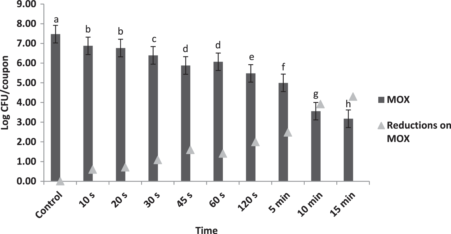
Mean (n=21) log recoveries and bacterial reductions of Listeria monocytogenes on stainless steel coupons on modified oxford medium (MOX) due to photohydroionization treatment. Bars not connected by the same letter are significantly different (p<0.05). CFU, colony-forming units.
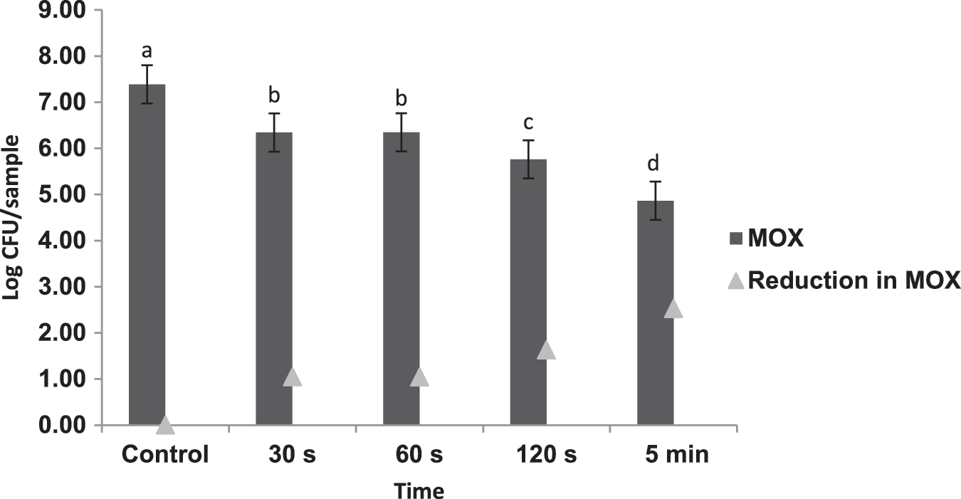
Figures 2 and 3 present mean recoveries and reduction in L. monocytogenes on sliced American cheese and RTE turkey after treatment with the PHI unit for 30, 60, and 120 s and 5 min compared with controls, respectively. The initial attachment of L. monocytogenes on the surface of sliced American cheese was 7.6 log CFU/sample. The treatment showed reductions (p<0.05) in bacterial populations with time; 30, 60, and 120 s and 5 min treatment of cheese with the PHI unit were 0.9, 1.03, 1.39, and 2.16 log CFU/sample, respectively. No difference was detected (p>0.05) between bacterial populations recovered on MOX and TALMOX, indicating that no additional injured cells were recovered after the treatment. The RTE turkey samples showed results similar to those for American cheese. Bacterial reductions (p<0.05) were seen over time with the treatment. Reductions up to 2.5 log CFU/sample were observed during the 5-min treatment time.

Mean (n=21) log recoveries and bacterial reductions of Listeria monocytogenes from sliced American cheese on modified oxford medium (MOX) due to photohydroionization treatment for 30, 60, and 120 s and 5 min. Bars not connected by the same letter are significantly different (p<0.05). CFU, colony-forming units.
Mean (n=21) log recoveries and bacterial reductions of Listeria monocytogenes populations from ready-to-eat turkey on modified oxford medium (MOX) due to photohydroionization treatment for 30, 60, and 120 s and 5 min. Bars not connected by the same letter are significantly different (p<0.05). CFU, colony-forming units.
Oxidative changes in the food products due ROS, the key components in the process, were also studied by performing TBAR analysis, which is widely accepted as evidence of oxidation and reflects oxidation of polyunsaturated fatty acids (Pimpa et al., 2009). Analyses performed on cheese and turkey samples for the two longest treatment times, 120 s and 5 min, indicated that the PHI treatment did not affect (p>0.05) TBAR values. The TBAR values for turkey samples for both the 120-s and the 5-min treatment were lower than those of the controls (Table 2). Similarly, TBAR values for cheese samples for 120-s and 5-min treatments were not significantly different from those of the controls. In comparison, previous research has reported chemical changes in food composition and deterioration of quality due to high or prolonged exposure to UV (Kolakowska, 2003).
There is no statistical difference (p>0.05) between the control group and each treated food group at two time points.
UV and ozone have continued to receive attention due to their antimicrobial effects. Ultraviolet-C (UV-C) is applied to fresh fruits and vegetables before storage to reduce microbial contamination on the surface. The FDA has approved UV treatments for pathogen reduction in water. UV treatment has shown effectiveness at reducing pathogens in apple cider (Hanes et al., 2002; Quintero-Ramos et al., 2004) and goat milk (Matak et al., 2005). Because of its oxidative ability, ozone has been used for many years in the food industry for odor control, disinfection, and water treatment, and has been used with mixed success to inactivate contaminant microflora on meat, poultry, eggs, fish, fruits and vegetables, and dry foods (Kim et al., 1999). Research has also shown 96.4%–99.9% reduction of several pathogenic forms of mold, fungi, bacteria, and viruses, which included methicillin-resistant Staphylococcus aureus, E. coli, Bacillus spp., and Stachybotrys chartarum, using photocatalysis and ozone treatment for 24 h (Ortega et al., 2007).
PHI with UV and ozone is a novel technology that is increasingly applied in food. Previously, studies have evaluated the combined effect of UV and ozone. UV treatment followed by ozonation showed a reduction in total heterotropic bacteria and total coliform count by 1 and 2 log CFU/mL, respectively, in a recirculating aquaculture system (Sharrer and Summerfelt, 2007). One of the advantages the UV-C ozone lamps offer is that the ozone that is produced as a part of the ongoing process can travel through the air and bring about the desired germicidal effect as opposed to conventional 254-nm germicidal UV in which air that passes only a few inches close to the UV lamps is treated. Short exposure times, low levels of ozone, and easy application in existing production capabilities are added advantages of the process.
To our knowledge, this is the first time PHI has been evaluated as a treatment to control surface contamination of food-contact surfaces and food products such as sliced American cheese and RTE turkey. Considerable reductions were seen as a result of use of this kind of advanced oxidation process. Reduction in L. monocytogenes populations was seen on stainless steel surfaces only after 15 min of treatment (Fig. 1). American cheese and RTE turkey showed reduction in bacterial populations after a short treatment time of 5 min (Figs. 2 and 3) without undesirable effects, such as lipid oxidation, on the product. Additional testing will have to be performed to ascertain the application of this technology at a commercial level by determining factors such as equipment size, load, and economic considerations. Furthermore, investigation of multiple factors that are included in food formulation and processing by utilizing the hurdle approach to control L. monocytogenes in food-processing facilities is warranted. Incorporation of hurdles such as using the PHI technology could play a synergistic role in order to achieve a safer food product. Further studies should also investigate the efficacy of this technology against varied types of strains of L. monocytogenes at different levels of inoculation that could pose a potential concern in food products and food-production facilities.
Conclusion
PHI technology was found to be effective in controlling L. monocytogenes on the surface of food-contact surfaces and RTE products (American cheese and oven-roasted turkey). It can be applied as a potential postlethality treatment (produced>1 log reduction in sliced American cheese and RTE turkey) or as a processing intervention in the integrated process to control L. monocytogenes contamination and ensure safe production of food. The PHI technology cannot be used as a kill-step in the production process but can be utilized as an effective processing aid or part of a hurdle approach to control L. monocytogenes.
Footnotes
Acknowledgments
The authors would like to thank the Food Microbiology Laboratory group and Food Chemistry group at Kansas State University for their help with this research.
Disclosure Statement
RGF Environmental Group Inc. did not provide any funding for this research, and the study was performed independently.