
Abstract
This article reports the prevalence and antibiotic resistance of the Bacillus cereus group isolated from different foods (milk and dairy products, spices, and rice salad) in Morocco. In total, 402 different food samples collected from 2008 to 2010 were analyzed by microbiological methods to isolate B. cereus. The strains were subjected to a polymerase chain reaction test in order to verify whether they belonged to the B. cereus group. Sixty-four of all isolates (15.9%) were found to be positive. Among the sources, B. cereus strains from milk and dairy products constituted the largest proportion of isolates (33/64; 51.6%) followed by spices (22/64; 34.4%) and salad with rice (9/64; 14.1%). The genetic diversity of the strains of B. cereus group was examined by pulsed-field gel electrophoresis (PFGE) of chromosomal DNA digested with SmaI. The enzyme restriction profiles showed a high degree of polymorphism among the strains. The results showed that PFGE analysis could reveal the genetic differences among B. cereus strains. Investigation of antibiotic-resistance profiles showed that isolates were resistant to ampicillin (98.4%), tetracycline (90.6%), oxacillin (100%), cefepime (100%), and penicillin (100%), and were susceptible to chloramphenicol (67.2%), erythromycin (84.4%), and gentamicin (100%). The results of this study indicated that B. cereus could be a significant etiological agent of food poisoning in Morocco because of its high prevalence. Also, we demonstrated that the majority of strains came from milk and dairy products. However, additional research involving cytotoxicity tests is needed to more evaluate this sanitary risk.
Introduction
F
B. cereus are Gram-positive and spore-forming bacteria. These microorganisms are widely distributed in nature because of the resistance of their endospores to various stresses and their long-term survival under unfavorable conditions. Their spores are hydrophobic and may attach to surfaces, germinate, and multiply in processing equipment, thus contaminating milk products. Many B. cereus strains cause food poisoning. Two main types of food poisoning linked to B. cereus have been described: emetic and diarrheal.
It is important to develop a rapid and reliable molecular subtyping method to epidemiologically trace B. cereus in order to monitor their presence in foodstuffs (Merzougui et al., 2013). Nevertheless, few studies have been conducted on the genotypic characterization of B. cereus isolates from Morocco in terms of foodborne poisoning concerns.
The present study was undertaken to evaluate the occurrence of B. cereus in foods in Morocco and to determine the genetic relationship between strains. In addition, the antibiotic resistance of isolates was also evaluated.
Materials and Methods
Sample collection
During the period from December 2008 to October 2010, a total of 402 different food samples were collected from hotels, restaurants, and private companies in several cities in Morocco, mainly from Casablanca (n=134), Tangier (n=78), Rabat (n=117), and Marrakech (n=73). Food samples are summarized in Table 1. Approximately 50 g of each sample (having no prolonged contact with the surroundings) was collected in a sterile plastic bag and transported to the laboratory for analysis.
Microbiological analysis
Ten grams of each food sample was suspended in 90 mL of 0.1% (wt/vol) buffered peptone water (BPW) from Bio-Rad (Marne-la-Coquette, France), and then homogenized for 2 min by a BagMixer stomacher (AES Laboratory, Combourg, France). After stomaching, 0.1 mL of homogenate was serially diluted (10-fold) in 0.85% saline, and then 0.1 mL of each dilution was inoculated onto polymyxin–mannitol–egg yolk–phenol red (Guinebretiere et al., 2003) followed by incubation at 30°C for 24 h. Plates that contain suspicious colonies were selected, and a maximum of three typical colonies from one sample were subcultured on nutrient agar (Difco, Detroit, MI). Among three subcultures per each sample, one colony was used for confirmation and further tests. Presumptive positives were confirmed biochemically by using API 50 CHB galleries (BioMérieux, Marcy l'Etoile, France).
DNA extraction
B. cereus strains were propagated for 16 h at 30°C on brain heart infusion–yeast extract (BHI-YE). Genomic DNA was extracted using the DNeasy 96 blood and tissue Kit (Qiagen, Courtaboeuf, France) as recommended by the manufacturer. Isolated DNA was used as a template for polymerase chain reaction (PCR) with the following protocols.
PCR
The PCR method targeted an sspE gene sequence adapted from Francis et al. (1998): Each reaction mixture (25 μL) contained 100 ng DNA template, 0.8 pmol of primers (BcFF2:GAGATTTAAATGAGCTGTAA and BcAPR1: CTT(C/T)TTGGCCTTCTTCTAA) (Sigma), 0.2 μL of Taq polymerase 1 U (Biolabs, Ipswich, MA), and 1 μL of deoxyribonucleoside triphosphate 0.2 mM (Biolabs). The amplification reactions were carried out in a BioRad-iCycler version 1.280 (BioRad, Marnes-la-Coquette, France) as follows: 5 min at 95°C, 30 cycles of 15 s at 95°C, 30 s at 52°C, and 30 s at 72°C, followed by a final extension step at 72°C for 2 min. After the amplification, 5 μL of reaction mixture was analyzed by electrophoresis on a 1% agarose gel in Tris-borate-EDTA (TBE) buffer (Tris 89 mM, boric acid 89 mM, EDTA 2 mM) at 80 V for 45 min. The gel was stained by ethidium bromide to check the size of the PCR products.
Pulsed-field gel electrophoresis (PFGE) genotyping
The protocol for the characterization of strains by PFGE is adapted and optimized (with modification) from our previous work (Gautier et al., 1996).
In brief, bacterial cells were grown in BHI-YE for 24 h at 30°C, and 9 mL of cell suspension was centrifuged for 10 min at 7000×g at 4°C. The supernatants were removed and the pellets were resuspended in 1 mL of TE buffer (pH 8; 10 mM Tris, 1.0 mg lysozyme, 1 M NaCl, and 50 mM EDTA), embedded in 500 μL agarose (Eurobio, les Ulis, France) and then digested with 2.0 mg of lysozyme (Sigma-Aldrich, St. Louis, MO). The plugs were transferred into a solution containing 0.1% sodium lauryl sarcosine in 0.5 M EDTA (pH 8), and 200 mg of proteinase K, and the mixture was incubated overnight at 44°C with gentle shaking. DNA was digested with the enzyme SmaI (20 U) following migration performed with the CHEF DRII BioRad system (BioRad) in a 1.5% agarose gel (Ultra-Pure; Gibco BRL, Paisley, Ecosse) in 0.5x TBE buffer at 14°C with a linear ramping time of 2–20 s over a period of 18 h, and a gradient of 200 V/cm. After migration, the gels were stained with 1 μg/mL ethidium bromide and photographed under UV light (Vilber Loumat, Marne-La-Vallée, France). PFGE patterns obtained were analyzed by the Bionumerics software (BioMérieux, Pearson correlation, optimization 1%). The relationship between two given strains was scored by the Dice coefficient of similarity, and strains were clustered by hierarchically clustering interstrain similarities based on the unweighted pair group method with arithmetic averages.
Antibiotic susceptibility test
The Kirbye-Bauer disc-diffusion method was used (Bauer et al., 1966; Drew et al., 1972). The disks used (Oxoid) and antibiotic concentrations were as follows: ampicillin (10 mg), penicillin (10 U), gentamicin (10 mg), tetracycline (30 mg), erythromycin (15 mg), chloramphenicol (30 mg), cefepime (30 mg), and oxacillin (1 mg). All B. cereus strains were classified as susceptible, intermediate susceptible, or resistant.
Results
Prevalence of B. cereus in food samples
Sixty-four of 402 (15.9%) food samples analyzed during 2008–2010 were found positive for B. cereus. Table 1 shows the prevalence of B. cereus isolates from a variety of food products. Among the sources, milk and dairy products constituted the highest contaminated foods (33/64; 51.6%), followed by spices (22/64; 34.4%) and salad with rice (9/64; 14.1%). More than 80% of food samples analyzed showed a high number of presumptive B. cereus colonies (exceeded 4 log colony-forming units/g).
Analysis of DNA by PCR amplification of the sspE gene showed that 76.1% (64/84) of the presumptive B. cereus isolates examined were confirmed to be of the B. cereus group. These results indicate that the biochemical identification is not sufficient to confirm the presence of B. cereus species.
PFGE analysis
The PFGE fingerprinting method was successful in separating the B. cereus strains isolated from milk and dairy products, spices, and rice salad. The results obtained showed a remarkable polymorphism existing among all strains (Fig. 1). A similarity was observed among all strains belonging to the same cluster.
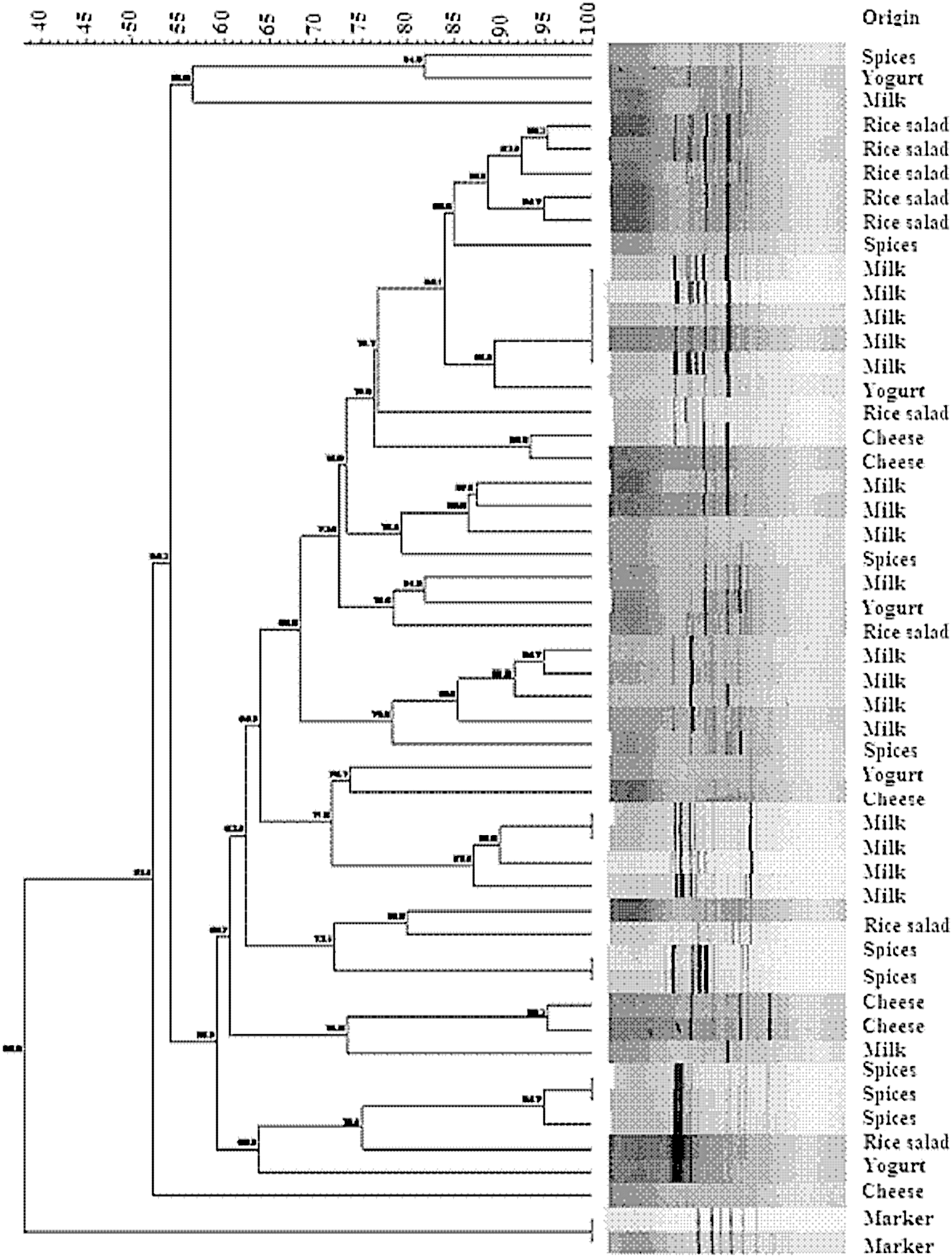
Dendrogram generated by Bionumerics software, showing the similarities of the patterns obtained from SmaI digestion patterns for the Bacillus cereus strains isolated from food (spices, rice salad, milk, and dairy products). The phenogram was constructed using the Dice coefficient and unweighted-pair group method with arithmetic mean analysis. The degree of similarity (%) is shown on the scale.
Resistance to antimicrobials
Antibiotic susceptibility of the tested strains is presented in Table 2 (Bauer et al., 1966). All strains were highly resistant to β-lactam antibiotics, including ampicillin, cefepime, oxacillin, and penicillin, while all of them were susceptible to other antibiotics (Table 2).
(%).
Discussion
B. cereus is an important food-poisoning organism because of its wide distribution in nature, and the presence of its spores in food products can be linked to the environment. It can be transmitted to food products when contaminated ingredients are used to formulate these foods. Heat-resistant endospores of B. cereus are known to survive cooking or frying processes, multiply rapidly, and produce sufficient amounts of toxin to cause food poisoning (Gilbert et al., 1974; Wayne et al., 2000).
The results of this study indicate that B. cereus strains from milk and dairy products constituted the largest proportion of isolates (33/64; 51.6%). At present, little is known about the extent of milk contamination by various B. cereus group organisms in Morocco. The incidence and contamination levels of B. cereus found in dried-milk products were similar to those reported by others (Bartoszewicz et al., 2008). The literature confirms that dried-milk products are known to be frequently contaminated with B. cereus, principally with its spores. The high level of nutrients in milk makes it an especially suitable growth medium for various bacteria, including those belonging to the families Enterobacteriaceae, Streptococcaceae, and Bacillaceae. In fact, these microorganisms can achieve high population densities following contamination during milk processing in dairy farms and in the dairy industry. In particular, contamination of milk by members of the B. cereus group is of significance, not only because of their spoilage capability, but also because of their potential to cause human diseases. Bartoszewicz et al. (2008) reported that 94 of 136 strains of B. cereus isolated from milk and cream exhibited human embryonic lung cell cytotoxicity when cultured in milk.
Spices are used extensively in Moroccan cuisine. In our study, the incidence and contamination levels of B. cereus in spices (32.8%) were similar to or lower than those previously reported (Powers et al., 1996; Cohen et al., 2006).
Results of our study demonstrate that biochemical identification is not enough to confirm the presence of the B. cereus group. Valero et al. (2007) reported that there was no difference in the number of B. cereus recovered from food when plated on mannitol egg-yolk polymyxin, PMYPA, and trypticase–soy–polymyxin blood agars. This result indicated the need for molecular analysis for confirmation of the presence of the B. cereus group (Valero et al., 2007).
In order to produce a higher discriminatory potential, PFGE was used to allow the comparison between different B. cereus groups. The protocol adopted in this study is reliable and has the advantage of being feasible in 4 days, which is very short compared with the protocol used for typing strains of this group by Harrell et al. (1995). The latter protocol, which was reused by Dubouix et al. (2005), in fact takes place in 8 days.
Isolates with identical, or almost identical PFGE patterns are considered to be of the same clonal origin (Tenover et al., 1995). In this study, there was a visible correlation between PFGE types and sources of isolates: the PFGE patterns of the same origin of strains were clustered closely together (Fig. 1). Recently, a study on the comparison between PFGE and other typing methods on the same sample used in this study showed that PFGE is a powerful tool to reveal genetic differences among B. cereus isolated from food (Merzougui et al., 2013).
With respect to the antibiotic susceptibility test, the results were consistent with many previous studies that reported resistance of B. cereus against β-lactam antibiotics including penicillin, oxacillin and cephalosporins (Andrews and Wise, 2002; Park et al., 2009).
Consistent with other countries, Morocco is experiencing newer consumer trends among traditional foods, with an obvious tendency to adopt international and more processed foods. Such a tendency combined with poor hygienic practices may contribute to increase the prevalence of pathogens in foods, thereby increasing the risk of foodborne disease for consumers (Badri et al., 2009).
The results of this study indicate that B. cereus could be a significant etiological agent of food poisoning in Morocco. Results also suggest that serious attention should be devoted to the sanitary and temperature conditions under which foods are prepared, cooked, and marketed. Therefore, the Moroccan government plans to adopt regulations enforcing the application of the hazard analysis critical control point system as a means to identify and control the hazards in foods.
Footnotes
Acknowledgments
The authors are grateful to all collaborators in this study. We would like to thank RIIP (Réseau International de l'Institut Pasteur) for financial collaboration.
Disclosure Statement
No competing financial interests exist.