
Abstract
The present study investigated the efficacy of single and combined treatment of both chlorine and thiamine dilaurylsulfate (TDS) on the reduction of Listeria monocytogenes biofilms in microtiter plate. The disinfectants used in this study were 50, 100, and 200 mg/L chlorine and 100, 500, and 1000 mg/L of TDS. Biofilm-forming index (BFI) and culturable cell count were used to evaluate the disinfectant assay. The highest BFI reduction was 0.80, achieved by the combination of 200 mg/L chlorine and 1000 mg/L TDS. In contrast, the highest culturable cell count reduction was 4.80 log colony-forming units/well by the combination of 200 mg/L chlorine and 100 mg/L TDS. The BFI was reduced in a concentration-dependent manner while culturable cell count was significantly reduced only when all chlorine concentration was combined with 100 mg/L TDS. However, when chlorine was combined with a higher concentration of TDS, the reduction decreased significantly. The result in this study showed that the combination of the 200 mg/L chlorine and 1000 mg/L TDS could be a practical application in removing L. monocytogenes biofilms from surfaces in food industry, and for the 200 mg/L chlorine and 100 mg/L, it can be used for killing the pathogen biofilms. However, more studies are still needed in order to show its efficacy on foods surfaces as well as to develop an even more effective treatment in both killing and removing biofilms.
Introduction
L
NaOCl is a widely used chlorine-based disinfectant in the food industry (Dychdala, 2001; Peng et al., 2002). It can ionize via hydrolysis into HOCl, which is a strong bactericidal agent (Fukuzaki, 2006). Additionally, thiamine dilaurylsulfate (TDS) is used as a synergistic agent with other bactericides in noodles in Korea. Many authors have reported its bactericidal activity and synergistic effect against foodborne pathogens (Kim et al., 2005; Lee et al., 2010; Kim et al., 2011), especially the combination of NaOCl and TDS (Lee et al., 2010; Kim et al., 2011). Studies concerning combined treatment (hurdle technology) have shown remarkable improvement in reduction value. Lee and Ha (2008) demonstrated that combined treatment of disinfectants with TDS shows better reduction of bacteria coliforms in rice. However, there is no study on the effect of combined treatment of disinfectants with TDS on L. monocytogenes biofilms yet. The purpose of this study was to evaluate the efficacy of combined treatment of sodium hypochlorite (NaOCl) with TDS on L. monocytogenes biofilm using a biofilm-forming index and culturable cell-count method.
Materials and Methods
Bacterial mix preparation
L. monocytogenes ATCC 19111 (serotype 1/2a, isolated from poultry), ATCC 19113 (serotype 3b, isolated from humans), and ATCC 19115 (serotype 4b, isolated from humans) were used as a mix in this study. The bacteria were recovered from −80°C frozen stock, and 100 μL of the stock was inoculated into 10 mL of tryptic soy broth (TSB; Difco, Detroit, MD). Then, the solution was incubated at 30°C for 24 h. After vortexing, 100 μL of the incubated culture was pipetted into 10 mL of fresh TSB followed by another 24-h incubation. The mix of the three strains was prepared with a 1:1:1 ratio.
Biofilm formation
Biofilms were formed in a microtiter plate following the previously described method by Stepanović et al. (2004) and Stepanović et al. (2007) with some modifications. Twenty microliters of the bacterial mix was inoculated into each well of the 96-well flat-bottom microtiter plate (Becton Dickinson Falcon, Cockeysville, MD) followed by 230 μL of sterile TSB. The plate was sealed and statically incubated at 37°C for 48 h. According to preliminary experiment, which compared the biofilm formation strength of different incubation temperatures and durations (data not shown), the strains used in this study made strong biofilm formation at 37°C with 48-h incubation, so this condition was used in the biofilm formation for the disinfection test. Two hundred fifty microliters of sterile TSB was used for negative control for the biofilm-forming index (BFI) assay. BFI was calculated according to procedures described previously (Jahid et al., 2013). In biofilm formation, TSB was used instead of Brain Heart Infusion (BHI) broth, since TSB gave higher BFI than BHI (author's observation in preliminary experiment, data not shown).
Disinfectants preparation
Chlorine disinfectant solution was prepared using 12% NaOCl (6% free chlorine, Yakuri Pure Chemicals Co., Ltd., Kyoto, Japan). The solution was diluted in sterile distilled water to get a chlorine concentration of 50, 100, and 200 mg/L. TDS, a vitamin B1 derivative, was first weighed and dissolved in 30% ethanol solution to obtain a concentration of 5%. Then, it was diluted in sterile distilled water or in the prepared chlorine solution to get a final solution with the concentration of 100, 500, and 1000 mg/L. All disinfectants were prepared and used right after dilution during disinfection test.
Disinfection assay by BFI
After 48-h incubation, the optical density (OD) of the total bacteria in the microtiter plate was measured at a wavelength of 600 nm (OD600nm; an optimum wavelength for estimating the concentration of bacteria; Matlock et al., 2011) with a microtiter plate reader (Spectra Max 190, Molecular Devices, Sunnyvale, CA). The planktonic cells and medium were removed, and each well was rinsed three times with 250 μL of sterile phosphate-buffered saline (PBS, pH 7.2) to remove the loosely attached cells. Then, chlorine and TDS were added individually or in combination for 1 min; instead of disinfectant, PBS was used for treating positive control (well with L. monocytogenes biofilm not subjected to disinfectant challenge) and negative control (well with TSB without L. monocytogenes biofilm). The disinfectant was discarded by pipetting and 250 μL of Dey/Engley (D/E) neutralizing broth (Difco) was introduced into each well for 5 min, including the control wells. Finally, the wells were rinsed three times with 250 μL PBS.
The biofilms were fixed with 250 μL of extra pure methyl alcohol (Daejung Chemicals & Metals Co., Ltd., Shiheung, Korea) for 15 min; the plate was dried overnight in an up-inverse position after pouring out methyl alcohol. Crystal violet dye 0.1% (CV; Sigma-Aldrich, St. Louis, MO) 250 μL was used to stain biofilms, positive control, and negative control wells for 15 min and removed by pipetting. The plate was rinsed with tap water until the washing water was dye free, and air-dried overnight. The bound dye was resolubilized in 95% ethanol for 30 min and transferred into a new well plate. The OD of the dye solution was measured at 570 nm (OD570nm).
The biofilm-removing efficacy of the disinfectants was compared using the BFI calculated with the following formula (Niu and Gilbert, 2004):
where OD570nm is obtained from the disinfected (treated) or positive control (biofilm treated with PBS) wells after staining and the ODC570nm is obtained from negative control wells (TSB wells treated with PBS) after staining. The OD600nm is obtained from the disinfected or positive control wells, and ODC600nm is obtained from negative control wells, after 48-h biofilm formation. These OD values only represented the presence of cells that still attached to the surface after the treatment. It did not show the biological state of L. monocytogenes.
Disinfection assay by culturable cell count
The biofilms were formed as stated previously. The test was conducted following the method previously described (Teh et al., 2010) with some modification. After 48-h incubation, all planktonic cells and medium were removed and all wells were rinsed three times with 250 μL of PBS. Each well was treated with disinfectants as mentioned above. After 1-min treatment, the disinfectant solutions were removed from the well and placed into 2 mL of D/E neutralizing broth (test tube A). Two hundred and fifty micro liters of D/E neutralizing broth were introduced into each well. Then, a sterile cotton swab was pressed to the bottom of the well and rotated 50 times clockwise and another 50 times anticlockwise. The swab and the remaining neutralizing solution in the well were also placed into test tube A. After leaving for 5 min, the tubes were then vortexed for 30 s each before serial dilution and spread-plating onto PALCAM Agar (Oxoid, Basingstoke, UK). All plates were incubated at 30°C for 48 h prior to counting.
Field emission scanning electron microscopy (FESEM)
Processing of FESEM samples of biofilms was performed according to the procedures described previously (Jahid et al., 2013) with some modifications. The biofilms developed in a 12-well microtiter plates and after a disinfectant test, biofilms were observed by using FESEM. Each well was rinsed three times with PBS, pH 7.2. The adhered cells were fixed in 2% glutaraldehyde (Sigma Aldrich) in PBS for 24 h and then washed three times for 15 min with PBS. The fixed cells were serially treated with ethanol (50% for 15 min, 60% for 15 min, 70% for 15 min, 80% for 15 min, 90% for 15 min, and 100% two times for 15 min) and were successively dehydrated by 33, 50, 66, and 100% hexamethyldisilazane (Sigma Aldrich) in ethanol for 15 min each. The dehydrated samples were coated with platinum and observed using FESEM. The FESEM was operated at an accelerating voltage of 5 kV and 5 mm working distance.
Statistical analysis
All treatments were conducted in three independent trials, with each having triplicate samples. Data were analyzed by one-way analysis of variance (Duncan's test) using the Statistical Analysis System (SAS; version 9.2, SAS Institute Inc., Cary, NC). Statistical significance was considered at p<0.05.
Results and Discussion
Effect of combined treatment of chlorine and TDS on BFI of L. monocytogenes
It has been observed that BFI is a relatively new approach, and it is being used in biofilm research to identify biofilm-forming bacteria as well as to categorize bacteria biofilm formation strength (Niu and Gilbert, 2004; Naves et al., 2008; Moriyama et al., 2009; Teh et al., 2010). However, it is not commonly used in the biofilms removal test even though it can be considered a good means for biofilm quantification on the surface, since biofilm is stained with crystal violet (CV), allowing the comparison of removal effect between the treated and untreated biofilm. In the disinfectants or surfactants test, it can be postulated that BFI can be used as an indirect evaluation of the chemical efficacy in removing biofilm from surfaces. This is because the BFI method allows the quantification of the amount of CV that is bound to the remaining biofilm on the surface after biofilms have been challenged with disinfectants or surfactants. In this experiment, 100 and 200 mg/L chlorine, and 500 and 1000 mg/L TDS as single treatment showed significant (p<0.05) removal of L. monocytogenes biofilm (Table 1). At 500 and 1000 mg/L, TDS removed 0.43 (≈32%), and 0.55 (≈41%) BFI, respectively; whereas 100 mg/L TDS showed little or no effect at all. It can be seen that the removal efficacy of chlorine or TDS was increased when they were mixed together. All chlorine concentrations, when used in combination with 1000 mg/L TDS, showed good biofilm-removal properties, which achieved a 0.67–0.80 BFI reduction, depending on the chlorine concentration. These values were significantly higher than those of chlorine-only treatment. The highest removal was by the combination of 200 mg/L chlorine with 1000 mg/L TDS, yielding 0.8 (≈60%) BFI reduction. Since TDS showed high BFI reduction, it can be speculated that TDS might hold an interfacial tension-lowering property, leading to removal of bacteria biofilm. TDS was also considered as a surfactant in another study (Fransisca et al., 2012).
Mean±standard deviation values denoted by different superscript letters in the same column are significantly different (p<0.05).
CFU, colony-forming unit.
Effect of combined treatment of chlorine and TDS on culturable cell count of L. monocytogenes
However, the disinfectants affect culturable biofilm cells differently. As shown by the results, chlorine 50 mg/L, 100 mg/L, and 200 mg/L could significantly (p<0.05) inactivate the cells by 3.27, 3.96, and 4.61 log colony-forming units (CFU)/well, respectively (Table 2). Up to now there have been no studies on the effect of chlorine and TDS on L. monocytogenes biofilm in microtiter plates; however, Ronner and Wong (1993) reported that chlorine 100 mg/L can reduce 4 log CFU/cm2 of L. monocytogenes biofilms on stainless steel surfaces after 2-min treatment. Mustapha and Liewen (1989) also observed a more than 4 log CFU/mL reduction of Listeria biofilm on stainless steel surfaces when treated with 200 mg/L more than 2 min. These results could be speculated to be comparable to that of this study. On the other hand, the results indicated that TDS alone is much less effective than treatment with only chlorine; the highest concentration (1000 mg/L) of TDS only reduced the cell count by 1.55 log CFU/well. Unexpectedly, the combined treatment reduced the chlorine effect in a TDS concentration-dependent manner. As illustrated in Table 2, the reduction was increased to 3.43–4.80 log CFU/well when chlorine was combined with 100 mg/L TDS. However, the reduction decreased significantly to 1.66–4.04 log CFU/well when chlorine was combined with 500 mg/L TDS. For the combination with 1000 mg/L TDS, the reduction decreased dramatically to 1.08–2.48 log CFU/well. The most effective combination for the reduction is 200 mg/L chlorine and 100 mg/L TDS. The results showed that chlorine and TDS might have an antagonistic effect in killing L. monocytogenes biofilm (The effect of combined treatment was less than the sum of the individual effect of TDS and chlorine) in a concentration-dependent manner since the reduction decreased as the concentration of TDS increased. There was no report on the effect of chlorine and TDS combined treatment on L. monocytogenes biofilm. These results were not consistent with other research in which the two compounds showed a synergistic effect (i.e., higher synergism with higher concentration) on the bacteria reduction in foods (Lee et al., 2010; Ha et al., 2012; Park et al., 2012). However, TDS is a derivative of vitamin B1, and it has been documented that vitamin B1 is destroyed by NaOCl (Dwivedi and Arnold, 1973; Bates, 2007); it is also unstable at neutral pH or can be destroyed at pH above 8 (oxidation) (Bates, 2007). This could be a possible explanation that could potentially explain the decrease in the reduction value of culturable cell counts in combined treatment.
Mean±standard deviation values denoted by different superscript letters are significantly different (p<0.05).
CFU, colony-forming unit.
The difference between BFI and culturable cell count method
It should be noted that the BFI method and the culturable cell count method could provide two completely different results. The BFI method allows an estimation of the cells that attach to a surface. It does not provide information on the biological state of the cells. Thus, the results from BFI represent only the cells remaining on the surface after the treatment including the dead, culturable, and viable but nonculturable cells. This method allows us to indirectly evaluate the removal effect of the disinfectant on the biofilm.
However, the culturable cell count method, used in this study, represents all the culturable cells after the treatment including the cells that were detached from the surface. Thus, this method provides the evaluation of the bactericidal effect of the disinfectants on the biofilm.
FESEM of L. monocytogenes treated with combined chlorine and TDS
The FESEM image showed that three-dimensional biofilms of L. monocytogenes were formed after incubation at 37°C for 48 h (Fig. 1A). After challenging with the combined treatment, the biofilms were removed and reduced to monolayer biofilms (Fig. 1B). The image showed that even though some cells were removed, the EPS still presented on the surface (Fig. 1C) as well as the damaged cells (Fig. 1D). It has been reported that if the removal of bacteria does not provide a bactericidal effect, the microorganism will be able to accumulate elsewhere in the industrial settings and reproduce biofilm (Gram et al., 2007). It is also suggested that because the bacteria biofilm is killed but not removed from the surface, it might act as an enhancer for bacteria attachment since pre-existing EPS will facilitate the adhesion (Flemming and Schaule, 1988; Tang et al., 2009). Therefore, it is necessary for researchers to study both the removal and bactericidal efficacy of disinfectant on biofilms.
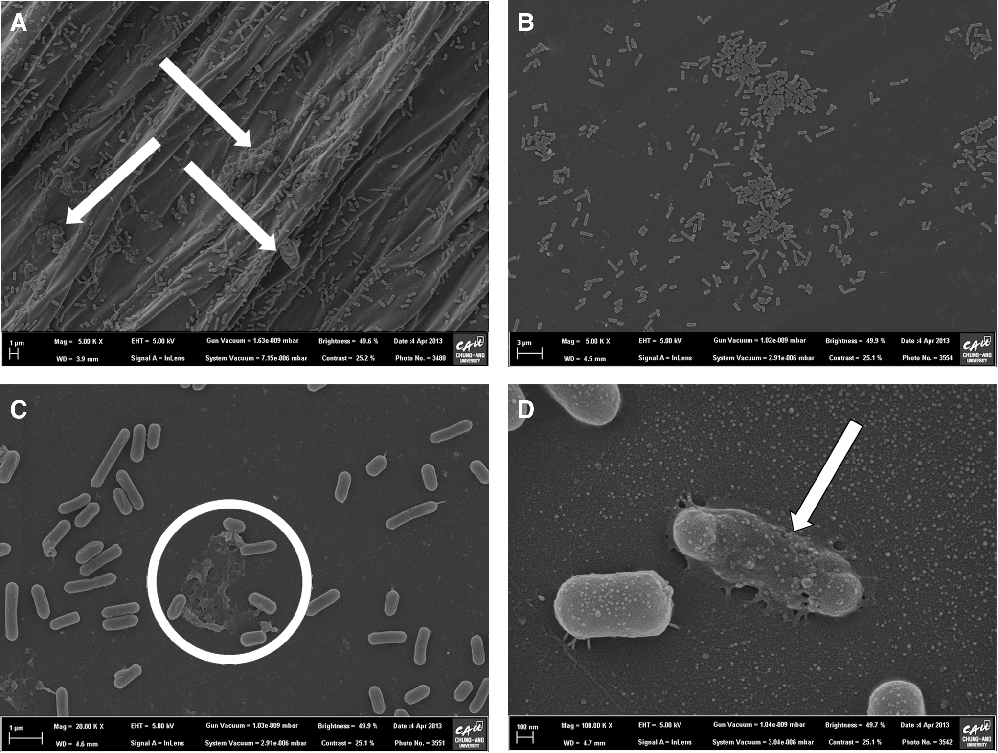
Field emission scanning electron microscopy images of Listeria monocytogenes biofilms in a microtiter plate.
Conclusions
In conclusion, the combination of 200 mg/L chlorine and 100 mg/L TDS achieved the highest culturable cell count reduction, when used to treat L. monocytogenes biofilms. However, the increase in reduction is relatively small (4.80 log CFU/well compared to single chlorine treatment 4.61 log CFU/well). The combination of 200 mg/L chlorine and 1000 mg/L TDS achieved the highest BFI reduction. It may be a practical application for biofilm removal (BFI reduction). Furthermore, future research should look into genes expression responding to chemical treatment in order to achieve a good synergism in combined treatment. In case of surface cleaning, both removal and bactericidal effect of the treatment should be evaluated, since it is important that the pathogens be removed and killed to prevent further contamination and biofilm formation.
Footnotes
Acknowledgments
This research was supported by funding from the 2012 grant (12162KFDA012) from the Korea Food and Drug Administration for studies on hazardous microbes and microbiological safety management of seafood.
Disclosure Statement
No competing financial interests exist.