
Abstract
Utensils and equipment from meat-processing facilities are considered relevant cross-contamination points of Listeria monocytogenes to foods, demanding tracking studies to identify their specific origins, and predict proper control. The present study aimed to detect L. monocytogenes in a beef-processing facility, investigating the diversity of serotypes and pulsotypes in order to identify the possible contamination routes. Surface samples from knives (n=26), tables (n=78), and employees hands (n=74) were collected before and during the procedures from a beef-processing facility, in addition to surface samples of end cuts: round (n=32), loin (n=30), and chuck (n=32). All samples were subjected to L. monocytogenes screening according ISO 11.290-1, and the obtained isolates were subjected to serotyping and pulsed-field gel electrophoresis. Listeria spp. were identified in all processing steps, in 61 samples, and L. monocytogenes was detected in 17 samples, not being found only in knives. Eighty-five isolates were identified as L. monocytogenes, from serotypes 1/2c (n=65), 4b (n=13), and 1/2b (n=7), being grouped in 19 pulsotypes. Considering these results, cross-contamination among hands, tables, and beef cuts could be identified. The obtained data indicated the relevance of cross-contamination in the beef-processing facility, and the occurrence of serotypes 1/2b and 4b in beef cuts distributed for retail sale is a public health concern.
Introduction
L
Due to the ubiquitous nature and adhesion ability of L. monocytogenes, the food industry environment is considered an important source of contamination of this foodborne pathogen (Carpentier and Cerf, 2011). Based on these characteristics, food industries have adopted control strategies to identify contaminated utensils and equipment, as well as cleaning procedures to eliminate L. monocytogenes. The beef-processing environment is usually associated with this foodborne pathogen, highlighting the relevance of beef products in the transmission of L. monocytogenes to humans (Barros et al., 2007; da Rocha et al., 2012).
The association between food and clinical L. monocytogenes strains is usually made by serotyping. The classical serotyping described by Seeliger and Höhne (1979) is routinely used to identify potentially pathogenic strains and to provide preliminary epidemiologic information (Liu, 2006). At present, 13 serotypes have been described (Allerberger, 2003), but only the serotypes 1/2a, 1/2b, and 4b have been reported to be responsible for 95% of listeriosis cases (Swaminathan and Gerner-Smidt, 2007).
To subtype L. monocytogenes isolates, different methods have been used. Pulsed-field gel electrophoresis (PFGE) is the standard method for L. monocytogenes subtyping, as it presents high reproducibility and discriminatory potential (Graves and Swaminathan, 2001). This method has proved to be very efficient for the surveillance contamination of L. monocytogenes in the beef-processing chain and listeriosis cases and outbreaks. The objective of the present study was to identify the diversity of serotypes and pulsotypes of L. monocytogenes in a beef-processing facility.
Materials and Methods
Sampling
A slaughterhouse located in the Minas Gerais state, Brazil, was selected for the present study. The process steps in the establishment starts with slaughtering of animals, and ending in the manufacture of meat products, and it was visited 13 times for sampling during the survey. In each visit, 400 cm2 surface samples of knives (n=26), tables (n=78), and the hands of employees (n=74) were collected before and during beef processing. In addition, 400-cm2 surface samples from beef cuts (round, n=32; loin, n=30; chuck, n=32) were also collected. Samples from each point were collected by swabbing 4 limited areas (100-cm2 sterile plastic templates) with sterile sponges (3M Microbiology, St. Paul, MN), previously moistened with 10 mL of buffered peptone water solution (at 0.1%, wt/vol, BPW, Oxoid Ltd., Basingstoke, England) and kept at 4°C until analysis. In some cases, more than 1 point (utensils, hands) was considered for sampling, in order to keep the pattern of 400 cm2 per sample.
In aseptic conditions, each set of 4 swabs from each sample was added to 160 mL of BPW (Oxoid) and homogenized, and 40-mL aliquots (representing 80 cm2 of each sample) were subjected to microbiological analysis to detect Listeria spp.
L. monocytogenes screening and serotyping
Protocol ISO 11.290-1 (ISO, 1996, 2004) was adopted in this step of the study. The 40-mL sample aliquots were centrifuged at 4°C for 15 min (1000×g), and after discarding the supernatant, the pellet was suspended with 10 mL of half-Fraser broth (Oxoid) and incubated at 30°C for 24 h. Then, 0.1-mL aliquots were transferred to Fraser broth (Oxoid) and incubated at 37°C for 24 and 48 h. The obtained cultures were streaked on Chromogenic Listeria agar (Oxoid) and Oxford agar (Oxoid), both incubated at 37°C for 24 and 48 h. Suspected Listeria spp. colonies were streaked and purified onto trypticase soy agar (Oxoid), incubated at 30°C for 24 h. Distinct colonies per sample were selected and subjected to biochemical identification using the protocols and interpretation guide described in ISO 11.290-1 (ISO, 1996).
Isolates identified as L. monocytogenes were subjected to a polymerase chain reaction to detect the hlyA gene as confirmatory step, using primers and the protocol described by Border et al. (1990). Isolates that were confirmed as L. monocytogenes were sent to the Laboratório de Zoonoses Bacterianas, Instituto Oswaldo Cruz (Fiocruz, Rio de Janeiro, Brazil) for serotyping using polyclonal somatic and flagellar antisera, as described by Seeliger and Höhne (1979).
PFGE
PFGE was conducted using AscI and ApaI (New England Biolabs Inc., Ipswich, MA), as described by Graves and Swaminathan (2001). Macrorestriction products were separated by electrophoresis in agarose gels (Agarose Seakem Gold 1%, wt/vol, buffer TE 0.5×) in CHEF-DR II (BioRad, Philadelphia, PA) adjusted with the following parameters: 6 V/cm, 4 s for initial switch time, 40 s for final switch time, and 20 h of running. The obtained gels were stained in GelRed (Biotium Inc., Hayward, CA) and the observed band patterns were analyzed using BioNumerics 6.6 (Applied Maths, Gand, Belgium) based on 5% Dice similarity of bands. Pulse Marker 50 (1000 kb; Sigma-Aldrich Corp., St. Louis, MO) was used as a reference.
Results and Discussion
L. monocytogenes, L. innocua, and L. seeligeri were detected in the tested samples, and their frequencies are presented in Table 1. Two hundred thirty-three isolates were identified by biochemical tests as Listeria spp., being preliminary identified as L. innocua (n=142), L. monocytogenes (n=85), and L. seeligeri (n=6). After a confirmatory step (hlyA detection), 85 isolates were identified as L. monocytogenes. The identified serotypes are presented in Figure 1.
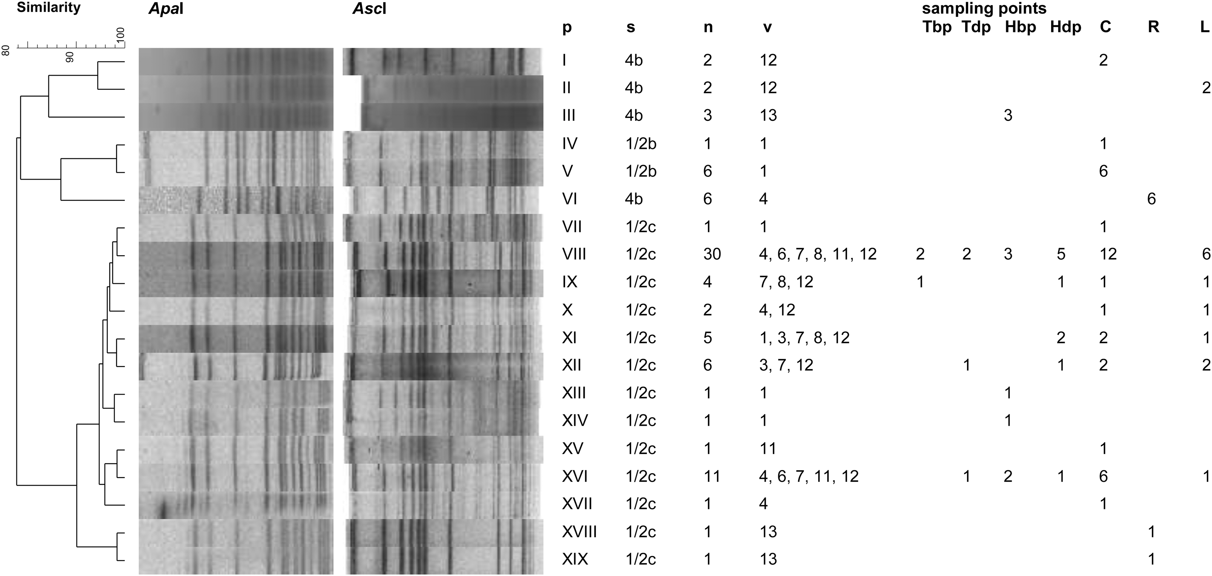
Pulsed-field gel electrophoresis patterns after macrorestriction of Listeria monocytogenes isolates obtained from a beef-processing facility. p, pulsotypes; s, serotype; n, number of isolates; v, visit; sampling points—Tbp, table before processing; Tdp, table during processing; Hbp, hands of employees before processing; Hdp, hands of employees during processing; C, chuck; R, round; L, loin. Similarities between pulsotypes were determined considering Dice coefficient (5%).
Two samples contaminated simultaneously by L. innocua and L. monocytogenes.
Three samples contaminated simultaneously by L. innocua and L. monocytogenes.
The occurrence of Listeria spp. in beef products is usually a consequence of contamination from the slaughtering environment, from the point of carcass handling and evisceration, to the production steps of the end products. The cross-contamination can occur among utensils and equipment (Fenlon et al., 1996; Yücel et al., 2005; Galvão et al., 2012). L. monocytogenes was detected in samples even before the beginning of beef processing, such as on tables and on hands of employees, indicating failures in sanitization and the previous occurrence of this pathogen in the environment (Table 1). In addition, some samples were contaminated simultaneously with L. monocytogenes and L. innocua (hands of employees and chuck cut of the carcasses, Table 1).
The serotype 1/2c prevailed (n=65), as observed by Ochiai et al. (2010) in pork-processing environments. Serotype 1/2c strains are usually recognized as possessing low virulence potential, due to the expression of truncated proteins, and demanding sequencing of specific genomic regions, such as inlA for proper characterization (Ragon et al., 2008; López et al., 2013). Raw foods, such as dairy and meat products, are sporadically implicated in listeriosis cases once they are usually consumed after heat treatment (Swaminathan and Gerner-Smidt, 2007); however, ready-to-eat products are considered the main vehicles for L. monocytogenes (Vit et al., 2007; Pichler et al., 2009; Fretz et al., 2010; Smith et al., 2011). Serotypes 1/2a, 1/2b, and 4b are responsible for more than 95% of listeriosis cases, and serotype 4b is usually associated with severe clinical symptoms (Swaminathan and Gerner-Smidt, 2007); some of the obtained isolates were identified as belonging to serotypes 1/2b and 4b, demonstrating their relevant presence in the beef-processing facility and beef cuts distributed for sale.
Based on macrorestriction analysis by PFGE, L. monocytogenes isolates were grouped in 19 pulsotypes (Fig. 1). The isolates were grouped in 2 major clusters, 1 composed by isolated from serotypes 4b and 1/2b (lineage I), sharing 78.5% of similarity (pulsotypes I–VI), and a second 1 composed by isolates from serotype 1/2c (lineage II), sharing 90.1% of similarity (pulsotypes VII–XIX). In the first cluster, isolates from pulsotype VI presented high similarity (86.8%) to isolates from pulsotypes IV and V, even belonging to different serotypes; this similarity can be considered as expected, once serotypes 4b and 1/2b belong to lineage I (Orsi et al., 2011). Also, in the same cluster, isolates from serotype 4b presented different restriction patterns, being grouped in different pulsotypes (Fig. 1), indicating a genetic diversity of isolates obtained from different visits (Galvão et al., 2012).
The second cluster grouped only isolates from serotype 1/2c (Fig. 1). In this group, it can be observed that isolates with identical pulsotypes were obtained from different samples in different visits (pulsotypes VIII, IX, X, XI, XII, and XVI, Fig. 1), confirming the cross-contamination among utensils, equipment, and end products. These results also indicate a possible persistence of serotype 1/2c in the studied environment, once identical pulsotypes were detected in different visits. L. monocytogenes isolates from lineage II, which includes serotype 1/2c, are known by their high capability of surviving and persisting in food-processing environments, leading to a usual contamination and explaining their high prevalence in foods (Ochiai et al., 2010; Orsi et al., 2011). As observed for isolates from lineage I, the pulsotypes diversity of isolates from lineage II indicates a continuous contamination by L. monocytogenes in the beef-processing facility (Fig. 1). L. monocytogenes is known for its ability to form biofilms in a diversity of surfaces, resulting in persistence of this pathogen in food-processing facilities (Jefferson, 2004; Møretrø and Langsrud, 2004), as observed in the present study.
Conclusions
Despite not being usually consumed without cooking, the presence of L. monocytogenes serotypes 1/2b and 4b in beef cuts is relevant due to the possibility of cross-contamination during handling by consumers, and thus may be a public health concern. The results clearly indicate the persistence and cross contamination of L. monocytogenes in this beef-processing facility, demanding special attention with respect to cleaning and sanitization procedures, aiming for the safety of products distributed for retail sale.
Footnotes
Acknowledgments
The authors thank CNPq (Conselho Nacional de Desenvolvimento Científico e Tecnológico), CAPES (Coordenação de Aperfeiçoamento Pessoal em Nivel Superior), and FAPEMIG (Fundação de Amparo à Pesquisa do Estado de Minas Gerais).
Disclosure Statement
No competing financial interests exist.