
Abstract
Listeria monocytogenes is a foodborne pathogen responsible for a severe disease known as listeriosis. The European Centre for Disease Prevention and Control (ECDC) coordinates a network of national public health laboratories (NPHLs) in charge of typing clinical strains. In food, it is the European Union Reference Laboratory for L. monocytogenes (EURL Lm), which manages a network of National Reference Laboratories (NRLs). A pulsed-field gel electrophoresis (PFGE) standard operating procedure (EURL SOP) has been used routinely at the EURL Lm since 2007. The EURL Lm has recommended that NRLs use the EURL SOP, whereas the Statens Serum Institut (SSI), under contract for ECDC, requested that NPHLs use Halpins' SOP (HSOP) published in 2010 for the PulseNet USA network. An update of Halpins' SOP (uHSOP) was published in 2013. To facilitate the exchange of profiles among human and food European reference laboratories, it is crucial to ensure that the PFGE profiles obtained with these different SOPs are comparable. The aim here was to compare the EURL SOP with HSOP and uHSOP. The panel comprised 114 well-characterized SSI/EURL strains. All were characterized at the EURL using both the EURL SOP and uHSOP. Seventy of the 114 strains were also characterized at the SSI using HSOP. The EURL SOP and uHSOP produced indistinguishable combined (ApaI/AscI) profiles for the 114 strains tested. The EURL SOP and HSOP produced indistinguishable combined profiles for 69 of the 70 strains tested. One strain displayed for the AscI profile an additional low-intensity band at 184 kbp with HSOP. For this strain, SSI and EUR Lm had already observed the same profile from NPHLs and NRLs. However, this deviation is minor as it accounted for about 1% of all the 114 combined profiles. This study should facilitate the exchange of reproducible PFGE profiles among human and food reference laboratories.
Introduction
L
In recent years, several European countries have reinforced their surveillance activities, leading to the development of a European surveillance network for L. monocytogenes. It is therefore crucial to encourage national laboratories to carry out molecular typing of circulating strains.
Although whole genome sequencing shows great promise for typing L. monocytogenes strains, it is not currently widely available for routine surveillance in reference laboratories. Pulsed-field gel electrophoresis (PFGE) still remains the “gold standard” method: It has been and still is invaluable for establishing epidemiological links during routine surveillance and outbreak investigations.
The US Center for Disease Control and Prevention (CDC, Atlanta) has initiated development of standard operating procedures (SOPs) for typing. The initial CDC PFGE SOP was published in 2001 (Graves and Swaminathan, 2001). It was optimized in 2010 by Halpin et al. (HSOP) in order to improve the quality of PFGE profiles and overall protocol performance (Halpin et al., 2010). In 2013, the CDC published an updated version of HSOP known as uHSOP (PulseNet-International, 2011;
The European Centre for Disease Prevention and Control (ECDC, Stockholm, Sweden) coordinates a network of national public health laboratories (NPHLs) in charge of typing L. monocytogenes strains isolated from national clinical cases. The Statens Serum Institut (SSI, Copenhagen, Denmark) has been under contract to ECDC since 2012 for L. monocytogenes typing: organization of external quality assessment (EQA) trials for the NPHLs, typing of strains, and providing expert advice for ECDC. This laboratory uses HSOP and encourages NPHLs to use this SOP (ECDC, 2014).
The ANSES Laboratory for Food Safety at Maisons-Alfort is the EU Reference Laboratory (EURL) for L. monocytogenes designated by the European Commission's Directorate General for Health and Consumers (DG SANCO). It coordinates a network of 35 National Reference Laboratories (NRLs) for L. monocytogenes in 28 Member States and Norway (Lombard, 2012), (EURL-Lm, 2014;
One of the EURL's activities is to evaluate recent molecular typing methods and/or SOPs and to integrate their use within the NRL network. Thus, the EURL has recently tested uHSOP. The EURL's ability to perform PFGE according to this SOP was assessed by successfully participating in two PFGE EQAs organized by the SSI: EQA-1 (ECDC, 2014) in 2012 and EQA-2 in 2013.
The ECDC has recently developed the European Surveillance System (TESSy) in order to share molecular epidemiological information and PFGE data on strains isolated from human cases (van Walle, 2013) in real time. In parallel, the EURL has recently set up a L. monocytogenes database that includes typing results as well as epidemiological information on strains isolated mainly from food samples (Felix et al., 2014). This database is shared by members of the NRL network. Exchanging and comparing PFGE profiles from human and food strains would be the optimal way to link cases from one country to another. This remains a challenge, however, given in particular the different versions of SOPs used throughout Europe.
The first aim of this collaborative study was to compare the EURL SOP and HSOP. For this purpose, PFGE data were compared by the SSI and EURL using the same panel of 70 strains isolated from clinical cases and various food channels. The second objective was for the EURL to compare its own SOP to uHSOP using a larger panel of 114 strains. The advantages and disadvantages of the three SOPs will be discussed, taking into account technical, financial, and time factors.
Materials and Methods
Description of the French NRL database
The EURL is also the French NRL for L. monocytogenes. As such, it has established a large collection of strains isolated from the main food production sectors throughout various French regions over the past 20 years. This collection also includes about 300 clinical strains mainly obtained during previous collaborative research projects, in particular with the SSI (Roussel et al., 2012). All the strains were typed by PFGE according to the EURL SOP (Félix et al., 2012b). All the strains in the database were also typed by agglutination serotyping and molecular serotyping using a multiplex PCR assay. This assay is based on the amplification of the same targets as described by Doumith et al. (2004): prs, lmo0737, ORF2110, lmo1118, ORF2819, and gene prfA, specific to the L. monocytogenes species (D'Agostino et al., 2004). Molecular serotyping helped to cluster isolates of lineages I, II, and III into six serogroups: IVb (4b, 4ab, 4d, 4e); IIa (1/2a, 3a), IIb (1/2b, 3b, 7), IIc (1/2c, 3c) IVa (4a, 4c), and Listeria (non-Monocytogenes species).
A molecular database (BioNumerics software, V 7.1; Applied Maths, Kortrijk, Belgium) centralizes all the epidemiological information, and genotype and phenotype data for 2196 strains. A combined PFGE pulsotype (defined by ApaI pulsotype/AscI pulsotype) and an identification group are assigned to each strain. The identification groups are composed of profiles with 80% similarity (unweighted-pair group method using arithmetic averages [UPGMA], Dice's coefficient, tolerance and optimization set at 1%) (Félix et al., 2012a). The 2196 strains were clustered within 72 distinct identification groups. Of these 72 groups, 55 included at least 2 strains and 17 only 1 strain (Table 1).
Identification group at 80% of similarity.
Identification groups including only one strain.
Strain panels
• Panel A (70 strains):
To compare the EURL SOP and HSOP, a panel of 70 strains common to the SSI and EURL was selected. Of these, 37 originated from the SSI collection, isolated from clinical cases of listeriosis in Denmark (35 strains) or the United States (2 strains). Seventeen strains were from the French NRL collection: 15 were isolated from foods and 2 from human cases. Sixteen strains labeled TS (“Testing study”) originated from the World Health Organization international multicenter L. monocytogenes subtyping study (Bille and Rocourt, 1996; Schonberg et al., 1996): 10 from human cases and 6 from food.
These 70 strains were selected to represent 15 of the 55 most frequent identification groups (Table 1). Some had previously been used in EURL PT trials (Félix et al., 2012b, 2013) and SSI EQAs (ECDC, 2014) (Table 2).
Molecular serogrouping performed according to the EURL method.
PT trial organized by the EURL in 2009, 2010, 2012 (Félix et al., 2012, 2013).
EQA organized by the ECDC-SSI in 2013 (ECDC, 2014).
Seven duplicated strains and 10 strains epidemiologically associated with 5 distinct groups (Bille and Rocourt, 1996; Schonberg et al., 1996) were added to assess the reproducibility of each SOP within the same laboratory (Table 3).
Serogrouping performed according to the EU Reference Laboratory method.
These two isolates are from the same patient who had two recurrent episodes of listeriosis (Bille and Rocourt, 1996).
n/a, not applicable.
• Panel B (114 strains)
To compare the EURL SOP and uHSOP, 114 strains were used. This panel included the 70 panel A strains along with 44 additional strains from the French NRL collection: 37 were isolated from food, 5 from processing-plant environments, 2 from human cases, and 1 from an animal (sheep). Moreover, three strains unable to be typed with the EURL SOP for the ApaI enzyme were selected in order to check whether these strains could be typed with uHSOP. For these 3 strains, the ApaI profiles were characterized by a single top band (1000 kb) with the EURL SOP (Fig. 1).
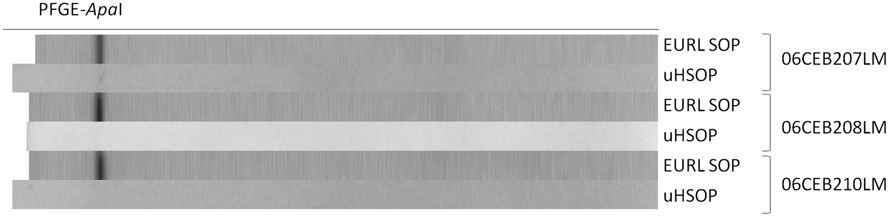
ApaI pulsed-field gel electrophoresis (PFGE) profiles from nontypable strains generated by the EU Reference Laboratory standard operating procedure (EURL SOP) and updated version of Halpin standard operating procedure (uHSOP).
Ninety-one distinct combined profiles (69 AscI profiles and 85 ApaI profiles) characterized the total strain panel (Fig. 2). The 114 strains were representative of 35 of the 55 most frequent identification groups (Table 1). The 35 different identification groups were representative of 95% of the total NRL molecular database (B. Félix, personal communication). The panel was also representative of the main 5 molecular serogroups (49 IIa strains, 15 IIb strains, 13 IIc strains, 32 IVb strains, and 5 IVa strains).
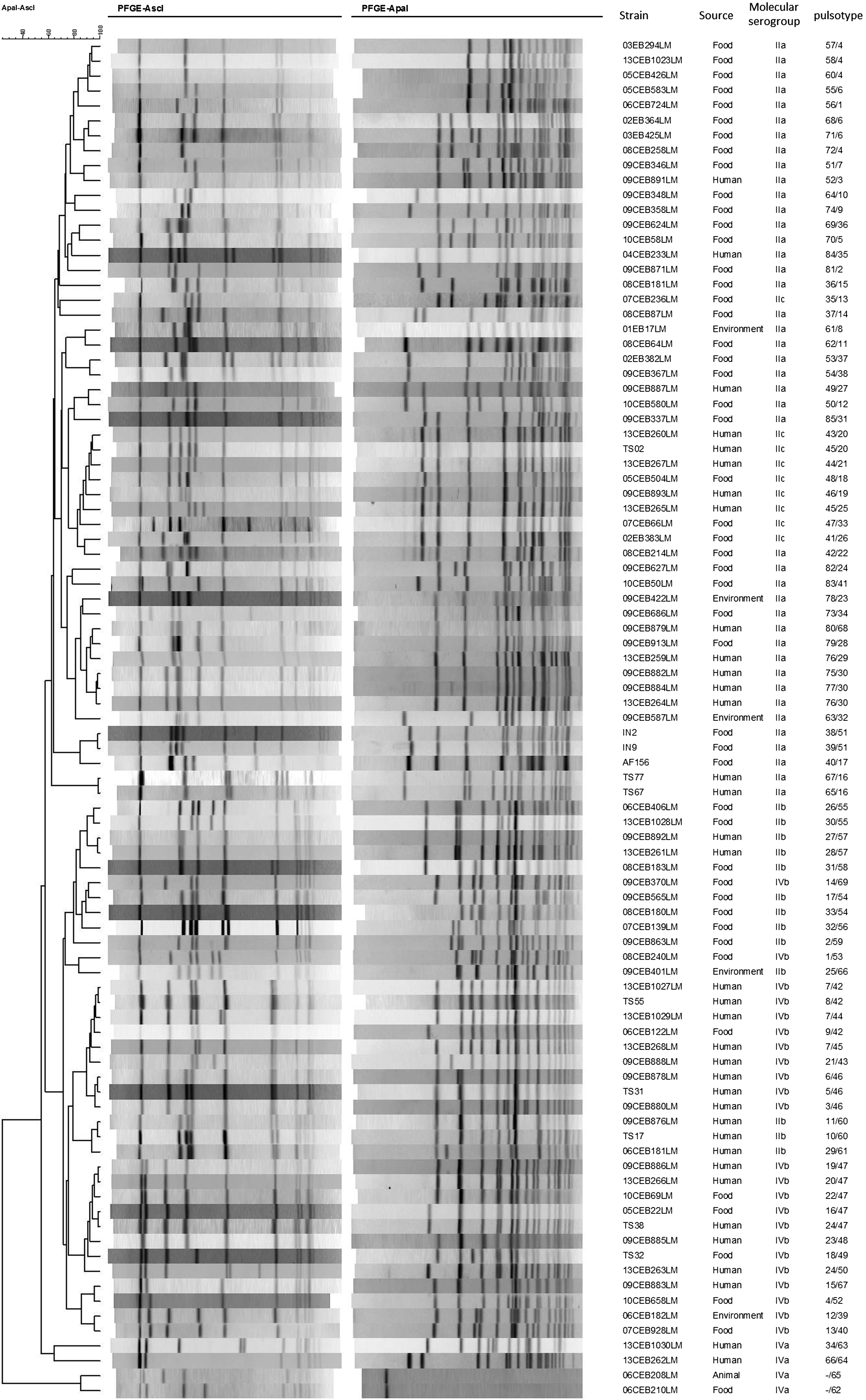
Dendrogram of similarity, for 91 Listeria monocytogenes strains based on the combined AscI-ApaI type obtained with the EU Reference Laboratory standard operating procedure, using the Dice coefficient and unweighted-pair group method using arithmetic averages with 1% of optimization and 1% of tolerance.
PFGE SOPs
The panel A strains were characterized with the HSOP at the SSI and with the EURL SOP (Félix et al., 2012b) at the EURL. The panel B strains were characterized at the EURL using the EURL SOP (Félix et al., 2012b) and uHSOP (PulseNet-International, 2011.
PFGE profile analysis
The profiles generated by the SSI were sent to the EURL in an XML file with the associated TIFF image. All the PFGE profiles obtained were centralized and compared within the same database using BioNumerics software V 7.1. A dendrogram was produced using the Dice coefficient and UPGMA, with a 1% tolerance limit and 1% optimization. These settings are similar to those used by PulseNet Europe in 2006 (Martin et al., 2006). Profiles were analyzed according to the EURL PFGE profile interpretation SOP (Félix et al., 2012a). PFGE profiles were classified as different if there was at least one different band. Each PFGE profile was arbitrarily assigned to a pulsotype number (Félix et al., 2012b).
Results
PFGE SOP comparison
Table 4 details the differences between the three SOPs at each step (e.g., reagent concentration and duration).
Including reference systems and extraction control needed. Only reagent cost is taken into account in the table as long as disposable equipment used is equivalent between the three SOPs.
Price for reagents purchased in France without special saving.
ALOA, Agar for Listeria according to Ottaviani and Agosti; BHIA, brain-heart infusion agar; BSA, bovine serum albumin; OD, optical density; PFGE, pulsed-field gel electrophoresis; SOP, standard operating procedure; TE, Tris EDTA buffer; TSYEA, Trypton soy yeast extract agar.
The cell suspension is obtained from plate culture in all three SOPs. All of them recommend the use of a nonselective medium (tryptone soy yeast extract agar, or brain-heart infusion agar). However, the EURL SOP includes the possibility of using a selective medium: agar for Listeria according to Ottaviani and Agosti. The incubation time for culture on a nonselective medium is shorter in both HSOPs.
The highest optical density of cell suspension is used in the EURL SOP. For the plug casting step, lysozyme is half as concentrated and the digestion temperature is significantly higher in HSOPs than in the EURL SOP. Proteinase K is fourfold more concentrated in both HSOPs. The concentration of agarose is lower in the HSOPs than in the EURL SOP. The lysis buffer volume and incubation temperature are higher in the HSOPs than in the EURL SOP.
For the restriction step, prerestriction—which consists of plug impregnation with a restriction buffer—is optional in HSOP, highly recommended in uHSOP, and required in the EURL SOP. Furthermore, bovine serum albumin (BSA) is not added in the prerestriction buffer of the HSOPs, while it is in the EURL SOP. The volume of the restriction solution in both HSOPs is twice that produced by the EURL SOP.
The amount of DNA restriction enzymes AscI and ApaI differs from SOP to SOP: In HSOP it is five- to eightfold more concentrated than in the EURL SOP. In uHSOP, these amounts are reduced but remain two-and-one-half to five times larger than in the EURL SOP. The incubation time for restriction is shorter in HSOP and reduced to 2 h in uHSOP.
For gel preparation, the HSOPs require the use of wide plug slices, while the EURL SOP requires the use of narrow plug slices. Wide plug slices generate 1-cm-width profiles and the dispatching of 15 plugs per gel, while narrow plugs generate 0.5-cm-width profiles and the dispatching of 30 plugs per gel. The two HSOPs recommend loading four large plug slices between two reference systems, compared to the EURL SOP, which recommends six narrow plug slices.
The EURL SOP is the only SOP to recommend the use of a control during analysis. The former reference system, L. monocytogenes H2446, previously used in the initial CDC PFGE SOP (Graves and Swaminathan, 2001), is still being used for extraction, restriction, and migration control in the EURL SOP (Félix et al., 2012b). The use of this control is optional in the EURL SOP, but highly recommended in order to figure out the origin of variations in quality within the profiles. The use of this control takes 2 wells in a gel and could be routinely used only in a 30-well gel.
Finally, HSOP and uHSOP are, respectively, 3.9 and 3.6 times more expensive than the EURL SOP. The cost calculation for the EURL SOP includes the extraction control.
Comparison of profiles obtained with the three SOPs
For each of the 3 SOPs, the 10 strains in the 5 epidemiological groups and the 7 strains in duplicate presented indistinguishable combined profiles. This demonstrated the reproducibility of each of the three SOPs within the same laboratory.
Comparison between the EURL SOP and HSOP
The ApaI profiles were indistinguishable for the 70 strains tested. The AscI profiles were indistinguishable for 69 strains. For strain 05CEB504LM (and its duplicate, strain 13CEB1026LM), the AscI profiles displayed a low-intensity band at 184 kbp with HSOP. This band did not appear on the profiles obtained with the EURL SOP (Fig. 3). These strains were tested three times with the two SOPs. The additional band was observed on all the profiles performed with HSOP.
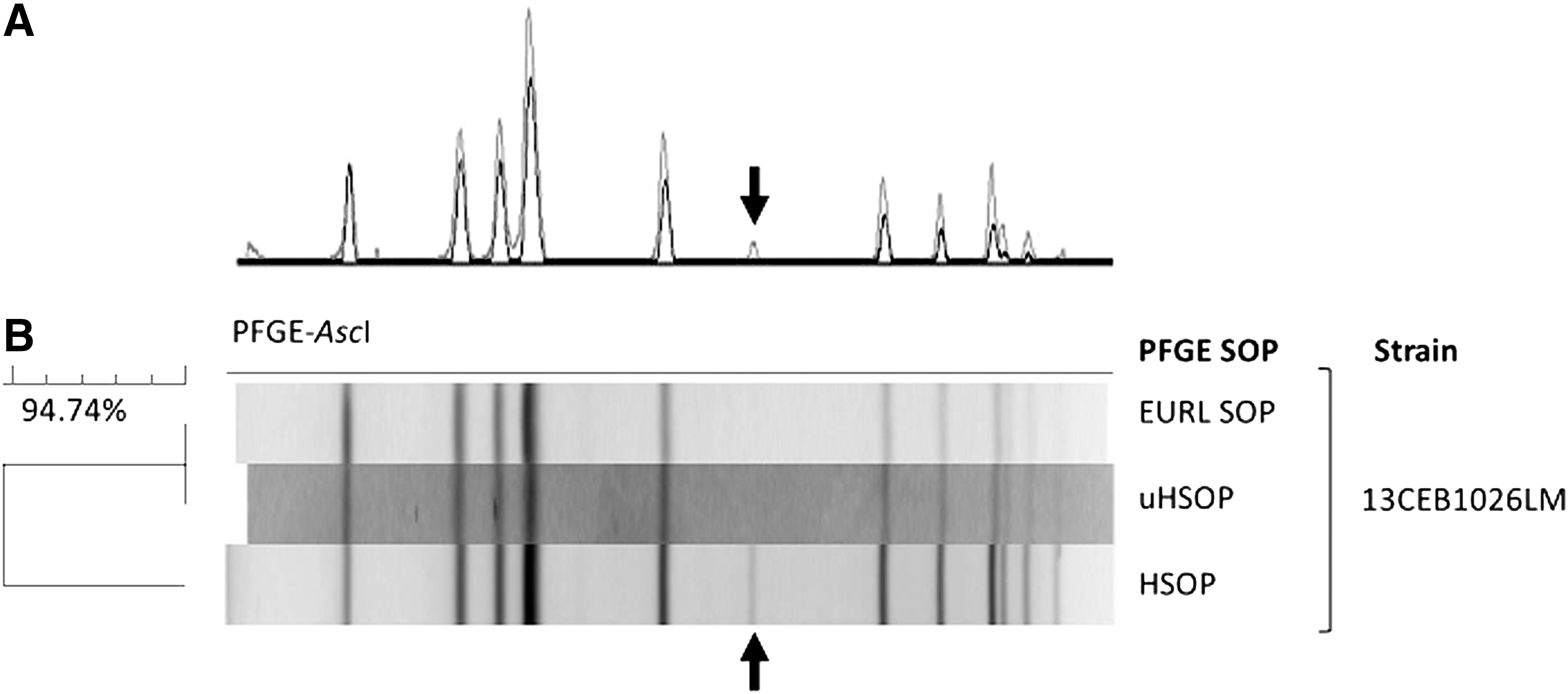
Pulsed-field gel electrophoresis (PFGE) profile differences observed among the three standard operating procedures (SOPs).
Comparison between the EURL SOP and uHSOP
All the AscI profiles were indistinguishable for the 114 strains. The AscI profiles were indistinguishable for strain 05CEB504LM. All the ApaI profiles were indistinguishable for the 111 strains typable with this enzyme. Three strains could not be typed by ApaI with the EURL SOP as well as with the updated Halpins' SOP (Fig. 1).
Discussion
The aim of our study was to compare three SOPs currently widely used in Europe: the EURL SOP, HSOP, and the more recent uHSOP. The strain panel used was representative of different molecular serogroups, different associated epidemiological data, and various combined PFGE profiles. This panel displayed the genomic diversity of L. monocytogenes observed within the French NRL molecular database.
The in-depth comparison of these SOPs revealed differences in the solution volumes, the concentration of reagents, and the incubation temperatures used. One of the main differences was the larger amount of restriction enzyme used in HSOP and uHSOP compared to the EURL SOP.
Another major difference between the three SOPs was the cost per strain. It is significantly lower with the EURL SOP. This difference is mainly due to the volume and concentration of the reagent and the size of plug slices. HSOP and the recent uHSOP require larger quantities of expensive reagents—Proteinase K, restriction enzyme, and agarose (Seakem gold). Furthermore, the Salmonella Braenderup H9812 reference system is more often inserted into the gel in HSOP and uHSOP, increasing the cost of these two protocols.
Regarding the duration of protocols (from bacterial cell culture to image acquisition), HSOP and uHSOP are 6 h shorter than the EURL SOP. Moreover, the culture incubation time could be reduced to 14 h in the HSOPs. Nevertheless, the growth culture for this incubation time is rarely sufficient (Donnelly and Nyachuba, 2007). In the three SOPs, the average time for PFGE analysis is around 48 h and the technical labor needed is similar.
Another difference is the use of plugs of different sizes. HSOP requires the use of wide plug slices, which are more difficult to manipulate. However, if a profile is covered by a spot or damaged by a blur, it is easier to avoid the spot or blur in a wider profile for which part of the signal remains exploitable. The EURL SOP requires the use of narrow plug slices, so there are twice as many profiles to run per gel.
All the profiles obtained in this study were similar in quality and indistinguishable for all but 1 of the 114 strains tested. The AscI profiles obtained for this strain using HSOP differed from those found with both uHSOP and the EURL SOP. This strain was previously used in the EURL PT trial 2009 and in the SSI's EQA-2 2013. Of the 14 NRLs having participated in the EURL PT Trial 2009, 1 laboratory (Lab 19) observed this additional band. Of the 18 NPHLs having participated in EQA-2, 6 laboratories (including the SSI) observed this same additional band.
The EURL had already observed an additional band for some profiles during each of the different PT trials it has previously organized (Félix et al., 2012b, 2013). For high-quality profiles, the main deviation observed was the presence of additional bands or the lack of bands in the profile. The origin of this deviation remains unexplained, but did not appear to be related to a given combined profile. Indeed, this deviation was observed for strains with different serotypes (1/2a, 1/2c, 3a) and combined profiles. This deviation likely is described as strain specific. Indeed, some strains are extremely sensitive to restriction digestion, and it may produce incompletely digested fragments depending on the procedures, reagents, or even on the experiments. The DNA methylation of the restriction site can explain some incomplete restriction. Moreover, the sensitivity of restriction enzymes to the methylated restriction site can vary according to the suppliers. Plasmid content (linear or nonlinear conformation) could also be the origin of this deviation (Barrett et al., 2006). However, it should be considered minor as it accounted for only 1% of all the combined profiles analyzed in each of the 3 PT trials set up in 2009 (90 combined profiles), 2010 (153 combined profiles), and 2012 (198 combined profiles). In this study, this deviation was also minor and accounted for about 1% of all profiles.
In conclusion, our results showed that PFGE profiles obtained with the different SOPs are both similar and comparable. This work should facilitate the exchange of profiles through the two NPHL/NRL networks. It will thus be possible to compare PFGE typing data from food and human reference laboratories. This research should also help improve the European surveillance of L. monocytogenes by facilitating the comparability of data from food and human sectors. Finally, this work should also contribute to the progress of the ECDC ELiTE (European Listeria Typing Exercise) project, which aims to compare typing results from clinical strains with those isolated from food in 2010 and 2011. This period corresponds to the European coordinated monitoring program on the prevalence of L. monocytogenes in certain ready-to-eat food categories retailed in EU Member States. A joint study between the ECDC, European Food Safety Authority, SSI, and EURL has therefore been initiated to compare the EURL SOP and the SOP used by the SSI to analyze PFGE profiles obtained during the ELiTE project.
Footnotes
Acknowledgments
We thank Dr. Laurent Guillier (ANSES) for his helpful advice on writing this article. This work was conducted as part of the activities of the European Union Reference Laboratory for Listeria monocytogenes and was supported by a grant from the European Commission's Directorate General for Health and Consumer Protection (DG SANCO).
Disclosure Statement
No competing financial interests exist.