
Abstract
Objectives:
Silver nanoparticles (AgNPs) as antibacterial agents are incorporated in many consumer products, while the use as antiviral agents is an ongoing area of research. We evaluated the antiviral properties of AgNPs of variable sizes (10, 75, and 110 nm) and doses (25, 50, and 100 μg/mL) at different contact time points against feline calicivirus (FCV), a surrogate for norovirus.
Materials and Methods:
Antiviral effects of the AgNPs were determined by comparing the infectivity of FCV, the appearance of cytopathic effects (CPEs), and the integrity of the viral capsid protein in viral suspension treated with AgNPs with the untreated controls.
Results:
The 10 nm AgNPs at 50 and 100 μg/mL concentrations inactivated the FCV beyond the limit of detection, resulting in a decrease of up to 6.5 log10 viral titer, prevented development of CPEs, and reduction in the western blot band signal of the viral capsid protein. No significant antiviral effect was observed for the 75 and 110 nm AgNPs.
Conclusions and Applications:
These results demonstrate that the antiviral effects of AgNPs are both size and dose dependent, thus potential applications of AgNPs as antiviral agents to prevent contamination of foodborne viruses need to consider size and dose effects.
Introduction
T
Among viral pathogens that cause foodborne illness, human norovirus (NoV) is the leading cause of sickness in about 267 million people, causing more than 200,000 deaths every year globally (Patel et al., 2009; Scallan et al., 2011; Debbink et al., 2012). The Centers for Disease Control and Prevention (CDC) estimates that NoV causes 19–21 million illnesses, resulting in more than 70,000 hospitalizations and nearly 800 deaths each year in the United States (
Control of NoV has also proven to be complicated as the virus persists in food, water, and in the environment for several days to months (Lopman et al., 2012) and can tolerate moderate heating, as is used in minimal food processing (Mormann et al., 2010). Possessing these characteristics of an infectious agent, NoV is considered the perfect human pathogen (Hall, 2012) and was classified as a Category B potential bioterrorism agent (Dancer, 2009).
Silver is increasingly used to prevent microbial contamination of food contact surfaces such as food packages (Martínez-Abad et al., 2013). Although ionic silver and AgNPs have been shown to kill viruses, several factors, including the effect of size and concentration on their antiviral efficacy, are still not clear (Martínez-Abad et al., 2013). We have recently shown that the 10 nm AgNPs disrupt the mucosa-associated gut bacteria and host immune response in orally gavaged rats (Williams et al., 2015). We have yet to understand whether such perturbations also occur with regard to the foodborne viruses as well as the community of gut viruses that are essential to host health (Duerkop and Hooper, 2013; Virgin, 2014). Due to the difficulty in cultivating human NoV in cell culture, feline calicivirus (FCV), which exhibits similar genome organization and structures, has been used as a surrogate for human NoV in several studies (Allwood et al., 2005; Doultree et al., 1999; Clay et al., 2006; Aboubakr et al., 2015; Shionoiri et al., 2015). In this study, characterized AgNPs of different sizes (10, 75, and 110 nm) and doses (25, 50, and 100 μg/mL) were used to investigate the antiviral effects of AgNPs against FCV based on the presence or absence of cytopathic effects (CPEs), virus infectivity, and western blot.
Materials and Methods
Virus propagation and determination of infectious virus titer
FCV strain 2280 (ATCC-VR-2057) and Crandell-Rees feline kidney (CRFK) cells (ATCCR-CCL-94™) were obtained from American Type Culture Collection (ATCC). The host cells were grown in Eagle's minimum essential medium (EMEM; ATCC-30-2003) supplemented with 8% horse serum, penicillin–streptomycin antibiotics, and fungizone. Virus propagation was achieved by infecting CRFK cells that formed 90% confluence in T-25 tissue culture flasks. The infected cells were maintained in EMEM (2% serum) at 37°C in a 5% CO2 incubator. Cells were observed daily under a microscope for the appearance of CPEs up to 96 h postinoculation.
The virus was harvested by two cycles of freezing and thawing, followed by low-speed centrifugation. The supernatant was filter sterilized and stored as stock at −80°C. Virus infectivity following the endpoint dilution method was used to determine the stock virus titer in CRFK cells grown in 96-well plates according to the Reed and Muench (1938) method and expressed as the median tissue culture infectious doses (TCID50/mL). The infectivity assay was repeated thrice and triplicates of each virus dilution were tested in each assay.
AgNP characterization
Citrate-stabilized AgNPs were procured from NanoComposix. The nanoparticles were characterized by the NanoCore Facility at the FDA-NCTR using transmission electron microscopy for particle size, shape, and aspect ratio, inductively coupled plasma mass spectrometry for silver mass and ionic concentrations, and dynamic light scattering for size distributions and dispersive index. Results of AgNP characterization matched the reports provided by the manufacturers.
Cytotoxicity
The presence or absence of visible cell toxicity and the maximum tolerated nontoxic dose (MTD) of the AgNPs were determined based on observations of morphological alterations and cell death following exposure of the CRFK cells to AgNPs. Briefly, double-fold serial dilutions of different sizes (10, 75, and 110 nm) and dosages of the AgNPs ranging from 500 to 0.98 μg/mL were used to inoculate monolayers of CRFK cells prepared in 96-well microplates, followed by incubation for 96 h at 37°C in a humidified 5% CO2 incubator, with twice per day microscopic examination similar to CPE observation. Lack of visible toxicity and cell survival was determined by comparing cells grown in the absence of the AgNP (negative control). The highest concentration levels that had no effect on the cells were selected as the MTD.
Antiviral efficacy of AgNPs
The antiviral properties of AgNPs of variable sizes (10, 75, and 110 nm) and doses (25, 50, and 100 μg/mL) were evaluated on FCV based on the presence or absence of CPEs and virus titer as endpoint measurements. Contact interaction of the virus with the AgNP was achieved inside the wells of sterile 24-well plates. Each AgNP was diluted in serum-free cell culture medium to 200, 100, and 50 μg/mL concentration. Equal volume (50 μL) of the virus suspension (9 log10 TCID50/mL) was mixed with the corresponding sizes and doses of AgNPs, followed by incubation at 37°C for 15, 30 min, 1, 2, and 4 h. The untreated virus suspension was similarly incubated in parallel with the AgNP-treated groups and used as positive control. After each contact time point, the positive control virus and the AgNP-exposed virus were eluted with 400 μL cell culture media supplemented with 10% serum, centrifuged, and the resulting supernatant (containing the virus) was serially diluted and used to inoculate CRFK cells grown to a 90% confluence in 96-well microplates. Mock cell controls receiving cell culture medium were run in parallel as negative controls. All cells were maintained in EMEM supplemented with 2% serum and observed daily under an inverted microscope for the appearance of CPEs up to 6 days.
The experiment was repeated thrice and triplicates of each virus dilution were tested in each experiment. Mean virus titers obtained as log10 TCID50/mL in positive controls were compared with the AgNP-treated groups to determine virus titer reduction as a result of the AgNP exposure at each contact time point. Statistical significance was determined using the two-sample t-test to compare the mean virus titer between the untreated positive control and each AgNP-treated group. In addition, the effects of AgNP exposure of the FCV on the development of CPEs were similarly monitored by inoculating AgNP-treated FCV and positive controls to monolayers of CRFK cells grown to a 90% confluence in T-25 tissue culture flasks.
Western blot analysis
Western blot analysis targeting the FCV major capsid protein (VP1) was performed using untreated FCV control and those treated with 10, 75, and 110 nm AgNPs at the 50 μg/mL dose. The FCV proteins were incubated overnight at 4°C with monoclonal antibodies specific for FCV VP1 protein (US Biological), immunoprecipitated with Dynabeads® Protein A (Life Technologies) before heat denaturation at 95°C for 5 min in sample buffer. Protein concentration was measured (Bradford, 1976) and equal amounts of the protein were run on 4–20% sodium dodecyl sulfate–polyacrylamide gel electrophoresis (SDS-PAGE) gels (Bio-Rad). The proteins were blot transferred using an iBlot (Invitrogen), followed by incubation with mouse anti-FCV monoclonal antibody specific for FCV VP1 protein (US Biological). The binding of the FCV antibody was visualized using a chemiluminescent substrate (GE Healthcare/Amersham). The relative intensities of western blot bands captured in X-ray films were determined for the untreated FCV control and those treated with the AgNP using NIH's ImageJ software package (
Results
Cell toxicity
Before determining the effects of AgNPs on FCV, the cytotoxicity of the different sizes (10, 75, and 110 nm) and dosages of the AgNPs ranging from 500 to 0.98 μg/mL to CRFK cells was evaluated to determine MTD. The AgNPs exhibited toxicity to the CRFK cells at concentrations ≥62.5 μg/mL, while no apparent toxicity was observed at concentrations ≤31.2 μg/mL. CRFK cells that received AgNPs at 62.5 μg/mL and greater concentrations showed signs of cell damage and became rounded, shrunken, and sloughed off from the bottom of the microplate well. No apparent cell damage was observed in control cells until 7 days of incubation. Therefore, the highest tolerable dose of AgNPs on CRFK cells was considered to be at 31.2 μg/mL. For the TCID50 infectivity assay, all the AgNP concentrations in the initial virus–nanoparticle elution used to inoculate CRFK cells were ≤25 μg/mL. Thus, we do not expect host cell toxicity to interfere with infectivity as the elution used for the TCID50 assay further underwent several 10-fold serial dilutions.
The antiviral effects of AgNPs against FCV
The antiviral effects of the AgNP on FCV are shown in Figure 1. The CRFK cells alone served as a negative control (Fig. 1a). CPE was observed in cells infected with FCV used as a positive control (Fig. 1b) within 48 h. This was characterized by rounding and shrinking of cell that followed complete detachment of cells in the monolayer. The infectivity of FCV was affected by the 10 nm AgNP (at 50 and 100 μg/mL) as evidenced by the absence of CPEs in CRFK cells inoculated with 10 nm AgNP-treated FCV (Fig. 1c). In all doses (25, 50, and 100 μg/mL), the 75 and 110 nm AgNPs (Fig. 1d, e) were not effective in inhibiting the development of CPEs.
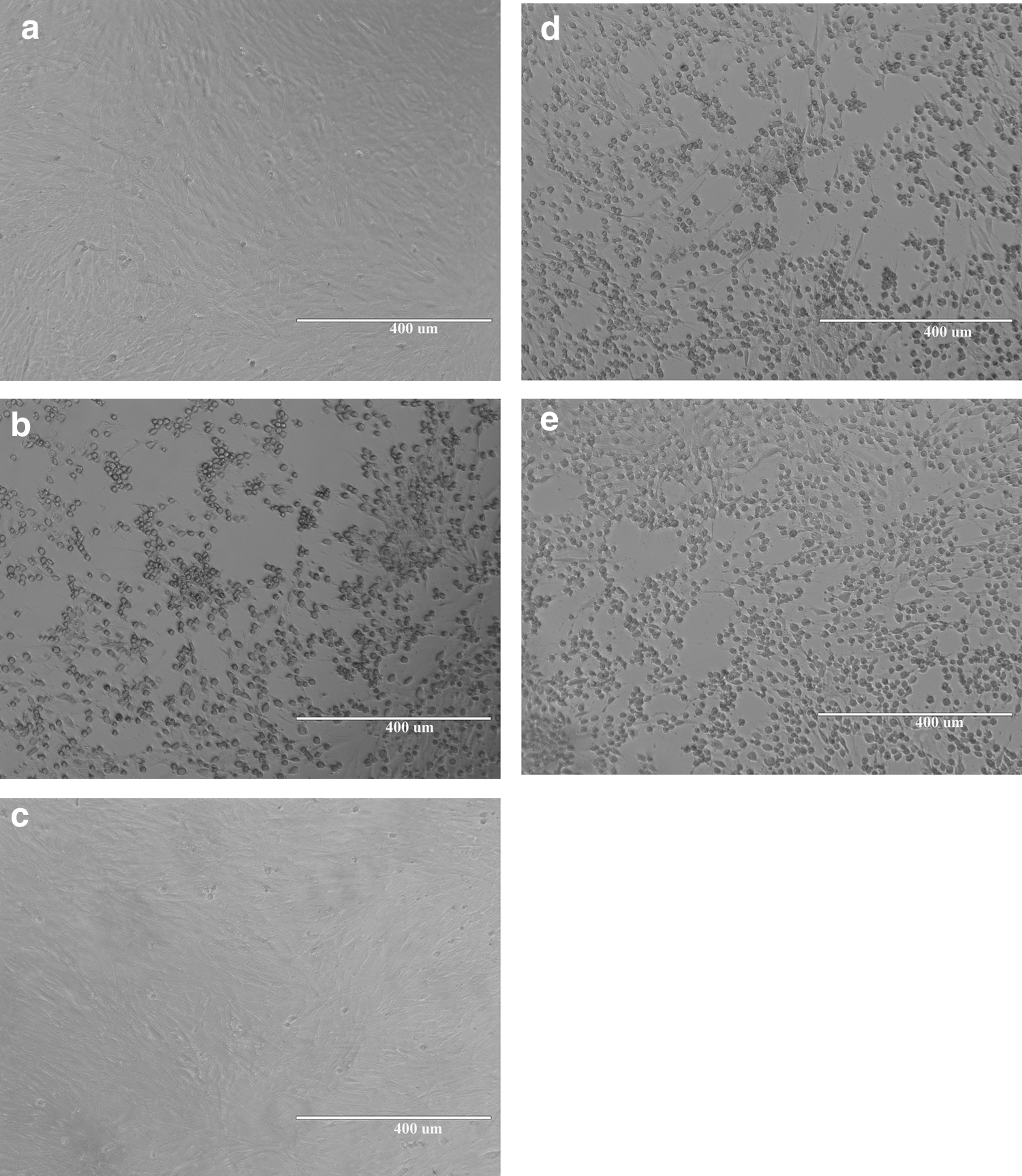
Appearance of CRFK cells infected with untreated and pretreated FCV with different sizes of AgNPs for 4 h at 50 μg/mL concentration. Mock cells that served as negative controls
The results of the AgNP effect on virus survival are shown in Figure 2. Similar to the CPE as seen in Figure 1, only the 10 nm AgNP was found to show significant reduction in virus titer (Fig. 2a). Compared with the viral titer (6.5 log10 TCID50) of the untreated positive control virus, the virus titer in the 10 nm AgNP (50 and 100 μg/mL) -treated virus showed a loss of 5.7 log10 TCID50 (p < 0.001) at 2 h postexposure with the AgNP. Similarly, compared with the viral titer (6.5 log10 TCID50) of the untreated positive control virus at 4 h, the 10 nm AgNP (50 and 100 μg/mL) -exposed FCV titer was significantly (p < 0.005) reduced beyond the limit of detection. Among the three AgNP sizes tested, only the 10 nm size (50 and 100 μg/mL) totally reduced the virus titer within 4 h, achieving a virus titer loss of 6.5 log10 TCID50. Such dose-dependent effect was also observed for the 10 nm AgNP at the earlier contact time points of 15 and 30 min. At the 50 μg/mL dose, the 10 nm AgNP showed a 2.3 log10 TCID50 (p < 0.001) titer loss, while the 100 μg/mL dose showed a slightly higher titer reduction of 3.2 log10 TCID50 (p < 0.001). The lowest dosage (25 μg/mL) of the 10 nm AgNP showed no appreciable titer loss at any time point (Fig. 2a). On the other hand, the 75 and 110 nm AgNPs did not exhibit significant (p > 0.1) virus inactivation at any of the three dosages used (Fig. 2b, c).
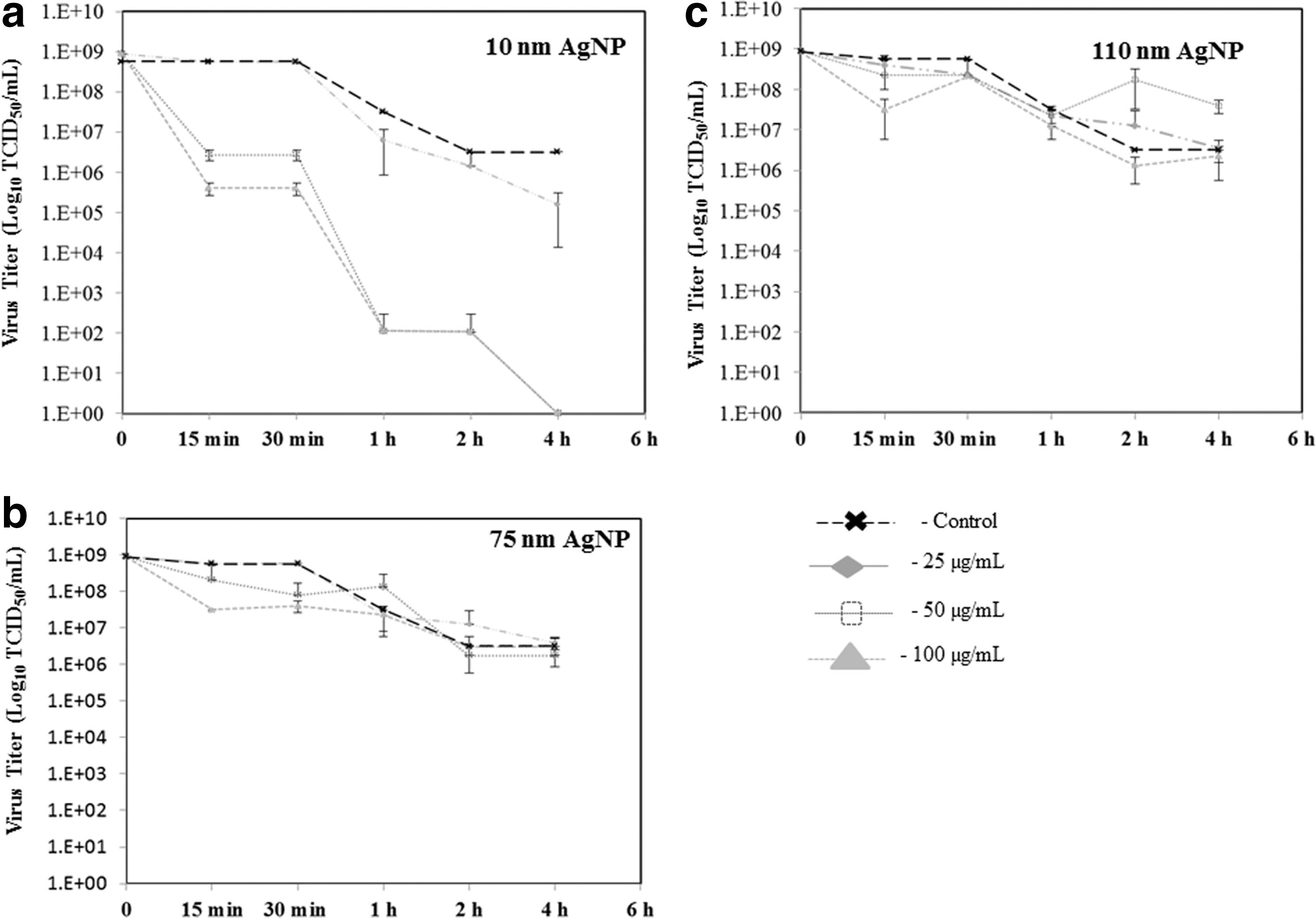
Effect of AgNPs on infectivity of FCV in CRFK cells. FCV at estimated titer of 9 log10 TCID50/mL was subjected to three doses (25, 50, and 100 μg/mL) and for each AgNP size:
SDS-PAGE analysis of AgNP-treated and nontreated FCVs showed a major capsid protein band of the expected size of 63 kDa (Fig. 3). Western blot analysis identified this band as an FCV major capsid protein (VP1). Compared with the nontreated positive control, the intensity of the protein band of the 10 nm AgNP-treated FCV was reduced by about 73%. Comparatively, no apparent effect was observed on the VP1 protein band of the 75 and 110 nm AgNP-treated FCV (Fig. 3).
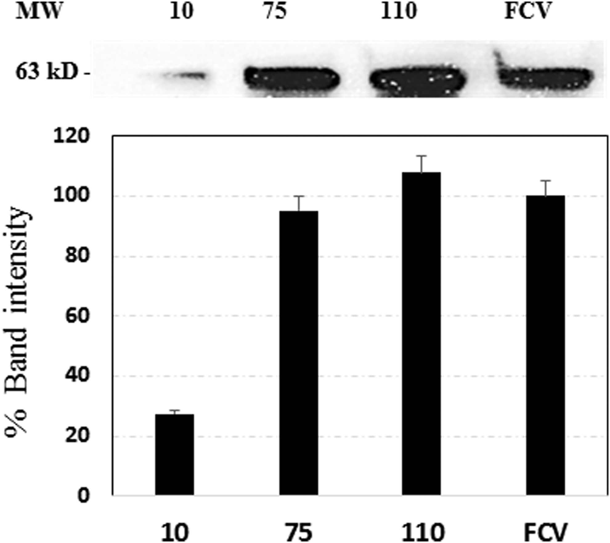
Detection of the FCV VP1 protein (63 kD MW) by western blot after treatment with AgNPs for 4 h at 50 μg/mL concentration. The numbers 10, 75, and 110 indicate the nanometer sizes of the AgNPs used to treat the virus, while FCV indicates the virus only used as positive control. The bar graphs represent the intensity of the western blot bands. AgNPs, silver nanoparticles; FCV, feline calicivirus.
Discussion
Silver has been used as a broad-spectrum antimicrobial agent in the form of metallic silver to keep food from spoilage, silver nitrate to prevent eye infections in newborns, and silver sulfadiazine for treating wounds. The use of AgNPs in food industry and the potential toxic effects following oral exposure have been extensively studied by Gaillet and Rouanet (2015). Although silver has antibacterial properties (Guggenbichler et al., 1999; Morones et al., 2005; Chen and Schluesener, 2008), the discovery of antibiotics and other disinfecting agents has significantly decreased silver use. The use of silver as antiviral agent, however, is a relatively recent effort largely linked to the increasing interest in nanotechnology. With regard to foods, silver-impregnated materials have been proposed to be used as antimicrobial packaging to reduce food contamination (Martínez-Abad et al., 2013). In this study, we evaluated the antiviral effects of AgNPs based on size (10, 75, and 110 nm) and dose (25, 50, and 100 μg/mL) against FCV, a commonly used surrogate for Caliciviruses, including human norovirus (Allwood et al., 2005; Aboubakr et al., 2015; Shionoiri et al., 2015).
The present study showed that only the 10 nm size AgNP was effective at significantly reducing FCV virus titer and achieving reduction of the FCV titer beyond the limit of detection. Since the size of FCV (27–40 nm) is comparable with the 10 nm AgNP, it is likely that there was nanoparticle and virus interaction based on size. Previously, such size-dependent virus–nanoparticle interaction has been suggested as the mode of antiviral action against HIV-1, in that only AgNPs in the 1–10 nm spectrums resulted in a loss of infectivity (Elechiguerra et al., 2005). Similarly, analysis of the antiviral effect of AgNPs ranging from 10 to 80 nm showed that reduction of monkey pox virus plaque-forming units was achieved only by the 10 nm AgNP (Rogers et al., 2008), while up to 50 nm AgNPs were reported to inhibit replication of hepatitis B virus (Lu et al., 2008).
AgNP–virus interaction studies on HIV-1 further confirmed that AgNPs in the 10 nm dimension preferentially bind to the viral gp120 glycoprotein and prevent the virus from binding to host cells (Lara et al., 2010). The western blot analysis in our study showed a reduction of the FCV VP1 viral capsid protein following the 10 nm AgNP treatment. It has been suggested that the FCV VP1 protein is important for attachment to functional receptor molecules in permissible cells (Bhella et al., 2008). Thus, the antiviral effect of the AgNP observed in this study may relate to the direct physical interaction with the FCV VP1 capsid protein. Although the amount of silver ions released by the AgNP was not determined in this study, previous work has shown that silver ions were less effective than AgNPs at reducing the infectivity of HIV-1 (Lara et al., 2010). Furthermore, there is an ongoing debate among nanomaterial scientists about the mass-based toxicity of nanoparticles and potential hazard assessment. We can speculate that both the ionic effect and mass-based inactivation of virus may contribute to the viral inactivation effect achieved by the 10 nm AgNP in this study.
The kinetics of virus inactivation also appears to be both time and dose dependent. At earlier time points of 15 and 30 min, the 50 and 100 μg/mL doses of the 10 nm AgNP resulted in the reduction of 2.3 and 3.2 log10 TCID50 of the infectious virus, respectively. In comparison, no titer loss was found as a result of the 25 μg/mL dose at 30 min of contact time. Compared with the virus titer in the untreated virus, the 50 and 100 μg/mL AgNP doses achieved significant loss of viral titer corresponding to 5.7 and 6.5 log10 TCID50 at 2 h, while no virus was detected at the 4-h contact time. However, the 25 μg/mL dose of the 10 nm AgNP effected only a marginal reduction of 1.7 log10 TCID50 virus titer at this time point and the surviving virus appears to be enough to induce cytopathic changes in the CRFK cells (Supplementary Fig. S1 and S2; Supplementary Data are available online at
The lack of antiviral activity from the larger sized AgNPs (75 and 110 nm) needs further investigation as nanoparticle size could influence agglomeration and rate of dissolution properties of the AgNP. Previously, we have seen similar lack of antibacterial activity of the 110 nm AgNP to the microbiota of rats orally gavaged with AgNPs (Williams et al., 2015), and based on the reports of Sintubin et al. (2011) who indicated agglomeration of large-sized nanoparticles, we speculated that the 110 nm AgNP used in our experiments may agglomerate. Taken together, these results indicate that efficient virucidal effect of AgNPs against FCV requires optimum size, concentration, and sufficient contact time of the AgNP. Maintaining a sustained dose is particularly important for AgNP-based products that are meant to continuously provide antiviral effect.
Conclusions
Overall, this study demonstrates that AgNPs have potential applications as antiviral agents to prevent viral contamination of foodborne viruses such as noroviruses; however, the antiviral properties are size, dose, and time dependent.
Footnotes
Acknowledgments
The authors acknowledge the support provided by Dr. Anil Patri and Dr. Angel Paredes from the NanoCore facility at NCTR for characterizing the nanoparticles used in this study and Drs. Syed Ali, Rajesh Nayak, Marli Azevedo, and Carl Cerniglia for their thoughtful critique of the manuscript. A.Z.B. is supported by the FDA Commissioner Fellowship Program and K.W. and K.G. by the Oak Ridge Institute for Science and Education.
Disclosure Statement
The results and opinions presented in this article are the views of the authors and do not necessarily reflect those of the FDA.
References
Supplementary Material
Please find the following supplemental material available below.
For Open Access articles published under a Creative Commons License, all supplemental material carries the same license as the article it is associated with.
For non-Open Access articles published, all supplemental material carries a non-exclusive license, and permission requests for re-use of supplemental material or any part of supplemental material shall be sent directly to the copyright owner as specified in the copyright notice associated with the article.