
Abstract
In the past, Listeria monocytogenes has been isolated from game feces and meat. However, less information is available on the occurrence of L. monocytogenes in other specimens originating from game animals. Hence, the aim of this study was to get an overview of the occurrence and distribution of L. monocytogenes in game animals by characterization of isolates from different matrices. For that purpose, samples were collected from red deer (Cervus elaphus), wild boars (Sus scrofa), and feed during the hunting season 2011–2012 in three different regions of Germany and Austria. Six samples from each animal were examined: tonsils, content of the rumen or the stomach, liver, intestinal lymph nodes, cecum content, and feces. Nineteen of 45 red deer and 12 of 49 wild boars were found to be positive for L. monocytogenes as well as 4 of 22 pooled feed samples. L. monocytogenes was isolated most frequently from the rumen of red deer (14 of 19) and the tonsils of wild boars (7 of 12). Serotypes 1/2a, 1/2b, 4a, and 4b were detected in samples of game animals and feed, and serotypes 1/2a and 4b were the most prevalent serotypes. The presence of L. monocytogenes serotype 4a had not yet been described in red deer. This might be due to the fact that it was only isolated from the content of rumen and that no other study has yet examined ruminal content. Pulsed-field gel electrophoresis showed a wide variety of strains. Some strains occurred in both species and feed samples, but one strain was dominant in one region. The results show that red deer and wild boars can be carriers of L. monocytogenes in different matrices, although the feces samples can be negative.
Introduction
L
In the past, L. monocytogenes has also been isolated from the feces of healthy game animals and their environment (Weis and Seeliger, 1975; Yoshida et al., 2000; Hayashidani et al., 2002; Lyautey et al., 2007; French et al., 2010; Wacheck et al., 2010; Obwegeser et al., 2012; Sasaki et al., 2013) and game carcasses and game meat (Kanai et al., 1997; Deutz et al., 2000; Jakšić et al., 2003; Apelt, 2007; Türck, 2008; Wacheck, 2008; Membré et al., 2011; Avagnina et al., 2012; Bortolas et al., 2012; Stüber, 2012). In a previous study, L. monocytogenes was found in the tonsils of wild boars (Wacheck et al., 2010), but no studies on the occurrence of L. monocytogenes in other organs of healthy game animals were published. For this reason, the aim of this study was to get an overview of the occurrence and distribution of L. monocytogenes in red deer (Cervus elaphus) and wild boar (Sus scrofa) populations and their feed in different regions by characterization of isolates from different matrices.
Materials and Methods
Samples
Between November 2011 and February 2012, samples originating from 45 healthy red deer and 49 healthy wild boars, which were hunted for consumption, were collected in three regions. The animals have been observed by the hunters before they got shot, especially for central nervous system disorders. When opening the carcasses, no abnormalities were recognized. Therefore, we assume that the animals were healthy. Region I is the Bavarian Forest National Park, Germany, established in 1970 and neighboring hunting ground (five areas A–E separated by an average 8 km). The deer were hunted in the hunting grounds (areas D and E) or shot in front of compounds (areas A, B, and C). Red deer were kept in these compounds over the winter and fed to prevent excessive damage to the forest. Of the ∼470 free-living red deer in the national park, 45 were examined. Additionally, four wild boars were shot in front of a compound in area C.
Region II is a park about 140 km southwest of region I in Germany, where about 1000 wild boars live, surrounded by a fence. In this park, game animals have been kept for hunting for more than 250 years. From this area, 25 wild boars were tested for the occurrence of L. monocytogenes.
Region III is a private hunting ground about 120 km southeast of region I in Austria. Since 1972, game animals have been kept there for hunting, also surrounded by a fence; 20 wild boars were examined from this area.
From each freshly shot animal (maximum 1 h postshooting), six specimens were collected and examined: tonsils, content of the rumen or the stomach, liver, intestinal lymph nodes, cecum content, and feces. To avoid contamination, sterile instruments were used for each animal. First, tonsils, liver, and intestinal lymph nodes were removed before the rumen, stomach, cecum, and rectum were opened. The content of the cecum and the feces were dripped in separate sterile bags. After opening the rumen and stomach, the content was taken by everting the sterile bags and using them like a glove. Additionally, 22 pooled feed samples from regions I and II from red deer feed (5 hay, 5 silage, 5 fodder beets, and 5 apple pomace pools) and wild boar feed (2 corn pools) were examined for the occurrence of L. monocytogenes. Sterile instruments were used for each sample. The pooled samples contained seven specimens for silage and five samples for corn pools, and for each of hay, fodder beets, and apple pomace, four samples were taken. Each sample material was put in a separate sterile bag and stored at +4°C for maximum 2 days until examination.
Isolation of L. monocytogenes
The bacteria were isolated and confirmed according to the horizontal detection method for food and feed (DIN EN ISO 11290-1:2005-01). This method was used to analyze the feed samples, and based on experience from previous studies (not published), it can also be applied to organ and fecal samples. To avoid contamination from the surface of the tissue, the surface of the intestinal lymph nodes, liver, and tonsils were flame treated using a Bunsen burner before taking the sample. The samples were diluted in Half-Fraser Broth and incubated for 24 ± 2 h at 30°C for enrichment. 0.1 mL of the incubated Half-Fraser Broth was transferred to 10 mL of Fraser Broth and incubated for 48 ± 2 h at 37°C. The samples were cultured from the Half-Fraser Broth and the Fraser Broth (Merck, Darmstadt, Germany) onto ChromID™ Ottaviani Agosti Agar (Biomérieux, Nürtingen, Germany) and Oxford agar (Merck) and incubated for 24 ± 2 h at 37°C. L. monocytogenes presumptive colonies were biochemically confirmed with API® Listeria test (Biomérieux). From each positive sample, one isolate was obtained and preserved for the following analyses.
Serotyping
The isolates were serotyped by slide agglutination tests using the scheme of Seeliger and Höhne (1979) and according to the manufacturer's instructions for antisera (MAST Group, Merseyside, United Kingdom). The molecular serotypes were determined by polymerase chain reaction (data not shown) according to Doumith et al. (2004).
Pulsed-field gel electrophoresis
Pulsed-field gel electrophoresis (PFGE) was performed using a protocol based on the standard operating procedure for PulseNet PFGE of L. monocytogenes (
The L. monocytogenes isolates were restricted with the enzymes ApaI (50,000 U/mL) and AscI (10,000 U/mL) (New England Biolabs, Frankfurt, Germany). The reference strain Salmonella Braenderup H9812 BAA-664™ (ATCC®, Manassas, VA), which served as a size standard, was digested by XbaI (20,000 U/mL; New England Biolabs). Strains were digested at +37°C for 5 h.
The pulse time was ramped from 4 to 40 s for more than 21 h at +14°C using the CHEF Mapper System (Bio-Rad, Munich, Germany). PFGE patterns were analyzed and compared with BioNumerics software version 7.3 (Applied Maths, Ghent, Belgium). For the dendrogram, the average of both experiments (ApaI and AscI) and the unweighted pair group matching algorithm (UPGMA) were used.
Results
Isolation of L. monocytogenes
In total, 19 of 45 red deer (42.2%) were found to be positive for L. monocytogenes in at least one sample. Of these positive verifications, the content of the rumen showed the most frequency (14 animals). Three red deer harbored L. monocytogenes in the tonsils, rumen, cecum, and feces. In seven cases, only the samples from the rumen were positive (Table 1).
n.t., nontypable by agglutination, PCR serogroup IIa.
Altogether, 12 of 49 wild boars (24.5%) were positive for L. monocytogenes in at least one sample. Here, the highest positive verifications were from the tonsils (seven animals). In one animal, L. monocytogenes was isolated from the liver, intestinal lymph nodes, content of the cecum, and feces. In five cases, only the tonsil samples were positive (Table 1).
In 4 of the 22 pooled feed samples, L. monocytogenes were detected. Two pooled samples of hay, one pooled sample of silage, and one pooled sample of fodder beets were positive.
In total, 58 L. monocytogenes isolates were obtained from red deer (35), wild boars (19), and feed samples (4). A higher number of colonies were isolated from Oxford agar than from ChromID Ottaviani Agosti Agar, especially in the cultivation from Half-Fraser Broth.
Serotyping
Altogether, 57 L. monocytogenes isolates were assigned to four different serotypes (Table 1). In one isolate (ruminal content, red deer #112), the serotype could not be determined by slide agglutination; however, the molecular serotype of the isolate was identical to the isolate with serotype 1/2a from feces of the same animal (IIa, data not shown). The isolates from red deer were positive for serotypes 1/2a, 4b, 1/2b, and 4a, while the isolates from wild boars were positive for serotypes 1/2a and 4b and those from feed samples for serotypes 1/2a and 1/2b.
Pulsed-field gel electrophoresis
Two red deer (#130 and 135) carried two and three L. monocytogenes isolates with different PFGE patterns, respectively. One wild boar carried L. monocytogenes isolates in the liver and cecum with different patterns (#30). The four isolates of L. monocytogenes from the wild boar #5 were distributed among two different patterns with two isolates each per pattern (Fig. 1).
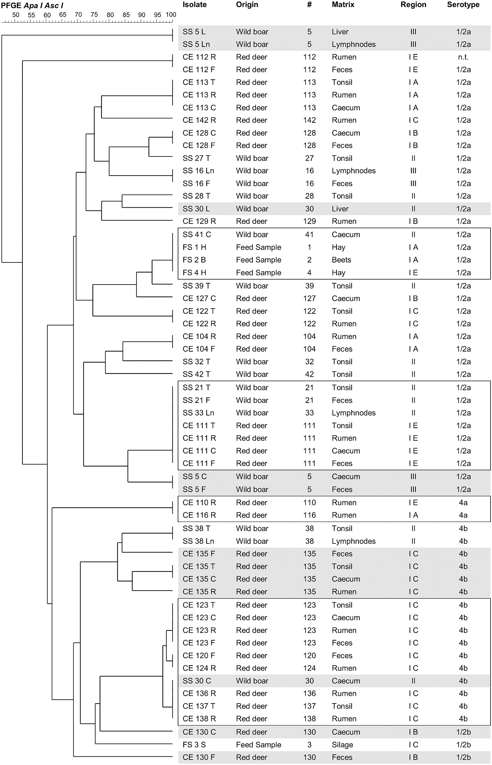
Dendrogram analysis of Listeria monocytogenes isolates from red deer, wild boars, and feed samples. The isolate names, the origin, and the serotypes are indicated. The tree was constructed by BioNumerics software v. 7.3 using the average of experiments and UPGMA on a matrix resulting from comparison of PFGE ApaI and AscI patterns. CE, Cervus elaphus (red deer); FS, feed samples; SS, Sus scrofa (wild boars). The scales at the top indicate the similarity indices (in percentages). Isolates from the same origin with distinguishable PFGE ApaI and AscI patterns are highlighted in gray. Isolates from different origins with indistinguishable or almost indistinguishable (more than 96.0% similarity) PFGE ApaI and AscI patterns are shown in boxes. PFGE, pulsed-field gel electrophoresis; UPGMA, unweighted pair group matching algorithm.
Seven red deer (#104, 111, 112, 113, 122, 123, and 128) and three wild boars (#16, 21, and 38) each possessed L. monocytogenes isolates with indistinguishable patterns in at least two different sample matrices.
Two isolates of different red deer (#110 and 116), originating from areas E and A in region I, showed indistinguishable patterns. The positive pools from hay and fodder beets from region I possessed L. monocytogenes isolates with indistinguishable patterns; this pattern was also identified in the isolate from the cecum of a wild boar (#41) from region II. There was no direct or indirect contact between the wild boar and the hay and the fodder beets.
The isolates from red deer (#111; region I) and two wild boars (#21 and 33; region II) had indistinguishable patterns. Furthermore, there was a group consisting of six red deer (#120, 123, 124, 136, 137, and 138; region I C) and one wild boar (#30; region II) whose isolates showed very closely related patterns (Fig. 1). Contact between wild boars from region II and red deer from region I could be ruled out. There was no direct or indirect contact between the animals.
Discussion
Wild animals have been described as reservoir and asymptomatic carriers of L. monocytogenes (Giovannini et al., 1988; Yoshida et al., 2000; Hayashidani et al., 2002; Lyautey et al., 2007; Wacheck et al., 2010). This study shows that L. monocytogenes is not only shed with feces, as described by many authors. Red deer and wild boars also carry the pathogen in different matrices, such as the rumen content, tonsils, intestinal lymph nodes, and liver. The distribution of L. monocytogenes in the different matrices could be due to oral intake and subclinical infection as none of the animals showed clinical symptoms. To our best knowledge, no work has been published on L. monocytogenes in different organs of game animals. This study is the first to examine all previously mentioned organs of healthy game animals for the presence of L. monocytogenes. It was shown that more animals were determined to be positive for L. monocytogenes by testing different matrices (19/45 red deer and 12/49 wild boars) than by examining only the feces (8/45 red deer and 3/49 wild boars).
In the literature, the serotypes 1/2a, 1/2b, and 4b have been described to occur in red deer, sika deer, fallow deer, roe deer, and chamois, with 1/2a and 4b as the main serotypes and 1/2b as a rare serotype (Weis and Seeliger, 1975; Eriksen et al., 1988; Tham et al., 1999; Yoshida et al., 2000; Schwaiger et al., 2005). In this study, the serotypes 1/2a and 4b dominated as well, but serotypes 1/2b and 4a were determined, too. The serotype 4a was only detected in ruminal content. As far as we know, serotype 4a has not yet been described in red deer. The reason may be that no ruminal content, but only samples of soil, feed, feces/intestinal content, and brain, has been examined in the literature. Maybe L. monocytogenes serotype 4a could also be found in other matrices but might be overgrown by other serotypes when it is cultured on agars.
In the few studies about L. monocytogenes serotypes from wild boars, the main serotypes were 1/2a and 4b, but 1/2b has also been described (Hayashidani et al., 2002; Wacheck et al., 2010). In this study, the isolates were all positive for either serotype 1/2a or 4b. The serotype 1/2b may not have occurred because of the low number of wild boars examined.
Two red deer and two wild boars each carried L. monocytogenes isolates with different PFGE patterns. This substantiates the fact that it is possible that one animal carries several L. monocytogenes strains (Tham et al., 1999; Ho et al., 2007).
Eight red deer and four wild boars carried L. monocytogenes isolates with indistinguishable patterns in different sample matrices. Contamination between samples taken from an animal would be possible but is unlikely because samples were always taken in the same order, and often, negative samples followed positive samples. Identical strains in different sample matrices can occur when L. monocytogenes amplifies. Nightingale et al. (2004) showed that L. monocytogenes ingested with feed multiplies in the intestinal tract of cattle and that the bacteria are reintroduced and dispersed into the environment. We believe that L. monocytogenes multiplies in the intestinal tract of red deer and wild boars as well and is thus disseminated to different tissues and organs of the host animal. Our study demonstrates that L. monocytogenes can also be found in lymphatic tissue of healthy red deer and wild boars and in liver tissue of healthy wild boars.
A continuous fecal oral enrichment cycle is claimed to exist in domesticated ruminants (Vázquez-Boland et al., 2001; Nightingale et al., 2004); the current findings suggest a similar phenomenon in case of game animals. The occurrence of L. monocytogenes–positive wild boars was clearly different in regions II (10/25) and III (2/20). The higher detection rate in region II might be due to enrichment in the soil. While wild boars have been living in region II for more than 250 years, they have only lived for 42 years in region III. This could lead to a lower contamination rate of the soil in region III compared to that of region II. Our data show the current distribution of L. monocytogenes in the investigated areas, but no statement can be made on the evolution of the strains. In the literature, a linkage between animals (farm animals or wild animals), their manure/feces and L. monocytogenes–positive soil samples, or feed samples has been described (Nightingale et al., 2004; Szymczak et al., 2014). For red deer, the geographical distribution of the sampled animals was too wide, so no conclusion on persistence of L. monocytogenes in soil can be drawn.
Isolates from different animals and feed samples from different regions showed indistinguishable patterns (Fig. 1). A similar wide distribution of L. monocytogenes has been reported with strains having been found in different matrices also (silage, hay, water, and soil) (Gudmundsdottir et al., 2004; Nightingale et al., 2004; Esteban et al., 2009). In this study, some L. monocytogenes strains occurred in both animal species (red deer and wild boars) and feed samples and wild boars from different regions. Furthermore, it is possible that the six red deer, of which a cluster of closely related L. monocytogenes strains was isolated, are part of a herd with a preferred location in which the related strains have been enriched over time or that they were infected by cross-contamination via feces of one animal in the herd. In contrast, there was no contact of the deer to the wild boar, which also carried a L. monocytogenes strain of the cluster of closely related strains.
Only 4 of the 22 pooled feed samples were positive, and the same isolates could not be found in feed and animals from the same region. We believe that the feedstuff fed in the compounds, where the animals are kept, is not the main source of infection. Research by Szymczak et al. (2014) demonstrated that L. monocytogenes was found in soil samples from land fertilized with manure, pastures fertilized with feces of domestic animals, and forests fertilized with feces of wild animals. Therefore, it seems more likely that the infections of the animals are due to the enrichment of L. monocytogenes in the soil. Further investigations should be undertaken on the occurrence of L. monocytogenes in the soil of the compounds.
Conclusion
It has been shown that red deer and wild boars are asymptomatic carriers of L. monocytogenes. Further studies to investigate the management of compounds and parks and their influence on the infection rate would be helpful. Red deer and wild boars are reservoirs for many different L. monocytogenes strains, and some of these strains seem to be widely distributed. These strains are maybe better adapted lineages, which can be found in many different animals and different places due to their broad distribution (Nightingale et al., 2004; Cantinelli et al., 2013). L. monocytogenes was not only found in the content of the gastrointestinal tract but also in the tonsils, liver, and intestinal lymph nodes of the animals.
Footnotes
Acknowledgments
We would like to thank the teams from the Bavarian Forest National Park, the Bavarian State Forest, and the other hunting grounds, especially M. Penn, R. Fischer, J. Knobling, F. Neudecker, and W. Höpfler. For the technical support with the PFGE, we thank V. Hohenester.
Disclosure Statement
No competing financial interests exist.