
Abstract
Campylobacter spp. are foodborne pathogens responsible for a significant portion of human cases of bacterial-mediated gastrointestinal disease. A primary method for the introduction of Campylobacter into the food supply is through poultry products. Reducing the number of Campylobacter on poultry products may reduce the incidence of human disease. Research has been conducted on the use of light to inactivate Campylobacter on poultry products and processing environments. More recently, the use of high intensity visible 405-nm light has been proposed for the elimination of pathogenic bacteria. This study investigated the ability of 405-nm light to reduce Campylobacter jejuni and Campylobacter coli in poultry products. Campylobacter in chicken exudate were placed onto chicken skin or food-grade stainless steel before treatment with 405-nm light. A range of 405-nm light doses were applied to cocktails of six C. jejuni or six C. coli strains in exudate at 10°C to minimize thermal effects. Little difference was observed between inactivation of C. jejuni and C. coli on poultry skin with only minor average reductions of 1.7 logs and 2.1 logs, respectively, at the maximal dose of 184–186 J/cm2. More noticeable differences were observed when the samples were placed on stainless steel and treated with a dose of 89 J/cm2, producing an average reduction of 3.0 logs for C. coli but only 1.1 logs for C. jejuni. The maximal dose (181–183 J/cm2) applied to Campylobacter on stainless steel produced significant (p ≤ 0.05) reductions for C. jejuni and C. coli of 4.9 logs and 5.1 logs, respectively. However, significant 405-nm-mediated reductions in Campylobacter numbers required exposure times to achieve necessary dose levels that might be impractical under processing conditions. In addition, the most potent exposure times likely produced secondary thermal effects by raising sample surface temperatures above 48°C.
Introduction
I
The objective to decrease Campylobacter numbers in poultry products has driven research on a variety of intervention technologies with the potential for achieving reductions (Gunther et al. 2015a, 2015b). Ultraviolet (UV) light has been extensively used to sterilize a range of different environmental surfaces as well as food products; this was observed to have significant activity against Campylobacter spp., reducing the number of Campylobacter on poultry and ham (Butler et al., 1987; Chun et al., 2009; Isohanni and Lyhs, 2009; Haughton et al., 2011). However, UV light may cause damage to the skin and eyes of workers (Matsumura and Ananthaswamy, 2004; Maclean et al., 2014).
Recent research has investigated the use of near UV (≤400 nm) and visible violet/blue light (≥400 nm) using inexpensive LED arrays as a potentially cheaper and safer method for eliminating microbes (Guffey and Wilborn, 2006; Maclean et al. 2009, 2013; Wasson et al., 2012; Bumah et al. 2013, 2015; Ghate et al., 2013; McKenzie et al. 2013, 2014; St Denis et al., 2013). Two articles have focused specifically on reducing Campylobacter by using either near UV or visible violet/blue light, but both used clear artificial media for Campylobacter suspensions in their experimental procedures. (Murdoch et al., 2010; Haughton et al., 2012).
It was our objective to evaluate the effectiveness of 405-nm light against multistrain Campylobacter jejuni and Campylobacter coli preparations in conditions that more completely approximate poultry products. We chose conditions that included the suspension of Campylobacter in chicken exudate that was alternately placed on chicken skin and food-grade stainless steel surfaces and with the experiments conducted at 10°C to reduce the influence of thermal effects from different doses of 405-nm light.
Materials and Methods
Bacterial strains
C. jejuni: poultry isolates RM1221, RM1188, and RM1285 and human isolates RM3194, RM1246, and RM1247 and C. coli: poultry isolates RM1182, RM1403, and RM1529 and human isolates RM4803, RM4796, and RM4780 were obtained from Dr. Robert Mandrell (USDA; Agricultural Research Service, Albany, CA) and maintained frozen (−80°C) until use. Three days before 405-nm light treatments, each strain was cultured on Brucella agar (1.5%) (Becton Dickinson, Sparks, MD) at 42°C in a microaerobic chamber (MACS-VA, Don Whitley, United Kingdom; 5% O2, 10% CO2, 85% N2) for 12 h. Individual isolates were cultured in Brucella broth at 42°C microaerobically for 24 h before treatment.
Sample preparation
For chicken skin and stainless steel experiments, 24 h cultures for each of the six individual C. jejuni or C. coli were removed from the microaerobic chamber. One hundred microliter aliquots were taken from each of the six C. jejuni cultures and combined to form a C. jejuni cocktail or from the six C. coli strains and combined to form a C. coli cocktail. The following procedures were identical for both C. jejuni and C. coli cocktails. Cells in the cocktails were microcentrifuged (7000 rpm, 5 min.) and the pellets were resuspended in 3 mL of sterile chicken exudate. Chicken exudate was collected from multiple commercially prepared whole broilers from a retail butcher. Exudate was irradiated to sterility (40 kGy) using a self-contained 137Cs gamma irradiator (Lockheed Georgia Company, Marietta, GA) at a dose of 0.073 kGy/min.
Fifty microliters of the Campylobacter exudate mixtures (∼108 CFU/mL) was placed on chicken skin medallions (10 cm2) or stainless steel medallions (12.6 cm2). Chicken skin medallions were cut from the skin from chicken thighs using a custom punch and irradiated to sterility, 20 kGy (0.073 kGy/min dose). Stainless steel medallions were laser cut from 16 gauge 304 mill finished stainless steel and sterilized by autoclave (121°C, 30 min). Chicken skin and stainless steel medallions inoculated with Campylobacter were placed at 4°C before treatment and during the experiment until all samples were treated.
For the light treatments of individual C. jejuni or C. coli strains, the strains were grown in Brucella broth for 24 h. For each strain, two 100 μL samples were placed in separate microcentrifuge tubes. Samples were pelleted (7000 rpm, 5 min) and supernatant was removed. Each strain had one pellet resuspended in 500 μL fresh Brucella broth and the other pellet was resuspended in 500 μL sterile chicken exudate. From each resuspension, 50 μL was placed in a 96-well plate for treatment and a second 50 μL aliquot in another 96-well plate as a control. All plates were placed at 4°C before treatment and during the experiment until all plates were treated.
405-nm light treatments
Treatment with 405-nm light was achieved using an LED Spot 100 high power lamp, powered by an LED powerdrive (Honle UV America, Inc., Marlboro, MA). The lamp was placed in a 10°C incubator 20.3 cm above the samples for treatment. A thermocouple probe was placed next to the samples and connected to an Easy View 15 data logger (Extech, Nashua, NH) to record changes in air temperature surrounding the samples during treatment (Table 1).
Temperature measured by thermocouple.
Temperature measured by IR camera.
All samples were photographed using a Ti32 IR camera (Fluke, Everett, WA) immediately before and after treatment to determine surface temperature of the samples (Table 1). Light treatments of the Campylobacter on chicken skin and stainless steel were conducted for 10 min with the LED powerdrive at 90% (306 ± 6mW/cm2/s), 65% (226 ± 5mW/cm2/s), or 42% (151 ± 3mW/cm2/s) power levels. Light treatments on the individual Campylobacter strains in 96-well plates were conducted for 5 min with the LED powerdrive at 42% power level. Irradiance (power density) was measured for each power level using a UV meter with a visible light sensor (Honle UV America, Inc.) in mW/cm2.
Irradiance values were multiplied by exposure times (seconds) to determine the doses, measured as J/cm2. Control samples were maintained in the same manner as treated samples except that they were never exposed to 405-nm light. Once all treatments were completed for an experiment, all samples were processed at the same time to determine the number of Campylobacter that survived treatment.
Enumeration of Campylobacter
Treated or control chicken skin or stainless steel medallions were placed in sterile Whirl-Pak stomacher bags (Nasco, Modesto, CA) containing 1 mL Brucella broth. Bags containing the chicken skin were mixed by Stomacher and the bags containing stainless steel were mixed by hand for 1 min. From each of the newly mixed samples, 300 μL was removed and serially diluted. Experiments investigating individual Campylobacter strains, suspended the strains in 50 μL Brucella broth or chicken exudate in 96-well plates. After light treatment, 250 μL of Brucella broth was added to each well, mixed, and serially diluted. Enumeration of viable Campylobacter was done by plating the serially diluted samples onto Brucella agar using the 6 × 6 drop plate method with six plating replicates per dilution (Chen et al., 2003).
Statistical analyses
Colony-forming units per milliliter were calculated for samples and expressed as log10 (CFU/mL). Data with 0 colony counts were transformed to random log10 values between 0 and 2 (experimental limit of detection, 100 CFU/mL). All studies were analyzed following a split-plot design model with the whole plot associated with different strains. Each study was replicated three times. Analysis of variance was performed for each study and mean separations were performed using the Bonferroni LSD technique (p ≤ 0.05 significance level; Miller, 1981; SAS Institute, Inc., 2004). Mean values along with standard errors were graphed for each analysis and letter values assigned to indicate mean values without letter values in common differed significantly. Where appropriate, second-order polynomial trend lines were calculated from the data and displayed along with their R2 values.
Results
The six C. jejuni strains suspended in chicken exudate were placed on chicken skin and exposed to varying doses of 405-nm light (Fig. 1). The maximum 405-nm light dose used in this set of experiments, 184 J/cm2, produced an average reduction in C. jejuni numbers of 1.7 logs. A 405-nm light dose of approximately half that, 92 J/cm2, produced an average reduction in C. jejuni numbers of 1.0 log that was not significantly different from the reductions produced by the 184 J/cm2 dose. The 184 J/cm2 light dose produced an average air temperature around the chicken skin samples of 36.8°C ± 1.4°C and an average surface temperature on the skin of 37.0°C ± 5.2°C (Table 1).
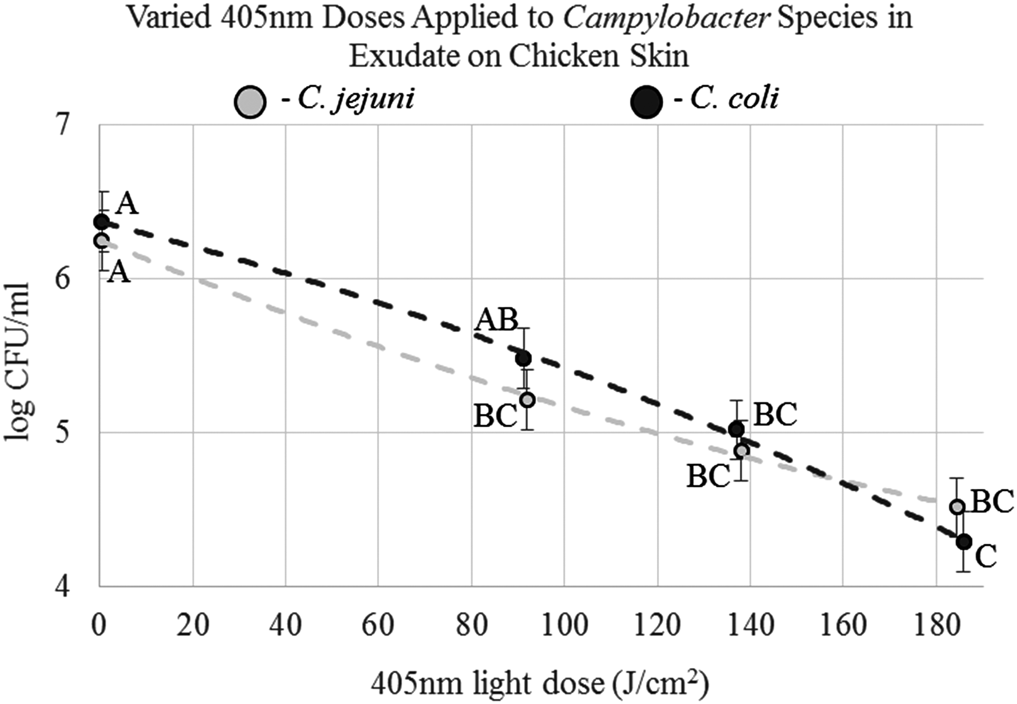
C. jejuni and C. coli strains suspended in sterile chicken exudate and placed (25 μLs total) on sterile chicken skin coupons. A range of different 405-nm light doses were applied to the samples and the numbers of Campylobacter surviving the light treatment were determined and recorded as mean log10 CFU/mL, with trend lines (R2 values 0.9986 [C. jejuni] and 0.9985 [C. coli]).
The six C. coli strains suspended in chicken exudate were placed on chicken skin and then exposed to varying doses of 405-nm light (Fig. 1). In these experiments, the maximum light dose used was 185.8 J/cm2 that produced an average reduction in C. coli numbers of 2.1 logs. When the light dose was approximately reduced by half, 91.2 J/cm2, the reduction in C. coli numbers was 0.9 logs, which was not significantly different from the reduction produced by 185.8 J/cm2. The 185.8 J/cm2 light dose produced an average temperature in the air around the chicken skin of 44.2°C ± 3.9°C and an average surface temperature on the skin of 44.3°C ± 4.2°C (Table 1). The differences between the average survival of C. jejuni versus C. coli strains at individual light doses were not significantly different (Fig. 1).
Next, the six-strain C. jejuni cocktail in exudate was placed on stainless steel and exposed to doses of 405-nm light similar to those used in the chicken skin experiments. As the light dose increased, the survival of C. jejuni on stainless steel decreased significantly (Fig. 2). The maximum light dose applied to C. jejuni on stainless steel, 183.4 J/cm2, produced an average reduction in C. jejuni numbers of 4.9 logs. When the light dose was approximately reduced by half, the average decline of C. jejuni numbers after treatment was reduced to 1.1 logs, which differed significantly from the average reduction of the maximum dose. The light dose of 183.4 J/cm2 produced an average air temperature around the experiment of 54.7°C ± 2.6°C and an average surface temperature of 56.3°C ± 3.2°C (Table 1).
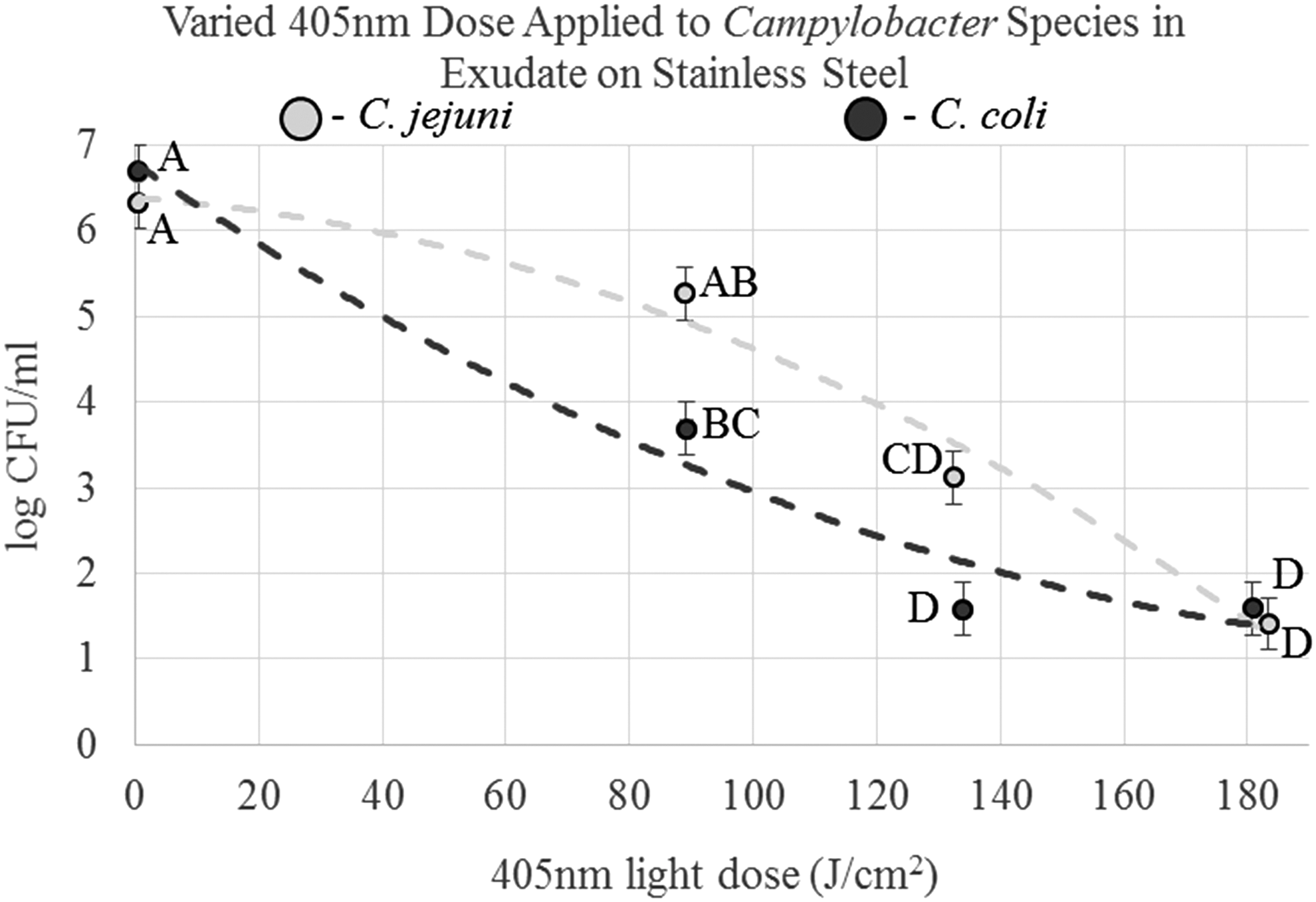
C. jejuni and C. coli strains suspended in sterile chicken exudate and placed (25 μLs total) on sterile stainless steel coupons. A range of different 405-nm light doses were applied to the samples and the numbers of Campylobacter surviving the light treatment were determined and recorded as mean log10 CFU/mL, with trend lines (R2 values 0.9800 [C. jejuni] and 0.9695 [C. coli]).
The six-strain C. coli cocktail was substituted for C. jejuni on the stainless steel and treated with 405-nm light (Fig. 2). When the maximum 405-nm light dose was used, 180.8J/cm2, an average reduction in C. coli numbers of 5.1 logs was observed. When the light dose was reduced to 89.2 J/cm2, the observed reduction in C. coli numbers was 3.1 logs, this reduction was significantly different from the average reduction achieved by the 180.8 J/cm2 dose.
The average air temperature around the stainless steel samples produced by the 180.8 J/cm2 dose was 51.1°C ± 6.9°C with the average surface temperature observed as 53.6°C ± 4.4°C. At the ∼89 and ∼133 J/cm2 light dose, the comparison of average survival of the C. jejuni strains with that of the C. coli strains failed to achieve significant differences by Bonferroni analysis (Fig. 2). A less conservative ANOVA (p < 0.05) contrast found that the Campylobacter species exhibited significantly dissimilar average survivals at both light doses.
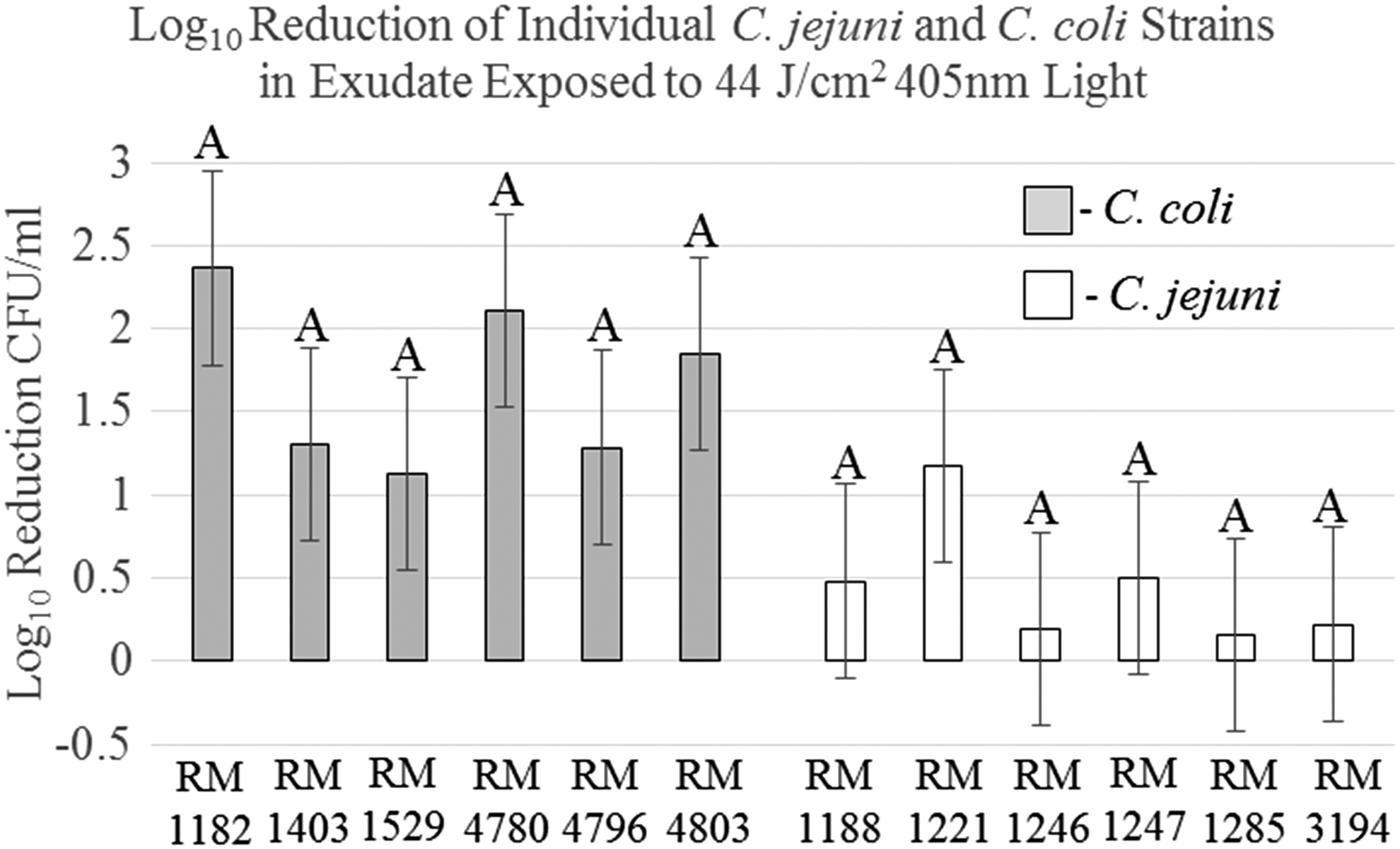
Individual C. jejuni and C. coli strains were suspended in Brucella broth or exudate and exposed to a 44 J/cm2 dose of 405-nm light. The 405-nm light treatments of C. jejuni strains produced larger average reductions in bacterial numbers in Brucella broth than in chicken exudate (Fig. 3). However, given the small reductions observed for the C. jejuni strains in Brucella broth or exudate, the differences in the reductions between the two environments were not significant for any C. jejuni strains. However, with the C. coli, four of the six strains had significantly greater treatment-mediated reductions in the Brucella broth than in chicken exudate (Fig. 3).
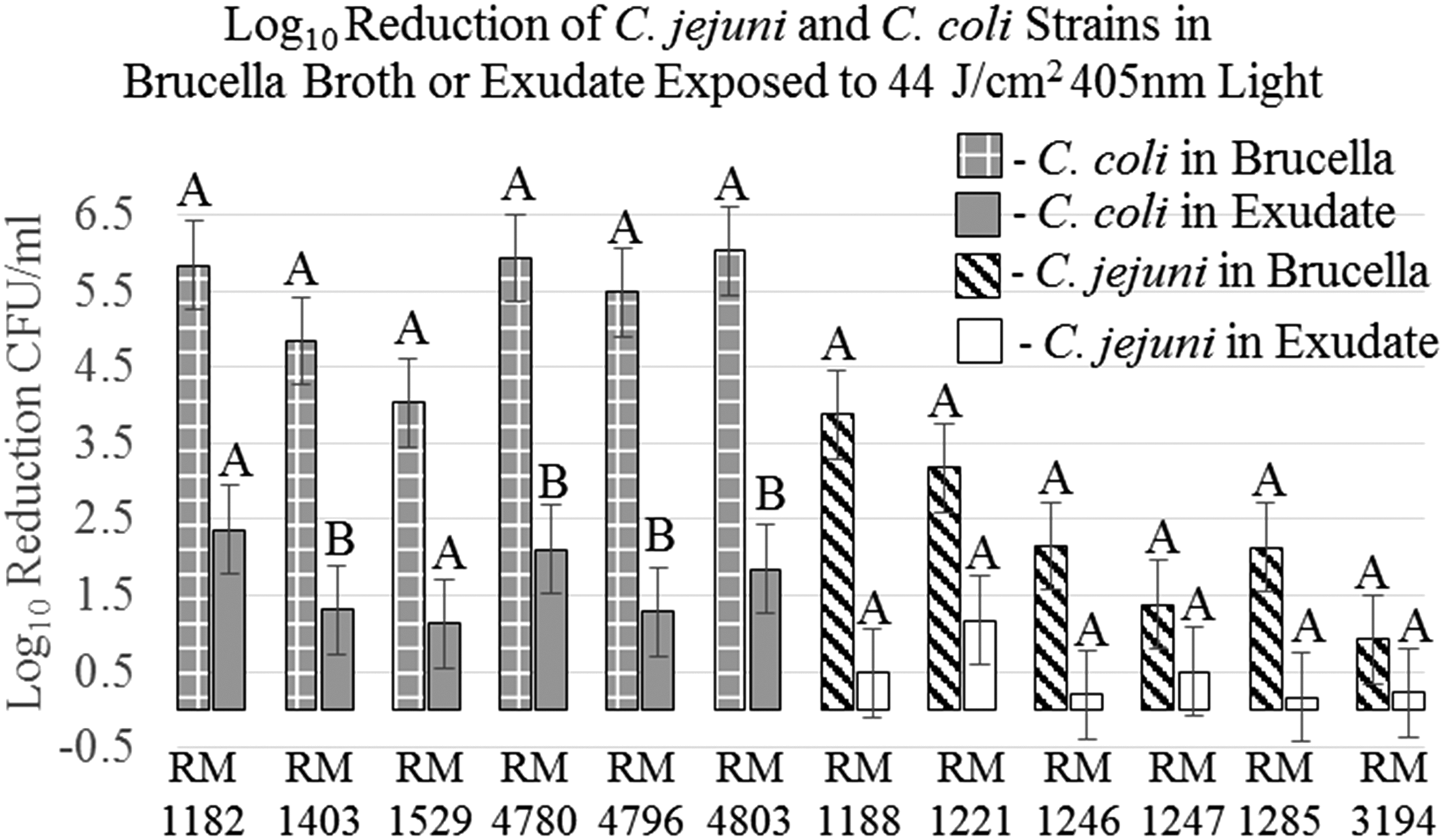
Results of six C. jejuni and C. coli strains comprising the cocktails used in the previous experiments suspended in Brucella broth or chicken exudate and treated with a 405-nm light dose of 44 J/cm2. The average log10 CFU/mL reduction of bacterial numbers from light treatment for the individual C. coli and C. jejuni strains was graphed along with the appropriate standard error to determine whether a statistical difference exists within an individual strain treated in Brucella broth versus exudate.
Although there was some variations between the reductions in treated C. jejuni numbers between C. jejuni strains in both Brucella broth and exudate, no variation was significant by Bonferroni analysis (Figs. 4 and 5). The reduction differences between strains RM1188 or RM1221 and RM3194 when treated in Brucella broth were significant when a less stringent ANOVA contrast was used (Fig. 4). C. coli strains did not have significant differences between the individual strains' mean reductions in bacterial numbers after light treatment in Brucella broth or exudate cultures (Figs. 4 and 5).
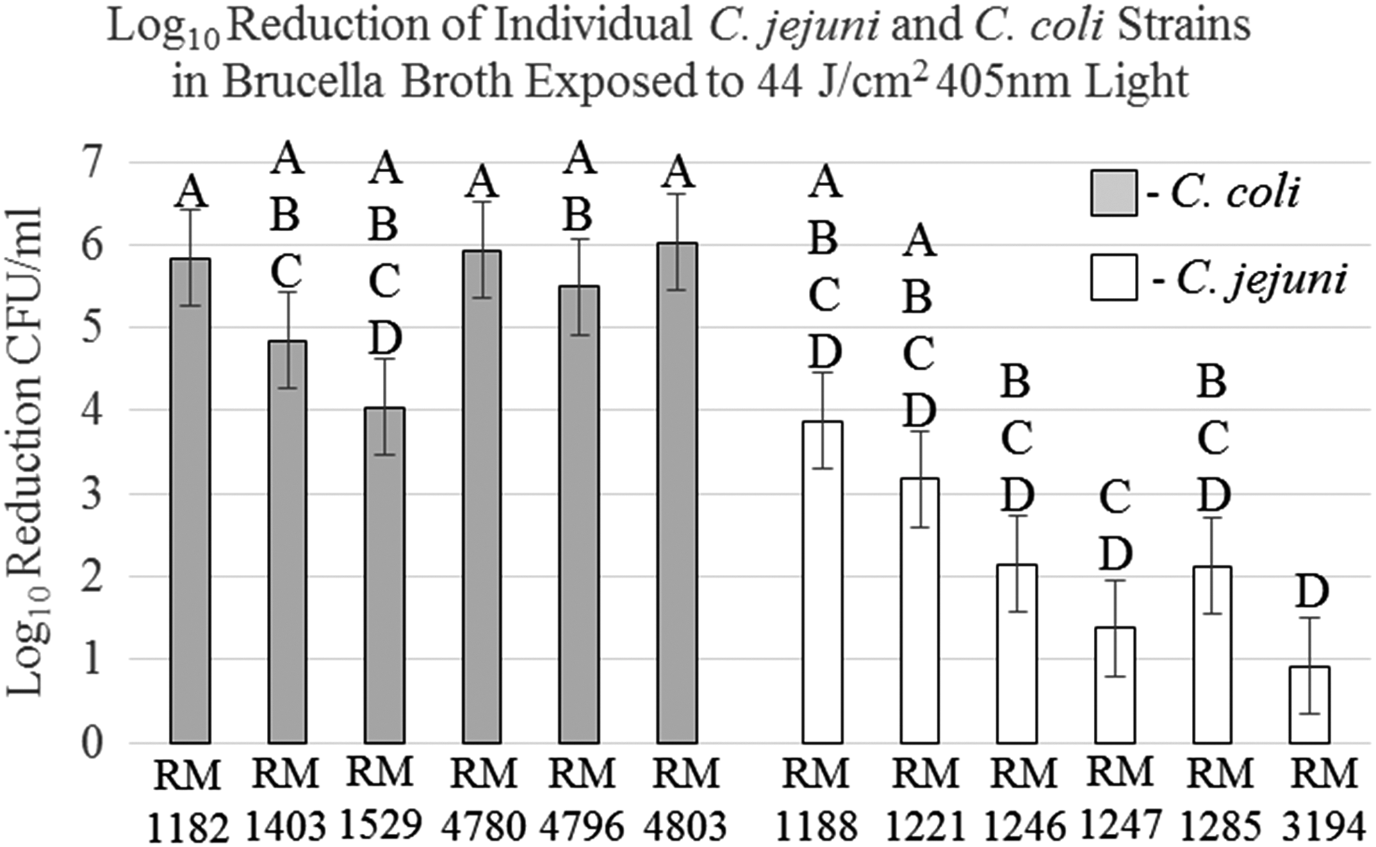
The average log10 CFU/mL reductions of the six C. jejuni and C. coli strains in Brucella broth graphed with the appropriate standard error and analyzed to determine whether a statistical difference exists between the strains when light treated in Brucella broth.
The average log10 CFU/mL reductions of the six C. jejuni and C. coli strains in exudate graphed with the appropriate standard error and analyzed to determine whether a statistical difference exists between the strains when light treated in exudate.
Significant differences were observed between the reductions in bacterial numbers after treatment of individual C. jejuni strains in Brucella broth compared with C. coli strains (Fig. 4). In Brucella broth, the average reductions of light-treated C. coli RM1182, RM4780, and RM4803 differed significantly from those of C. jejuni RM1246, RM1247, RM1285, and RM3194. In addition, C. coli RM4796 differed significantly from both C. jejuni RM1247 and RM3194, whereas RM1403 only differed significantly from RM3194. Significant differences in the reduction of bacterial numbers were not observed between light-treated Campylobacter species when suspended in chicken exudate (Fig. 5).
Discussion
In this study, we used chicken exudate as the primary environment for Campylobacter as opposed to an artificial growth media to test the effectiveness of 405-nm light treatment when the bacteria containing exudate were in contact with chicken skin or stainless steel (Gunther IV, 2010; Gunther IV et al., 2011). The use of conditions more relevant to poultry products led to a different appraisal of the effectiveness of 405-nm light against C. jejuni and C. coli.
A previous study applied 405-nm light to C. jejuni suspended in phosphate buffered saline (PBS) and was able to achieve a 5-log reduction in C. jejuni numbers using only 18 J/cm2 (Murdoch et al., 2010). In our experiments, to achieve anything close to a 5-log reduction using C. jejuni or C. coli in exudate on stainless steel required more than 180 J/cm2 and 134 J/cm2, respectively (Fig. 2). We only achieved a reduction of ∼2 logs for C. jejuni and C. coli in exudate on chicken skin when applying a dose more than 180 J/cm2 (Fig. 1). We believe that we were only able to achieve the ∼5-log reduction on stainless steel because of secondary thermal effects from the >50°C temperatures of the steel surfaces during treatment, given that Campylobacter are subject to heat inactivation above 48°C (Table 1).
The differences between the data in this article and previous research are likely the protective effect against 405-nm light that the exudate appears to provide. The exudate is more opaque than PBS and would reduce the level of light penetration. This is supported by the observation that when some individual C. coli strains were suspended in either exudate or Brucella broth and exposed to the same dose of 405-nm light, there were significantly greater reductions in the Brucella broth than in exudate suspensions (Fig. 3). The Brucella broth is also less opaque than exudate. The chicken skin surface may supply additional protection by providing shelter for the Campylobacter from the light treatment, given the skin's irregular surface and presence of feather follicles (Chantarapanont et al., 2003).
The reduced effectiveness of 405-nm light treatment in our research suggests that this technology may face challenges when applied to poultry products. This becomes obvious when trying to eliminate C. jejuni or C. coli on chicken skin. In our research, it took ∼90 J/cm2 to reduce the bacteria by 1 log, and doubling that dose to ∼180 J/cm2 only increases the reduction an additional 1 log. The system used in our studies had to be used at 90% power for 10 min to achieve a dose of ∼180 J/cm2, which leaves little room for increasing the irradiance of the system or the practical exposure length. In addition, the temperature of the chicken skin after treatment had already risen to approximately between 37°C and 44°C; therefore, longer exposure times or greater irradiance would likely result in adverse thermal effects.
When the 405-nm light was applied to stainless steel surfaces, the reductions of C. jejuni and C. coli were larger (Fig. 2). An average of 5-log reduction was achieved in C. coli numbers with a dose of 134 J/cm2and in C. jejuni with a dose of 183.4 J/cm2. These reductions may be even larger given that any surviving bacteria fell beneath the limit of detection of the growth assay. Despite the more promising result in the treatment of the stainless steel, problems with possible thermal effects remain as the doses of 134 J/cm2 and 183.4 J/cm2 resulted in surface temperatures of 44.2°C ± 1.4°C and 56.3°C ± 3.2°C, respectively.
Finally, Figure 4 (and to a lesser extent Fig. 2) suggests that there exists a difference between C. jejuni and C. coli strains with regard to their sensitivity to 405-nm light. When exposed to equal doses of light (44 J/cm2), most of the C. jejuni strains in Brucella broth showed significantly smaller reductions in cell survival than most of the C. coli strains (Fig. 4). In the varied dose light experiments on the cocktails of C. jejuni or C. coli strains on stainless steel, light dose of ∼133 and 89 J/cm2 produced differences between the reductions of C. jejuni and C. coli numbers that approached significance under conservative analysis (Fig. 2).
An explanation for these observations may be found in previous research that implicated production of reactive oxygen species as the bactericidal mechanism of 405-nm light; the following additional research suggested that there are differences to the mechanisms for resistance to oxidative stresses between C. jejuni and C. coli (Purdy et al., 1999; Maclean et al., 2008). Since there exists a testable hypothesis to explain these observations, it may be worth further investigation.
Conclusions
Visible 405-nm light treatments have shown promise in the reduction of Campylobacter species under ideal conditions. However, under conditions in this study that more closely approximate the poultry products in which Campylobacter are commonly found, the efficacy of 405-nm light may be hindered. Prohibitory high irradiance levels and long exposure times resulting in secondary thermal effects may be problematic for the adoption of this technology to eliminate Campylobacter in poultry.
Footnotes
Acknowledgments
We would like to thank Aisha Abdul-Wakeel and Joseph Sites for technical assistance on this project.
Disclosure Statement
No competing financial interests exist.