
Abstract
Background:
Toxoplasmosis is caused by the protozoon Toxoplasma gondii, which is one of the most widespread parasites that infect animals and humans worldwide. One of the main routes of infection for humans is through the consumption of infected meat containing bradyzoites in tissue cysts. Pork is one of the foremost meat types associated with outbreaks of acute toxoplasmosis in humans.
Materials and Methods:
Sixty blood samples were collected from finished pigs at slaughter and their sera was evaluated by an indirect-IgG ELISA. Matched muscle samples were obtained from the tongue and loin. Whole blood and tissue samples were evaluated to search for T. gondii DNA using a nested-polymerase chain reaction.
Results:
Seroprevalence of T. gondii was 96.6% (58/60) of sampled pigs. Meanwhile, T. gondii DNA was present in 23.21% of tongue tissue samples (13/56), 7% of loin tissues (4/57), and 0% in blood samples (0/44), respectively. Two pigs were serologically indeterminate.
Conclusion:
This is the first report of the presence of T. gondii DNA in tissue samples obtained from finalized pigs. Results from the present study suggest a high exposure to T. gondii in pigs intended for human consumption from the tropical region of Mexico. Thus, the consumption of some undercooked pork meat meals typical from the southern region of Mexico could represent a significant risk for acquiring infection for the human population.
Introduction
T
The consumption of meat infected with the parasite is considered the main route of transmission to humans, if raw or undercooked meat is consumed (Dubey and Jones, 2008; Dubey et al., 2012). Pork meat is the most important reservoir associated with T. gondii tissue cysts (Dubey, 2009). Several reports of acute toxoplasmosis in humans have been associated with the consumption of infested pork (Choi et al., 1997; Vitale et al., 2014). The main groups at risk for negative consequences of toxoplasmosis are women who acquire the primary infection during pregnancy and infected individuals who suffer some immunosuppression (Weiss and Dubey, 2009).
In the United States, seroprevalence of T. gondii in pigs reared under controlled conditions has been reduced to 2.7% due to the improvement in management practices (Hill et al., 2010). In Mexico, seroprevalence of T. gondii in pigs varies from 13% to 100%, with highest proportions associated to areas with hot tropical climate, raising pigs outdoors, and the presence of cats in the farm (Alvarado-Esquivel et al., 2011, 2014, 2015; Ortega-Pacheco et al., 2011, 2013).
Although the seroprevalence of T. gondii in the pig population is important to understand, it is essential to identify the presence of the parasites in the edible tissues as destined for human consumers. The sole published report was conducted in the west of Mexico (Jalisco state), where a 2.1% of positivity was found using inoculated mice (Galván-Ramirez et al., 2010). The study of the presence of T. gondii in pork meat could help to identify potential risks to human health in relation to T. gondii infections.
The objective of this study was to determine the presence of T. gondii specific IgG antibodies in the serum of finalized pigs ante-mortem and the presence of T. gondii DNA in tissue samples obtained postmortem from the same pigs in an abattoir from a tropical region of Mexico.
Material and Methods
Study area
A cross-sectional study was conducted in a slaughterhouse in the central region of Yucatan State, southeastern Mexico. Origins of pigs were not provided, but were from different farms from all over the central zone of the Yucatan State. The study period is considered from October 2014 to June 2015. The climate in the region is tropical subhumid with summer rains (Aw), an average annual temperature of 24°C–28°C, and an annual range of precipitation total 400–2000 mm (INEGI, 2013).
Study population and sample collection
The sample size was calculated using EpiMuestra (Segura, 2008), considering an expected 10% prevalence in pork by polymerase chain reaction (PCR), a confidence level of 90%, and population size of 150 pigs slaughtered annually at the slaughterhouse studied. The sample size consisted of 60 finished pigs (Sus scrofa) destined for human consumption. All the pigs included in the study were males of not more than 6 months old, raised in semitechnified farms. Blood samples were obtained during bleeding at the time of the daze before slaughter time. Individual animal blood samples were obtained from the anterior cava vein (Vacutainer® tubes without anticoagulant), together with whole blood samples (1 mL). After the pigs were slaughtered, tissue samples from the tongue (50 g) and loin (25 g) were collected, these tissues were chosen because T. gondii shows a marked tropism to the tongue, and loin tissue was selected because it is highly consumed in the study region.
Determination of IgG antibodies
Blood samples were centrifuged for 10 min at 448 g to obtain the serum. Serum samples were individually labeled and stored at −20°C until further processing. The presence of IgG specific antibodies against T. gondii was determined using an indirect ELISA test (Human-GmbH, Wiesbaden, GER). The technique was adapted using a secondary antibody anti-swine IgG-HRP (Catalog no. J3007, Santa Cruz, Inc., CA). Serum samples were evaluated at a concentration of 1:100, and IgG secondary antibodies were used at a dilution of 1:5000 (Ortega-Pacheco et al., 2013). A commercial normal swine serum (catalog no. B2613; Santa Cruz, Inc., CA) was used as a negative control and a serum from pig previously tested by nested-PCR (nPCR) as positive (in tissue sample), and ELISA seropositive was used as positive control. The optical density was measured at 450 nm using a spectrophotometer (xMark™, Bio-Rad).
DNA extraction from tissue samples
The DNA from whole blood samples was obtained following the protocol reported by Jalal et al. (2004). Samples of tissues (tongues and loins) were predigested in pepsin solution following the methodology reported by Dubey (1998). Then, the protocol of the commercial kit, DNeasy Blood and Tissue (QIAGEN GmbH, Hilden, Germany), was followed. Once purified, the DNA samples were stored at −20°C until further analysis. To assess the absence of endogenous inhibitors for PCR amplification, a housekeeping gene glyceraldehyde 3-phosphate dehydrogenase (GAPDH) was analyzed in each DNA sample included in the study using the primers fwd 5′ GACTTCAACAGCAACTCCCAC 3′ and rev 5′ TCCACCACCCTGTTGCTGTA 3′.
Detection of T. gondii DNA from tissue samples through a nPCR
A nPCR was performed to amplify a fragment of 390 bp, the SAG1 gene (encoding main surface protein of T. gondii), using a 96-well Veriti thermocycler (Applied Biosystems). Amplification was performed with the external primers fwd 5′ GTTCTAACCACGCACCCTGAG 3′ and rev 5′ AAGAGTGGGAGGCTCTGTGA 3′ (Su et al., 2010). In the second amplification, the internal primers used were fwd 5′ CAATGTGCACCTGTAGGAAGC 3′ and rev 5′ GTGGT TCTCCGTCGGTGTGAG 3′. The first amplification reaction was performed with the following conditions: 1X PCR buffer (Promega) with an adjusted concentration of 2 mM MgCl2; 0.8 mm dNTPs; 0.5 μM for both forward and reverse primers; 1.5 U Taq polymerase; and 2 μL DNA sample in a final volume of 25 μL. The second PCR had the same characteristics as the first, except that in this case, the concentration used for forward and reverse primers was modified to 0.3 μM and as a template for the second round was used 2 μL of the product from the first round of PCR. The nPCR conditions in the first run were 95°C for 5 min, followed by 30 cycles of 94°C for 30 s, 55°C for 1 min, and 72°C for 2 min. In the second run, it was 95°C for 5 min, followed by 35 cycles of 94°C for 30 s, 60°C for 1 min, and 72°C for 1.5 min. DNA from T. gondii axenic tachyzoite culture was used as positive control; in contrast, a reaction without DNA was used as negative control in each round of nPCR. The amplification products were visualized on agarose gel 1.5%, which were stained with ethidium bromide (10 mg/mL en H2O).
Statistical analysis
The prevalence of positive pigs (presence of T. gondii IgG antibodies or DNA) was calculated using the formula p = Σy/n, where p is the prevalence and Σy is the sum of positive samples.
Results
The seroprevalence of IgG antibodies against T. gondii in pigs was 96.6% (58/60); two pigs were serologically indeterminate (Fig. 1). GAPDH gene was amplified in 56/60, 57/60, and 44/60 of the tongue, loin, and blood samples, respectively. Meanwhile, the presence of T. gondii DNA was detected in 23.2% (13/56) of the tongue tissue samples and 7.0% (4/57) of the loin samples (Table 1) and an example of SAG1 gene amplified in an agarose gel and stained with 1.5% ethidium bromide is shown in Figure 2. All blood samples tested were nPCR negative for T. gondii. An indeterminate case of serology was found in the presence of T. gondii DNA in two different tissues (tongue and loin).
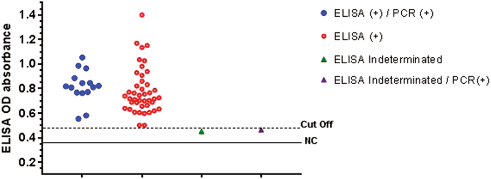
Serum anti-IgG absorbance by indirect ELISA assay with results of pig tissue positive to nPCR (tongue/loin or both). Cutoff OD value was 0.497 (pig negative control value plus 15%), negative OD value was 0.368. OD, optical density. nPCR, nested-polymerase chain reaction. Color images available online at
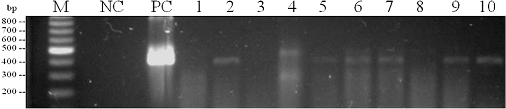
Agarose gel electrophoresis (1.5%) of nPCR product of the Toxoplasma gondii SAG1 gene (390 pb). Lanes: M: molecular weight marker (100 bp DNA Ladder, reference: G210A, Promega); NC: negative control (mastermix without DNA); PC: positive control (DNA of tachyzoites of T. gondii from axenic culture); 2, 5, 6, 7, 9, and 10: samples of tissue positive to T. gondii; 1, 3, and 8: samples negative for T. gondii tissues; 4: sample of tissue negative to T. gondii with nonspecific amplifications.
NA, does not apply; O.D., optical density; CC, negative control value plus 15%; nPCR, nested-polymerase chain reaction.
The lack of GAPDH gene amplification in the different samples analyzed may be due to a number of factors: First the type of sample, in this case blood samples had the highest number of inhibitors. The blood of pigs has a significant amount of fat that could interfere with the amplification of internal controls. In addition, the method of extraction used might have influence on the amount of DNA recovered, since extraction was performed from 1 mL of whole blood.
Discussion
A high seroprevalence of IgG antibodies against T. gondii was found in the surveyed pigs (96.6%). This result is similar to earlier reports in the same tropical zone of Mexico (Ortega-Pacheco et al., 2011, 2013) where a high proportion of pigs were seropositive indicating that they had contact with the parasite before the end of their fattening period (≤5 months of age). Environmental conditions in hot tropical regions seem to play an important role in the oocyst survival, allowing them to remain infectious for long periods of time (Dumétre and Dardé, 2003; Dubey, 2010) infecting a wide variety of reservoirs such as rodents. Although all pigs included in this study came from semitechnified farms, they seemed to lack the appropriate biosecurity measures for the control of T. gondii dispersion (i.e., control of the presence of non spayed cats in the pens, strict rodent control, maintaining feed in close containers, etc.). Furthermore, high seroprevalence found in the present study indicates the presence of a possible common source of infection for pigs in the different farms of origin. It has been described that the possible transmission paths in pigs are through ingestion of food and contaminated water with oocysts excreted by infected cats inhabiting the farms. Likewise, pigs are considered carnivorous and they can consume rodents containing tissue cysts, representing another source of infection (Thiptara et al., 2006). The presence of T. gondii in the pork tissues is an important finding. However, it is necessary to conduct more studies to understand the risk that pork can be represented in the study area, where there have been reported high seroprevalences of T. gondii in the human population (Zavala-Velazquez et al., 1989; Góngora-Biachi et al., 1998; Jiménez-Coello et al., 2011; Vado-Solis et al., 2013; Hernández-Cortazar et al., 2016). In the present study, a higher frequency of T. gondii DNA was found in the tongue tissue samples (23.2%) compared to the loin samples (7.0%) of finished pigs. In pigs, the most common T. gondii infected tissues are the tongue, brain, heart, retina, lungs, and dorsal muscles (Dubey et al., 1986; Yai et al., 2003; Juránková et al., 2014). Belfort-Neto et al. (2007), mentioned that T. gondii is found to be inconsistently distributed among the organs and muscles, but overall, tongue is more heavily infected than other tissues. Several isolates of T. gondii obtained from tissues samples in naturally infected pigs are reported worldwide. However, those isolates were obtained mainly through bioassays using mice (Dubey, 2010). In the present study, the difference found between tongue and loin tissues may be due to the initial amount of the sample. The tongue sample consisted of 50 g, whereas only 25 g of loin muscle was used. Such difference in the quantity of sample used resulted from restrictions imposed at the abattoir and the commercial value of the loin meat that led to difficulties in the process of collection. To increase the probability of detecting T. gondii tissue cysts, a bigger amount of loin sample should be tested. The presence of T. gondii DNA in the muscle samples from pigs indicates the existence of the agent on a cystic form representing a risk to public health. In southern Mexico, pork meat is common in local typical meals and can be consumed even on a daily basis and during family and religious festivals. Pork loin is highly consumed (SAGARPA, 2009), and pork tongue and many other organs (guts, heart, liver, spleen, brain, etc.) are also commonly consumed. Although the study area is not common for consumption of raw pork meat, there are meals highly ingested such as grilled pork, pork meat at “Pastor” (similar to kebab) that because of the speed of preparation sometimes is not properly cooked, representing a risk for consumers. Considering that one pig infected with T. gondii with a market weight of 100 kg can produce more than 600 individual pieces of meat, it represents an important source of infection for human (Dubey et al., 2012).
Two pigs with indeterminate serological results were found in the present survey. However, a pig had T. gondii DNA in both tissue samples (tongue and loin). Serological undetermined or seronegative cases in pigs with the presence of T. gondii DNA in tissue samples have been reported before, attributing to a recent infection (Dubey et al., 2002, 2012). In this study, all blood samples were PCR negative for T. gondii, but some cases were found just by nPCR in tissue samples. In the study area, it has been reported that pigs are infected when entering to fattening period at an early age (Ortega-Pacheco et al., 2013); therefore, it is probable that finished pigs were not presenting the acute phase of infection, since parasitemia does not last more than a couple of weeks (Klun et al., 2011).
The high seroprevalence indicates a wide circulation of the agent in the environment. Serological and molecular results provide clear evidence that T. gondii is present in finished pigs intended for human consumption. Given the high pork consumption in the study area, the presence of T. gondii in finished pigs represents a major risk of infection, especially when pork is consumed undercooked. However, it is recommended that more studies be further conducted in the area to improve the conclusion given.
Footnotes
Acknowledgments
The authors gratefully acknowledge CONACYT (Consejo Nacional de Ciencia y Tecnología) for grant funds for 259166 project and also for the financial support of a PhD student from a PNPC National Program.
Disclosure Statement
No competing financial interests exist.