
Abstract
We report data on the Toxocara seroprevalence evidenced in 2015 from samples of 40 children and 298 adults of the population living in different areas of Serbia, and on possible association of certain variables with infection. Detection of specific antibodies was performed using an enzyme-linked immunosorbent assay; all ambiguous results and part of the positive and negative sera were further analyzed by confirmatory Western blot test. An overall 23.5% seroprevalence was noticed, which was confirmed in 13.0% of the examined population with no significant difference regarding the age (children = 10.0%; adults = 13.4%) or by country area (East = 18.2%; North = 15.5%, Southeastern = 9.5%; p = 0.005). In contrast, the group of adult women proved more reactive than men (p = 0.001), and subjects both who spend spare time in square/parks (p = 0.041) and with positive onychophagy (p = 0.001) habit turned out more exposed to the infection. Possible reasons of these differences were analyzed, and the medical, veterinary, and economic impact of this soil-transmitted zoonosis were discussed.
Introduction
D
Humans become infected through the accidental ingestion of embryonated eggs present on soil/vegetables (Despommier, 2003), or of larvae present in under-cooked/raw meat of vertebrate and/or invertebrate paratenic hosts (Woodhall et al., 2014), or, finally, through the transplantation of infected organs. Patients infected by human immunodeficiency virus (HIV) or treated with immunosuppressive drugs are predisposed to the activation of silent infections in the past acquired or recently received with an organ transplant (Eid et al., 2015).
When humans ingest embryonated eggs or visceral larvae, released larvae penetrate the small intestinal wall and migrate via the bloodstream to the liver, lungs, muscles, eye, and central nervous system (Wiśniewska-Ligier et al., 2012). Most infections remain asymptomatic (Macpherson, 2013), whereas several of them are clinically evident as visceral or ocular larva migrans syndrome (Považan et al., 2011), neurotoxocariasis and covert (in children), or common toxocariasis (in adults) (Rubinsky-Elefant et al., 2010).
The fact that most positive cases of this zoonosis, especially the asymptomatic forms, remain undiagnosed is the main reason why it is very difficult to assess the prevalence of toxocariasis (Woodhall et al., 2014). However, it is a cosmopolitan zoonosis that occurs more frequently in the tropical and subtropical areas of the world and whose prevalence and incidence, due to infectious ways, are higher among children, especially from socioeconomically disadvantaged populations (Macpherson, 2013).
The diagnostic procedure starts with serological tests aimed at detecting specific antibodies by commercial enzyme-linked immunosorbent assays (ELISA), followed by the application of confirmatory immunoblot analyses Western blot test (WB) (Fillauxa and Magnavalc, 2013). Direct methods to detect the presence of migrating/blocked larval stages are invasive and vain, with extremely low sensitivity.
Despite the fact that the postwar period in Serbia potentiates the paying of more attention to the so-called neglected infections, there are insufficient data of human toxocariasis prevalence in the country. Moreover, a small number of laboratories offer diagnostics of toxocariasis. On the contrary, available literature data from personal researches showed that Serbia is hyperendemic for toxocariasis in dogs (Kulišić et al., 1998), which contributed to the high contamination of soil; moreover, children as well as patients with visceral and ophthalmological problems were proved as highly reactive to the parasite antigens (Lalošević et al., 1993, 2001; Považan et al., 2011). These findings urged us to investigate the seroprevalence of asymptomatic toxocariasis in individuals living in different areas of Serbia, with the aim to evaluate the risk factors that could be important for the strategy of preventive measures in the country.
Materials and Methods
Study design
The cross-sectional epidemiological study was carried out at the Department of Public Health and Infectious Diseases, “Sapienza” University of Rome (laboratory- and serology-based analyses) and Medical faculty, University of Niš (epidemiological- and statistical-based analyses). The Ethics Committee of the University of Niš, Faculty of Medicine (Decision No 01-2625-23/2014), approved this research.
Study involved subjects living in different areas of Serbia: North Serbia (Pančevo, Novi Sad), East Serbia (Zaječar, Pirot), and South-eastern Serbia (Leskovac, Vranje, Niš), between 42.33°–45.15°N and 19.50°–22.17°E. The study was conducted in 2015 from March 1 to May 31.
Inclusion criteria were approvals of all subjects and absence of infectious diseases, which can influence immunodeficiency. Exclusion criteria were the presence of HIV infection, diabetes, pregnancy, immunodeficiency, or viral infection. Blood samples were collected from all subjects. A semistructured questionnaire was used to collect descriptive data and data about risk factors (age, place of living—urban or rural region, place of residence—apartment or house), occupation (working outdoors or not), way of spending spare time (attendance square/park or not), data about exposure (keeping dogs or cats), and habits (onychophagy, geophagy).
Serological analysis
In serological analysis, serological tests were performed on blood samples to measure anti-T. canis antibodies using a commercial enzyme-linked immunosorbent assay working on excretory antigens of the parasite larval stages (Toxocara Ab ELISA, Cypress Diagnostics, Langdorp, Belgium), with declared 100% sensitivity and 98.4% specificity. According to the manufacturer's instructions, positive controls, negative controls, and samples (all in duplicate) were analyzed and the results were determined photometrically at 450 nm by a microtiter reader (BioRad, France). The ratio (R) between the mean optical density (OD) evidenced for each subject and the calculated cutoff gives the negative (0–0.89), doubtful (0.9–1.1), or positive (>1.1) result.
All ambiguous results as well as part of the positive (≈80%) and negative (≈10%) sera were further analyzed by the confirming western blot test (Toxocara WB IgG; LDBIO Diagnostics, Lion, France) following the manufacturer's instructions.
The diagnostic efficacy of the used immunoenzymatic kit was obtained in comparison with results of WB test.
Statistical analysis
In statistical analysis of the collected, systematized, and encrypted data, statistical calculator within the program Epi Info (Ver.6.04) and the statistical package SPSS (16.0 for Windows) were used. To compare values between two groups, the t-test was performed; for comparison of different frequency distribution, the chi-square test with/or without Yates's correction was used as well as the Fisher's exact test. Cohen's K index value was used to estimate the agreement between the two serological tests (ELISA and WB). The forward stepwise logistic regression analysis was used to evaluate the impact of multiple variables on seropositivity to T. canis. The p-value <0.05 was considered as statistically significant.
Results
The serological survey involved 338 (222 males and 116 females) randomly selected subjects, 2–86 years old (average age of 52.80 ± 17.96). The study group consisted of 40 children (2–14 years) and 298 adults.
A total of 76 sera (22.5%, CI 18.05–26.95%) showed reactivity to Toxocara antigens in ELISA, with R-values ranging from 0.92 to 4.55, and 10 of whom with borderline values. Of the total number of patients, 43 men and 33 women were positive for anti-T. canis IgG (4 children and 72 adults). Western blot confirmed 44/61 (72.1%, CI 60.84–83.36%) results as positive, concerning 19 men and 25 women. In detail, about all subjects with R > 3.10 proved positive to 2–5 proteins of 24–35 kDa, whereas 77.7% (CI 50.51–104.89%) of those with R > 2.11, and 73.1% (CI 56.06–90.14%) of those with R > 1.11 had confirmed their positivity. Interestingly, one half of the people (50.00%, CI 23.81–76.19%) with borderline ELISA R values were confirmed as positive and none out of the negative subjects was proven as positive (Table 1).
Ratio = optical density/cutoff.
Number of positive sera/number of examined sera.
Number of positive sera/number of examined sera.
Statistical analyses showed substantial agreement (k = 0.631, p < 0.001) between the results obtained in two testing. In addition, ELISA showed 100% sensitivity and negative predictive value, lower specificity (63.83%, CI 53.96–73.71%), positive predictive value (72.13%, CI 62.92–81.34%), and efficiency (81.32%, CI 73.31–89.31%) compared with the WB confirming test.
No difference in reactivity to antigens was found between children (10.0%, CI 3.96–23.05%) and adults (13.4%, CI 10.01–17.76%). The highest number of seropositive subjects was found in East Serbia (18.2%, CI 8.89–27.51%), followed by North part of country (15.5%, CI 8.51–22.49%), whereas the lowest one was detected in South-eastern Serbia (9.5%, CI 5.08–13.92%) (Fig. 1). Differences in reactivity to Toxocara antigens were not found by area (p = 0.134).
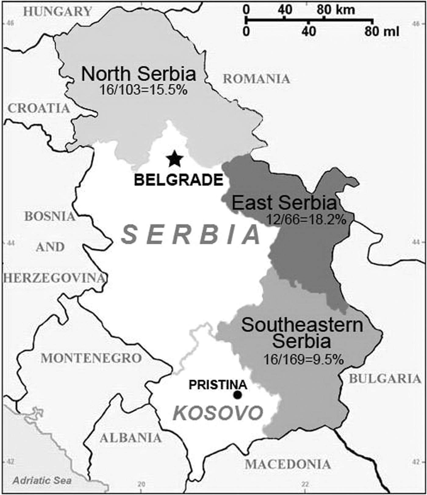
Human toxocariasis seroprevalence evidenced in the year 2015 in the Serbian population, by area.
In addition, as shown in Table 2, seropositive findings were significantly more frequent in women (p = 0.001), in subjects who practise onychophagy (p < 0.001), whereas no significant differences were evidenced by age, urban/rural place of residence, owning pets, or outdoor activity.
n, number of subjects.
Significance difference p < 0.05.
In the youngest population, seropositive findings were significantly more frequent in children younger than 3 years (p < 0.001), and in children with geophagy (p < 0.001), whereas no significant differences were evidenced by gender, residence, owning dogs, or outdoor activity.
Table 3 presents exposure regarding age, gender, residence, pet ownership, outdoor work, attendance square/park, and onychophagy associated with Toxocara seropositivity based on the univariate analysis and multivariate analysis. Statistically, it was found that onychophagy (OR 3.91, CI 1.81–8.43), gender (OR 2.52, CI 1.27–4.99), and square/park attendance (OR 1.94, CI 1.02–3.67) had a significant influence on the occurrence of infection.
n, number of positive results.
N, number of tested.
Univariate logistic regression.
Multivariate logistic regression.
Significance difference p < 0.05.
Significance difference p < 0.001.
OR, odds ratio; 95% CI, 95% confidence interval.
In the multivariate model, onychophagy was shown to be the best predictor of seropositivity (OR 4.57, CI 2.10–9.94). The forward stepwise logistic regression analysis was used to evaluate the impact of multiple variables on Toxocara seropositivity. In the final model, two variable effects on Toxocara seropositivity are described by the following equation:
Discussion
Toxocariasis is a neglected zoonosis that requires more veterinary and physician attention. Indeed, being mostly asymptomatic (Cong et al., 2014) or without specific characters (Cassenote et al., 2014), it should be successfully taken on only through a multidisciplinary approach (Traversa, 2012).
Globally, the widest reactivity to T. canis antigens has been detected in rural areas of tropical regions (Van Den Broucke et al., 2015). Findings herein reported suggest a current common environmental condition either in rural or in urban areas since we did not obtain a significant difference in seroreactivity in subjects living in different areas. As opposed to our results, previously published data in Europe reported higher (14–36%) seroprevalence in rural areas than in urban ones (2–5%) (Uhlíková and Hübner, 1998; Stensvold et al., 2009; Mughini-Gras et al., 2014).
The reactivity to T. canis antigens proved in 22.5% (CI 18.05–26.95%) and confirmed in 13.0% (CI 9.43–16.61%) of people from different Serbian areas classifies the country among hyperendemic regions. This finding fits in with researches so far conducted on Serbian dogs and on the soil, the results of which had evidenced very high prevalence of the infection (30.5–33.3%) and high contamination level with infective eggs (33.5%), respectively (Kulišić et al., 1998; Nikolić et al., 2008; Čolović-Čalovski et al., 2014).
Serological results herein reported are in general agreement with the available literature that refers North Serbia as a territory characterized as natural habitats ideal for the life cycle of Toxocara nematodes (Simin et al., 2014). Very high Toxocara reactivity was also detected in Eastern Serbia, from where we did not have much information about the prevalence of any parasitosis, toxocariasis included. Lower reactivity levels (9.5%) were proved in the population resident in South-eastern Serbian areas.
As for the association of certain variables with infection, children, due to their behavior (eating soil, putting objects in their mouths, and/or their poor hygiene), are considered the most exposed to toxocariasis (Macpherson, 2013). Apart from low seroprevalence (1–4%) in the past observed in Spain (Guerra et al., 1995; Fenoy et al., 1996), the first European epidemiological studies in this age class have reported seroprevalence ranging from 7% to 23% (Jarosz et al., 2010; Maizels, 2013), consistent with our finding (10%). However, no difference was found when compared to the adult class (p = 0.052), whereas the seropositive findings were significantly more frequent in children younger than three years (p < 0.001) and linked to geophagy (p < 0.001), which represents a significant risk for this infection. Based on higher reactivity against Toxocara antigens, statistical analyses in adults proved female gender, although underrepresented in the surveyed population, to be an important risk factor (p = 0.001). This evidence could be due to the fact that a woman's nails, usually longer than that of man, could easier capture and detain the soil, a possible source of infection with Toxocara eggs.
In addition, subjects with positive onychophagy, and who spend spare time outside, in urban recreational spaces, were also more reactive. The first finding was expected because the abovementioned bad hygienic habits are well-known associated variables for many soil-transmitted infections. As for subjects who spent time in urban recreational spaces, which are usually frequented by dogs to defecate, they are exposed to infection because these environments gradually become catchments areas of Toxocara eggs that here find the ideal conditions to became infectious, as demonstrated by the results of many investigations carried out in public spaces, parks, and playgrounds (Khademvatan et al., 2013; Čolović-Čalovski et al., 2014; Traversa et al., 2014). Moreover, as expected and like found in other studies (Traversa, 2012), pet owners are not more seroreactive than other people, since the animals disseminate unembryonated, therefore not yet infectious, Toxocara eggs, whose infectivity is necessarily dependent on a long stay on the soil under favorable conditions of temperature and humidity (Jimenez et al., 1997). In addition, the Government of Serbia in harmonization of laws with the EU regulation recently issued the acts to register private dogs and cats with intent for implementation of chipping, vaccinations, and deworming of animals. This preventive measure could influence the decrease of toxocariasis prevalence in pets. Based on data collected from the questionnaire, neither age nor urban/rural place of residence or working outside was found to be significantly associated with positive serology, probably due to the similar level of environmental contamination.
As for the serological tests applied, our findings indicate that the application of a western blot confirming test is always recommended (Fillaux and Magnaval, 2013). Confirmed substantial agreement of serological tests (k = 0.631), 100% sensitivity of the applied ELISA kit, and its proved specificity in 63.83% of the results, suggest that all laboratories that start with toxocariasis diagnostics in our country have to include the confirming WB test in the procedure. False-positive results are expected in case of cross-reaction with nematodes belonging to the same taxonomic group of Ascaridida. Further research which should include false ELISA positive sera for Toxocara could examine the possibility of cross-reaction due to Dirofilaria infection for which the high seroreactivity was determined, or to in country low prevalent Ascaris lumbricoides infection (Nikolić et al., 1998; Tasić-Otašević et al., 2014). As for T. cati infection, standardization or improving the commercial kits could enable research that will complete information of the population exposure to antigens specific for this species (Zahabiun et al., 2015).
Studies of geographical distribution and prevalence of toxocariasis, soil, and perhaps foodborne zoonosis are one of the primary tasks for veterinarians and physicians who can significantly contribute to the strategy of prevention. Legal regulations with determined preventive measures, including deworming of animals, could influence the decrease of toxocariasis prevalence in dogs, contamination of soil, and spread of infection to humans. However, very high toxocarosis seroprevalence established in this study throughout the country suggests the necessity for (i) better monitoring of this zoonosis in humans, (ii) performing the WB as confirmatory test in serological diagnosis and (iii) enhancing the medical record system for patients with Toxocara infection.
In addition, results of possible association of certain variables with infection, which demonstrated that onychophagy is significant risk factor, showed the need for constant education of people, especially children who could be more exposed in terms of hand hygiene.
This first information of asymptomatic toxocariasis seroprevalence in Serbia points to the need of one health approach and further research that will include all the Toxocara species involved in the sanitary problem.
Footnotes
Acknowledgments
This article was suported by the Serbian Ministry of Education and Science grant No III41007. The authors are grateful to Prof. Nataša Miladinović-Tasić and Aleksandar Tasić, DVM, PhD, for their precious assistance in collection of samples.
Disclosure Statement
No competing financial interests exist.