
Abstract
The current study was conducted to evaluate the ability to recover Salmonella from shell egg contents by culture methods. A total of 4,000 eggs were obtained from a grading and packing center located in the Gyeonggi Province of South Korea, and 200 samples were created by pooling 20 broken eggs. The pooled samples were held at room temperature for 4 d before a 25-mL aliquot of each pool was added to 225 mL of modified trypticase soy broth (mTSB) and incubated at 35°C for 24 ± 2 h. A loopful of the culture was streaked onto chromogenic Druggan–Forsythe–Iversen (DFI) agar and incubated at 36 ± 1°C for 18–24 h. In addition, 1 mL and/or 0.1 mL of the mTSB cultures were added to 10 mL of Muller–Kauffmann tetrathionate with novobiocin (MKTTn) or Rappaport-Vassiliadis (RV) broth, and they were incubated for 24 ± 2 h at 35 ± 2°C or 42 ± 0.2°C, respectively. A loopful from these cultures was streaked onto Brilliant Green (BG), xylose lysine deoxycholate (XLD), and bismuth sulfite (BS) agar plates, respectively. Directly streaking onto DFI agar revealed the presence of Salmonella in 14 out of the 200 pooled samples (7%); whereas the combination of RV medium and BG, XLD, and BS agar detected the pathogen in only 9 (4.5%), 7 (3.5%), and 3 (1.5%) of the pooled samples, respectively. When MKTTn broth was used, Salmonella was detected in 7 (3.5%), 2 (1%), and 0 (0%) of the samples when streaked onto BG, XLD, and BS agar, respectively. The results indicate that direct plating onto DFI agar without enrichment was the most suitable among the methods evaluated in this study for detecting Salmonella in raw shell egg contents with a low microbial load.
Introduction
F
Culture-based methods are recognized as the gold standard for detecting Salmonella in food, because they are sensitive, reliable, and remain the only way to obtain a live isolate (Carrique-Mas and Davies, 2008; Velusamy et al., 2010). Standard culture-detection methods recommended by the U.S. Food and Drug Administration (FDA), the United States Department of Agriculture, and the International Organization for Standardization include pre-enrichment and selective-enrichment procedures for detecting Salmonella in shell egg samples (Hammack et al., 1999; Hammack et al., 2001; U.S. FDA, 2012). Therefore, culture-based detection of Salmonella from whole shell eggs can become a lengthy and labor-intensive procedure, requiring several selective-enrichment and plating media after the pre-enrichment step (Beckers et al., 1987; Manafi, 2000; U.S. FDA, 2012). Numerous studies have compared the different media recommended for detecting Salmonella (Manafi, 2000; Rall et al., 2005; Maciorowski et al., 2006; Schonenbrucher et al., 2008; Carrique-Mas et al., 2009; Pal and Marshall, 2009); however, the sensitivity and specificity of Druggan–Forsythe–Iversen (DFI) selective agar has not been evaluated. DFI agar is a chromogenic, selective, and differential agar that is generally used for isolating Cronobacter sakazakii. Hydrogen sulfide-producing Enterobacteriaceae, such as Salmonella, appear black on this medium due to incorporated sodium thiosulfate and ferric ammonium iron (III) citrate (Iversen et al., 2004). Selectivity is achieved by including sodium desoxycholate and crystal violet in DFI selective agar, which serves to inhibit the growth of most gram-positive microorganisms (Iversen et al., 2007).
The objective of this study was to compare the FDA Bacteriological Analytical Manual (BAM) methods with the direct streaking method by using DFI chromogenic agar for detecting and isolating Salmonella spp. from whole shell eggs.
Materials and Methods
Sample preparation
Shell eggs were purchased from a grading and packing center in the Gyeonggi province of South Korea between November 2010 and March 2012. Shell eggs were processed according to the BAM methods recommended by the FDA (2012; Fig. 1). Briefly, any visibly cracked eggs were discarded, and any adherent materials on the shell surface were removed. To disinfect the shells, eggs were soaked in a mixture of 70% ethyl alcohol (3 parts) and iodine/potassium iodide solution (1 part) for at least 10 s and then air-dried at room temperature. Eggs were cracked aseptically and the contents of 20 eggs were pooled, according to the pooling criteria described in the FDAs BAM. The contents of each bulk pool were mixed manually until the yolks and albumens were completely mixed (Seo et al., 2003; Valentin-Bon et al., 2003). The homogenized egg pools were incubated at room temperature (20–24°C) for 96 ± 2 h.
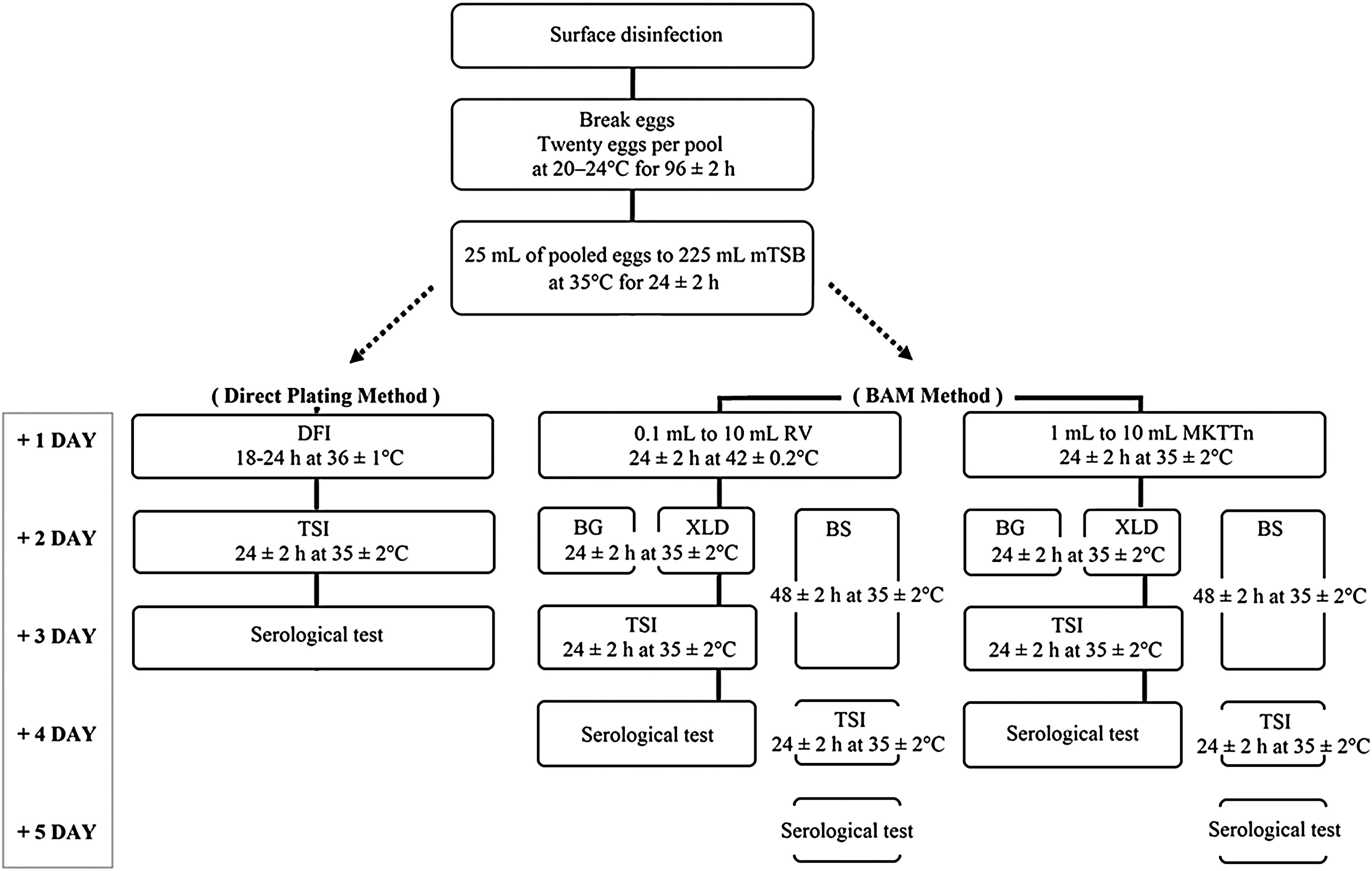
Flow chart illustrating the procedures used for isolating and detecting Salmonella in pooled shell egg samples by using the direct plating method with DFI medium and the BAM method. BAM, bacteriological analytical manual; BG, Brilliant Green agar; BS, bismuth sulfite agar; DFI, Druggan–Forsythe–Iversen agar; MKTTn, Muller–Kauffmann tetrathionate with novobiocin broth; mTSB, modified tryptic soy broth with FeSO4; RV, Rappaport–Vassiliadis broth; TSI, triple sugar iron agar; XLD, xylose lysine desoxycholate agar.
Salmonella isolation
The procedure used for Salmonella enrichment and detection in whole shell eggs is shown in Figure 1. Two hundred samples were prepared by pooling the contents of 20 shell eggs for each sample. The samples were left at room temperature for 96 ± 2 h before 25-mL aliquots of the pooled samples were transferred to 225 mL of modified trypticase soy broth (mTSB; BD Difco, Detroit, MI) that was supplemented with ferrous sulfate (35 mg of FeSO4/L TSB) and incubated at 35°C for 24 ± 2 h. A loopful of the culture was streaked onto DFI agar (Oxoid Ltd., Basingstoke, UK), and the plates were incubated at 36 ± 1°C for 18–24 h.
For Salmonella isolation by the BAM methods, 0.1 or 1 mL of the mTSB cultures were transferred to 10 mL of Rappaport-Vassiliadis (RV; bioMerieux, Marcy l'Etoile, France) or Muller–Kauffmann tetrathionate with novobiocin (MKTTn; bioMerieux) broth, and they were incubated at 42 ± 0.2°C or 35 ± 2°C for 24 ± 2 h, respectively. A loopful of the RV- and MKTTn-based enrichment cultures were streaked onto bismuth sulfite (BS; BD Difco), Brilliant Green (BG; BD Difco), and xylose lysine deoxycholate (XLD; Oxoid) agar plates. All selective agar plates were incubated at 35°C for 24 ± 2 h, and the BS agar plates were further incubated for an additional 24 ± 2 h (48 ± 2 h total). Presumptive colonies on the selective agar plates were transferred to triple sugar iron agar slants (BD Difco) and subjected to biochemical testing with the VITEK system (bioMerieux).
Salmonella serotyping
Serotyping of Salmonella isolates was performed by using commercially available antisera against Salmonella somatic O and flagellar H antigens (BD Difco, Sparks, MD). The agglutination properties were identified based on the antigenic classification observed, according to the Kauffmann–White scheme (Bale et al., 2007).
Real-time PCR
Presumptive Salmonella isolates were examined for the presence of the ttr gene (GenBank Accession No: AF 282268), which is associated with the tetrathionate reductase complex of Salmonella, by using an ABI 7500 Real-Time PCR instrument (Applied Biosystems, Foster City, CA). The primer and probe sequences, and the thermocycling conditions used for detecting Salmonella were previously described (Malorny et al., 2004). The sequences of the primers and probes used are as follows: forward primer (5′-CTC ACC AGG AGA TTACAA CAT GG-3′), reverse primer (5′-AGC TCA GACCAA AAG TGA CCA TC-3′), and probe (5′-Cy3-CACCGA CGG CGA GAC CGA CTT T-BHQ3-3′).
For DNA extraction, each colony was suspended in 1 mL of PBS and centrifuged at 16,000 × g for 3 min at 4°C. The cell pellets were resuspended in 200 μL of PrepMan™ Ultra Sample Preparation Reagent (Applied Biosystems) and boiled for 10 min. After cooling at room temperature for 2 min and centrifuging again at 16,000 × g for 3 min at 4°C, bacterial DNA was extracted from the supernatants and used as the template for real-time PCR. The supernatants (5 μL of extracted DNA) were transferred to 20 μL of PCR reaction mixture consisting of TaqMan Universal PCR Master Mix (12.5 μL; Applied Biosystems), 300 nM forward primer (2.5 μL), 900 nM reverse primer (2.5 μL), and 200 nM TaqMan probe (2.5 μL). The microwell plates were sealed and placed in an ABI 7500 Real-Time PCR instrument. The thermocycling conditions consisted of a 2-min hold at 50°C, followed by a 10-min hold at 95°C, and then 40 cycles of 95°C for 15 s, and 60°C for 60 s. The threshold cycle was calculated by using the ABI 7500 software (Applied Biosystems).
Statistical analysis
Contingency tables were prepared for the comparison of categorical variables between the standard FDA BAM culture methods and the direct DFI-plating method. p Values were derived by performing Fischer's exact test by using InStat™ software, version 3.05 (GraphPad Software, San Diego, CA), and p < 0.05 was considered statistically significant.
Results and Discussion
Salmonella was isolated from 14 out of 200 pooled samples (7%) when directly streaked onto DFI agar, whereas Salmonella was isolated from 0 to 9 (0–4.5%) samples by using the BAM culture methods (Table 1). All 14 black colonies observed on DFI selective media were confirmed as Salmonella spp. by PCR. Among the standard BAM culture methods, Salmonella was detected the most efficiently by using a combination of RV selective-enrichment medium and BG selective agar (9 of 200 pooled samples, 4.5%), whereas 7 out of 200 pooled samples (3.5%) scored positive for Salmonella by using BG selective agar after selective enrichment in MKTTn broth and after streaking RV-broth cultures on XLD plates. Thus, direct plating onto DFI agar plates resulted in significantly more positive samples (14/200 pooled samples, 7%) than did the methods involving MKTTn enrichment plus XLD or BS agar plates (p < 0.05, Table 1). Also, BS agar did not perform as well as DFI (p < 0.05), regardless of whether MKTTn or RV enrichment was carried out. The lowest isolation rate of Salmonella was achieved by using a combination of MKTTn enrichment plus BS plating, despite the additional 24-h incubation period (total selective enrichment: 72 h).
Salmonella isolation from shell eggs was performed through 4 trials, by using 50 tested pools per trial.
Different superscripts in rows and columns indicate significant differences in the Salmonella-detection frequency (p < 0.05).
BAM, bacteriological analytical manual; BG, Brilliant Green agar; BS, bismuth sulfite agar; DFI, Druggan–Forsythe–Iversen agar; MKTTn, Muller–Kauffmann tetrathionate with novobiocin broth; RVS, Rappaport–Vassiliadis broth; XLD, xylose lysine desoxycholate agar.
The method consisting of MKTTn and BS performed poorly compared with the RV-XLD and RV-BG methods, with the latter method performing better than all the other BAM methods, although it was not able to recover Salmonella from all positive samples. On the other hand, Salmonella was recovered 100% by direct plating onto DFI agar employed in this study (Table 2). Namely, none of the FDA BAM culture methods was successful in detecting Salmonella from the 14 pooled samples that tested positive by the DFI method, whereas the DFI method detected Salmonella in all samples that tested positive by the BAM methods. Overall, the agreement of positive results obtained from the BAM culture methods for identifying Salmonella-positive samples was considerably low, with a consensus detection rate of only 21.4% (Table 2). These results suggested that direct plating onto DFI agar is superior for detecting Salmonella from egg-content pools.
+, positive result; −, negative result; BAM, bacteriological analytical manual; BG, Brilliant Green agar; BS, bismuth sulfite agar; DFI, Druggan–Forsythe–Iversen agar; MKTTn, Muller–Kauffmann tetrathionate with novobiocin broth; RV, Rappaport–Vassiliadis broth; XLD, xylose lysine desoxycholate agar.
The FDA recommends performing multiple culture methods in parallel to detect Salmonella in egg-content samples (Stephenson et al., 1991), but this approach involves increased costs and labor. Based on the present study, even multiple culture and plating approaches might not result in the detection of Salmonella in some samples, since only 71.4% of the samples that tested positive with the DFI method were positive when combining the results from the multiple BAM culture methods used in this study.
The reason that the DFI method was superior in detecting Salmonella in the shell egg samples could be that it does not incorporate the same level of inhibitory substances designed to suppress background microorganisms. These agents are bound to also have some inhibitory effect on Salmonella growth, but this effect is outweighed by the need that Salmonella is not outcompeted. When background microorganism counts are low, such as in shell eggs, the need for background suppression is not as pressing and so Salmonella will be able to compete even when agents that inhibit background microbial populations are absent or not as potent. Therefore, elimination of the selective-enrichment steps appears to be a means to enhance the detection of Salmonella in whole-egg contents; however, selective enrichment is likely still required for many environmental samples or egg pools with high levels of competitive microflora, such as processed liquid egg products.
Conclusions
In this study, we showed that culture methods differ significantly in their performance regarding the isolation and detection of Salmonella from intact shell eggs. The direct-plating method with DFI agar after pre-enrichment in mTSB was more efficient and sensitive in detecting Salmonella from the contents of shell eggs. These results indicated that selective-enrichment broths greatly impaired the sensitivity and specificity of plating media. Prudent selection of an enrichment medium, combined with an optimal selective agar, will enable improved detection of foodborne pathogens in shell eggs by culture methods. In this study, the direct-plating method onto DFI agar provided more rapid results and superior selectivity in detecting Salmonella from egg-content pools, although further research is required to determine the most sensitive culture methods for analyzing shell egg contents.
Footnotes
Acknowledgments
This article was supported by Konkuk University in 2015.
Disclosure Statement
No competing financial interests exist.