




Abstract
The current trend in reducing the antibiotic usage in animal production imposes urgency in the identification of novel biocides. The essential oil carvacrol, for example, changes the morphology of the cell and acts against a variety of targets within the bacterial membranes and cytoplasm, and our in vitro results show that it reduces adhesion and invasion of chicken intestinal primary cells and also biofilm formation. A trial was conducted to evaluate the effects of dietary supplementation of carvacrol at four concentrations (0, 120, 200, and 300 mg/kg of diet) on the performance of Lactobacillus spp., Escherichia coli, Campylobacter spp., and broilers. Each of the four diets was fed to three replicates/trial of 50 chicks each from day 0 to 35. Our results show that carvacrol linearly decreased feed intake, feed conversion rates and increased body weight at all levels of supplementation. Plate count analysis showed that Campylobacter spp. was only detected at 35 days in the treatment groups compared with the control group where the colonization occurred at 21 days. The absence of Campylobacter spp. at 21 days in the treatment groups was associated with a significant increase in the relative abundance of Lactobacillus spp. Also, carvacrol was demonstrated to have a significant effect on E. coli numbers in the cecum of the treatment groups, at all supplementation levels. In conclusion, this study shows for the first time that at different concentrations, carvacrol can delay Campylobacter spp., colonization of chicken broilers, by inducing changes in gut microflora, and it demonstrates promise as an alternative to the use of antibiotics.
Introduction
S
Food-borne pathogens, including Campylobacter spp. and Escherichia coli, are a concern for the poultry industry. Campylobacter je juni, a microaerophilic bacterium, is well known for its ability to cause severe gastroenteritis and life-threatening diseases in humans and is considered a commensal in poultry (Crushell et al., 2004). The consumption of improperly cooked chicken meat is considered the main source of infections in humans. The positive effect of carvacrol, in vitro, against Campylobacter spp. has been shown at concentrations of 7.8–800 μg/mL (Aslim and Yucel, 2008) but the direct effect on virulence has only been described in INT-407 cells and using C. jejuni 108, a human isolate (van Alphen et al., 2012). Based on its antimicrobial proprieties, using carvacrol to modify the microbiota and to reduce the presence of Campylobacter spp., in broilers cecum has gained increasing interest (Ozogul et al., 2015). Meat quality can benefit from the inclusion of oregano oil in broiler diets, and it has been reported that carvacrol can inhibit lipid oxidation in meat at concentrations of 50–100 mg/kg feed (Luna et al., 2010). However, the industry is reluctant in relation to its applicability due to the fact that the literature lacks information in this area (Lillehoj et al., 2011). Recent data show that the inclusion of encapsulated carvacrol, thymol, and limonene (up to 100 mg/kg) can improve performance as well as apparent ileal digestibility of nutrients in broilers (Hafeez et al., 2015).
The present article describes the effect of carvacrol feeding on the microbiological composition of the cecal content in naturally colonized chicken broilers and investigates the dose effect of carvacrol on Campylobacter spp., E. coli, and Lactobacillus with a focus on key poultry performance indicators as well as on meat quality.
Materials and Methods
Broilers, diet, and experimental design
This study was carried out by using a total of 600 Ross-308 male chicken broilers that were divided into four treatment groups (Control, T1, T2, and T3), with pens containing 50 birds/pen. The four treatment groups were fed with 120, 200, and 300 mg/kg feed of carvacrol (Table 1) (Sigma, United Kingdom).
p, Phosphorous.
Analysis of poultry growth and performance
The performance parameters investigated were as follows: body weight (BW), feed intake, feed conversion ratio, and broiler mortality rate. To analyze the economic efficiency of growth, we have also calculated the European Broiler Index (EBI) and European Production Efficiency Factor (EPEF) (Broiler Management Manual Ross-308).
Plate count enumeration of Campylobacter spp. and E. coli in broilers ceca
For Campylobacter, the enumeration method was based on those described in the British Standard BS EN ISO 10272:2006 and the enumeration of E. coli was based on the British Standard BS EN ISO16649-2:2001.
DNA and RNA extraction
Cecal DNA was extracted by using the QIAamp DNA Stool Mini Kit according to the manufacturer's instructions. Total RNA was isolated from the cecum, the large and small intestine by using Qiagen RNA extraction kit according to the manufacturer's protocol.
16S rRNA amplification and sequencing
The 16S metagenomic sequencing library preparation was constructed by using Illumina guidelines (Illumina, USA). The 16S ribosomal primers used were V3 (tcgtcggcagcgtcagatgtgtataagagacagcctacgggnggcwgcag) and V4 (gtctcgtgggctcggagatgtgtataagagacaggactachvgggtatctaatcc) (Klindworth et al., 2013). A second polymerase chain reaction (PCR) step was performed to attach dual indices and Illumina sequencing adapters by using the Nextera XT Index kit (Table 2). Sequencing was performed on an Illumina MiSeq by using a v3 150 bp paired-end kit. Initial data quality was assessed in FastQC (S A, 2010). Data were uploaded onto BaseSpace and analyzed by using the Qiime preprocessing and visualization apps (Caporaso et al., 2010).
qPCR for quantification of lactic acid bacteria
The relative abundance of intestinal Lactobacillus in DNA isolated from broiler cecum was measured by quantitative polymerase chain reaction (qPCR) on a 7900 Fast Real-Time System. The PCRs were set by using SYBR Green Master mix (Applied Biosystems) and bacterial 16S group-specific primers (all Lactobacillus forward 5′-AGGGTGAAGTCGTAACAAGTAGCC-3′ and all Lactobacillus reverse 5′- CCACCTTCCTCCGGTYYGTCA-3′).
Mucin mRNA analysis
The RT-PCR was carried out as previously described (Smirnov et al., 2005). Briefly, chicken mucin primers (F 5′-GCTGATTGTCACTCACGCCTT-3′, R 5′-ATCTGCCTGAATCACAGGTGC-3′) and primers from the Gallus gallus 18S ribosomal RNA gene F: 5′-CGATGCTCTTAACTGAGTGT-3′ and R: 5′-CAGCTTTGCAACCATACTC-3′ were used.
Histology
Gastrointestinal tract samples (colon, small intestine, and cecum) were placed in Carnoy's solution at 4°C until processing. After fixation, the tissue samples were stained with hematoxylin (7 min) and eosin (3 min). The stained slides were dehydrated (70%IMS-1 min, 95% IMS-2 min, 100% IMS-2 min), cleared in xylene (30 min), and mounted in Distyrene Plasticizer Xylene (DPX) medium. Slides were analyzed under a brightfield microscope (Leica DMLB). Images were acquired by using a Leica DFC300x camera and the IM50 imaging software (Pircalabioru et al., 2016).
Biofilm assay
The biofilm assay was performed as previously described (Reuter et al., 2010). Briefly, C. jejuni RC039 was grown in Mueller Hinton medium containing 120, 200, and 300 mg/mL carvacrol; diluted in ethanol; added to the growth medium in polystyrene flat-bottomed 6-well plates; and incubated for 48 h at 42°C. One milliliter of a 1% crystal violet solution was added, and the wells were incubated at room temperature for 60 min. Unbound crystal violet was washed off with water, and the plates were dried at 37°C. Bound crystal violet was dissolved in 20% acetone in ethanol for 10 min and was then poured into cuvettes, and the A590 was measured.
Infection of chicken primary intestinal cells
The gentamicin protection assay (Corcionivoschi et al., 2009) was used to determine the effect of carvacrol on the virulence of C. jejuni RC039. Briefly, chicken intestinal primary cells were isolated as previously described (Byrne et al., 2007). Plate grown C. jejuni RC039 was washed and re-suspended in tissue culture medium at an OD600 of 0.4. Cells were washed with phosphate-buffered saline, and 2 mL of fresh culture medium containing DMSO or DMSO+carvacrol was added to each well (120, 200, and 300 mg/mL) (Qiu et al., 2010). The error bars represent standard deviations for three separate wells. The significance of differences in adhesion and invasion between samples was determined by using the Student t-test. A p-value of <0.05 was defined as significant.
TBARS
Lipid oxidation was evaluated by determining the thiobarbituric acid reactive substances as previously described (Cherian et al., 2002). The meat sample (5 g) was homogenized with 15 mL of deionized distilled water for 10 s. To the meat homogenate, butylated hydroxyanisole (50 μL, 10%) and thiobarbituric acid/trichloroacetic acid (TBA/TCA, 2 mL) were added. The absorbance of the resulting supernatant solution was determined at 531 nm against a blank containing 1 mL of double-distilled water and 2 mL of TBA/TCA solution. The amounts of ThioBarbituric Acid Reactive Substances (TBARS) were expressed as milligrams of malondialdehyde per kilogram of meat.
Gas chromatography for fatty acid analysis
For gas chromatography (GC) analysis of fatty acids, 1 g of meat sample was mixed with 3 mL methanol and 0.7 mL 10 N KOH and incubated at 56°C oven overnight. An internal standard (Tridecanoic acid) was added to check recovery. The sample was allowed to cool before adding 0.58 mL 24 N H2SO4, followed by a 90 min incubation, with occasional mixing. Once cooled, 3 mL of hexane was added and the sample was mixed. The extract was run for 91 min to ensure all FAMEs (fatty acid methyl esters) were recovered. These were then identified and analyzed accordingly by using the GC (Varian 3800 GC).
Results
Effects of carvacrol on cell invasion and biofilm formation in vitro
To reduce pathogen colonization of the broiler gastrointestinal tract, any antimicrobial used will have to reduce the capacity of this bacterium to adhere to and invade the gastrointestinal mucosa. Therefore, we have first investigated, in vitro, the efficacy of carvacrol (120, 200, and 300 mg/kg feed) in preventing the colonization and infection of a C. jejuni chicken isolate to infect chicken primary intestinal cells. Our results show for the first time (Fig. 1A, B) that after the gentamicin protection assay, both the adhesion (p < 0.0001) and the invasion (p < 0.0001) of C. jejuni RC039 to chicken intestinal primary cells were reduced significantly when inoculated in the presence of carvacrol. Moreover, carvacrol also significantly reduced the ability of C. jejuni RC039 to form biofilms (Fig. 1C).
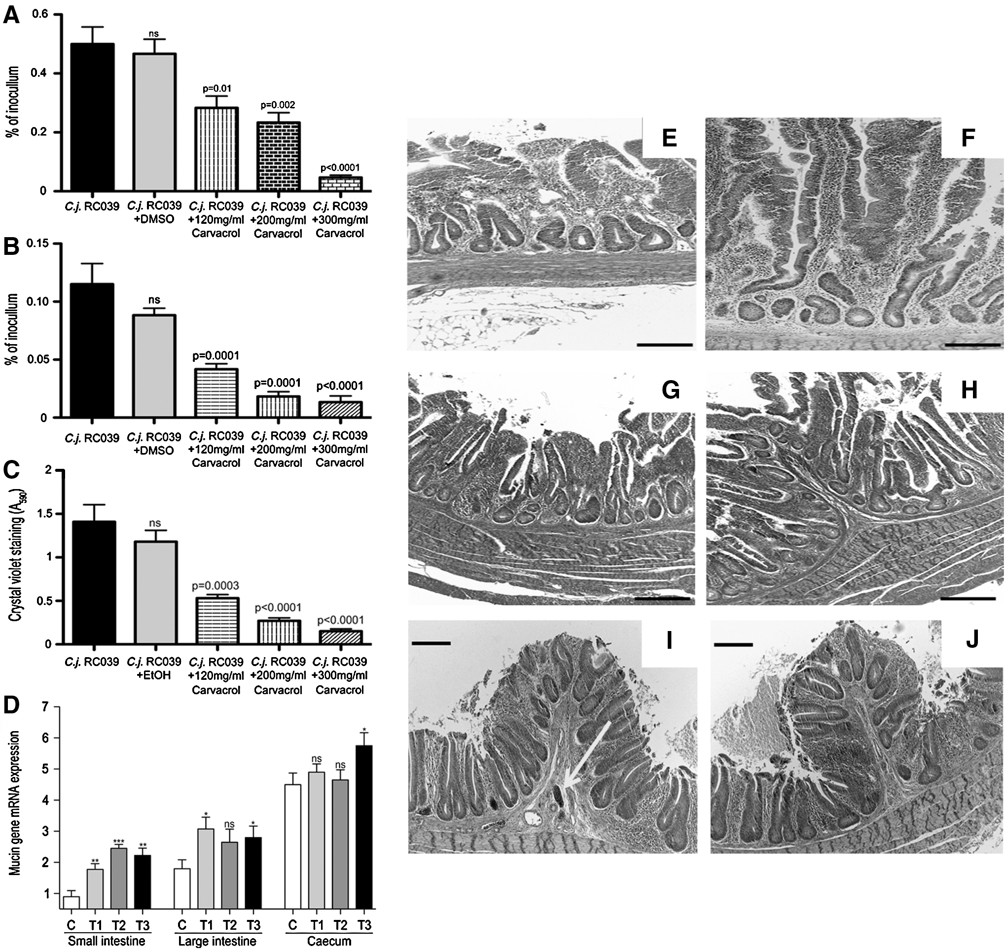
Adhesion, internalization, biofilm formation in vitro, and in vivo mucin expression.
Carvacrol effect on the chicken gastrointestinal compartments
Next, we investigated the effect of carvacrol on the integrity and development of the intestinal surfaces directly involved in bacterial colonization and nutrient absorption. The histologic analysis at slaughter indicates an increase in small intestinal villus height in all carvacrol groups (Fig. 1). Similar investigations performed on tissue harvested from the large intestine also revealed healthier epithelial surfaces in the experimental groups compared with the control (Fig. 1G, H). Changes were observed in the cecum; however, clear erythrocyte infiltrations [as indicated by the yellow arrow in Figure 1I] were observed in the control group and absent in the experimental group (Fig. 1J). To investigate whether the increase in epithelial surface in the experimental groups was associated with increased mucus production, we have investigated the presence of mucin mRNA (Fig. 1D). The expression of mucin mRNA increased both gradually and significantly in the experimental groups compared with the control group in both large and small intestines. In the cecum, similar increases were observed in mRNA expression; however, significance was only detected in experimental group T3. These results suggest that carvacrol can increase the epithelial surface and the production of the inner mucus layer.
Carvacrol delays Campylobacter spp. detection in naturally colonized chicken broilers
Our results show (Fig. 2D) that during the starter (0–10 days) and grower periods (11–21 days), the relative abundance of Lactobacillus spp. in broilers cecal content is significantly increased compared with the control group, and that the E. coli presence (Fig. 2C by plate count) is significantly reduced in all three experimental groups compared with the control. This increase in Lactobacillus presence is also associated with a lack of Campylobacter spp. detection at 10 and 21 days in all the experimental groups (Fig. 2A, B). The presence of Campylobacter spp. in the treatment groups (T1, T2, and T3) only occurs at day 35 when the abundance of Lactobacillus sp. decreases below the levels of the control group. Our results suggest that carvacrol can stimulate the increase in abundance of probiotic bacteria in broilers cecum and reduce Campylobacter spp. presence by as much as 31% at levels of supplementation of 120 mg/kg feed.
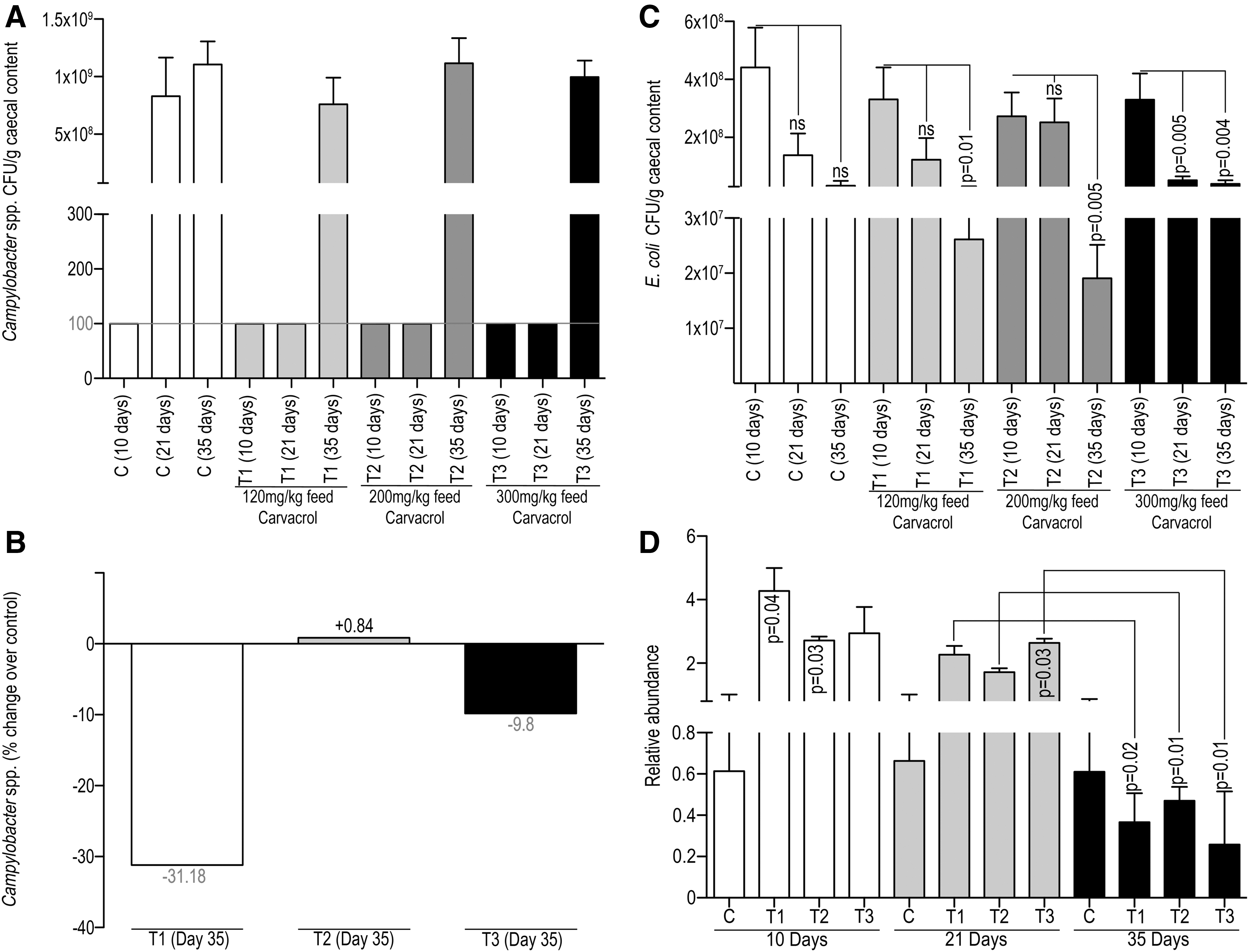
Campylobacter spp., Lactobacillus spp., and Escherichia coli quantification in cecal content at 10, 21, and 35 days.
Carvacrol induces changes in chicken cecum microbiota
The chicken cecum microbiome was assessed at days 10, 21, and 35. The major phyla were the Firmicutes (65.49%), Proteobacteria (28.24%), and Bacteroidetes (6.13%) (Figure 3). In day 10 Carvacrol samples, T1 (89.9%), T2 (83.7%), and T3 (82.7%) displayed a higher percentage of Firmicutes when compared with day 10 control samples (65.5%). Analysis of the day 21 samples displays the presence of the three major Phyla; however, the percentage of Bacteroidetes has increased in all day 21 samples: C (47.2%), T1 (39.4%), T2 (34.9%), and T3 (55.8%). At day 35, taxonomic analysis at the class level further identified differences between the samples. Further investigation of the Firmicutes identified a higher percentage of Bacilli within the Carvacrol samples. Further analysis of the Proteobacteria distribution identified the presence of Epsilonproteobacteria in day 35 control and Carvacrol samples (T1, T2, and T3). The day 35 control samples contained Campylobacter spp. at 10.52%. This was higher than day 35 Carvacrol samples T1 (6.43%) and T2 (7.85%); however, the day 35 Carvacrol T3 percentage Campylobacter spp. was noted to be higher (13.86%) than the respective day 35 control sample.
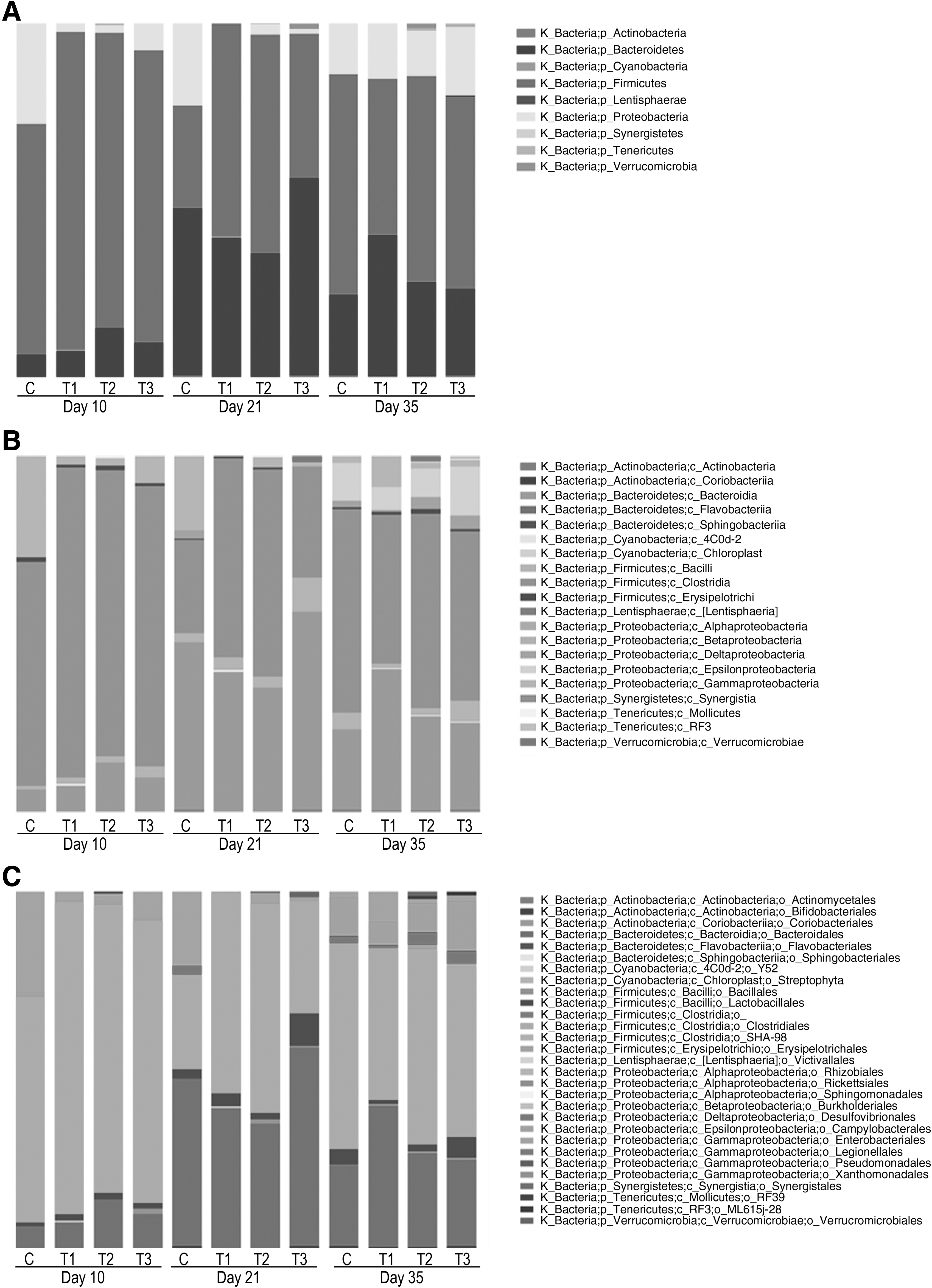
Plot bar charts of bacteria classified as phyla
Carvacrol improves production parameters at slaughter
The feed intake (Fig. 4A, B) for the experimental broilers in group T1 was slightly higher (+1.58%) compared with the control, but the increase was not statistically significant. The feed conversion rates were also reduced by 6.7% in experimental group T1 (non significant [NS]), by 24.8% (p = 0.04) in T2, and by 17.5% (p = 0.09) in T3 (Fig. 4C, D). As shown in Figure 4E and F at day 35 (slaughter), a 5.45% increase in BW was recorded for experimental group T1 (p = 0.02), a 5.10% increase for T2 (p = 0.03), and a 4.08% increase for T3 (p = 0.02). The experimental group T2 reduced its feed intake by 12.9% (p = 0.006) and T3 reduced its feed intake by 6.29% (p = 0.04) compared with the control at 35 days.
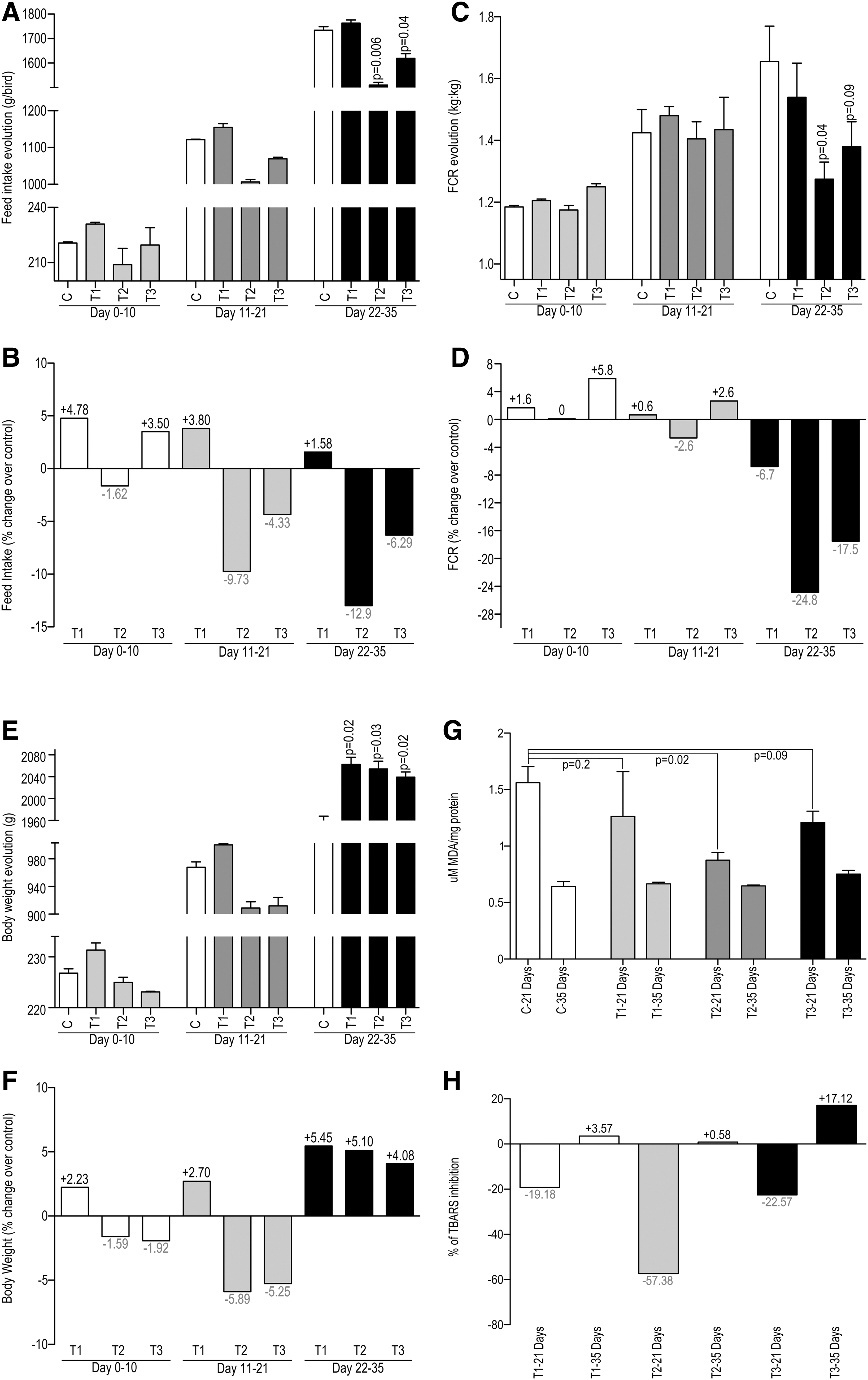
The effect of Carvacrol on the production parameters of naturally colonized chicken broilers.
Lipid oxidation and fatty acid composition of broiler thigh muscle
Across the carvacrol treatments, the TBARS values decreased significantly only at 21 days as shown in Figure 4G and H. Treatment T1 showed a 19.18% decrease (p = 0.2); at treatment T2, the TBRAS were reduced by 57.38% (p = 0.02) and they were reduced by 22.57% at T3 (p = 0.09). At day 35, compared with the control group, there were an 8.68% increase in total ω3 fatty acids, a 9.34% increase in ω6, a 13.77% increase in ω7, and an 8.43% increase in ω9. The total mono-unsaturated fatty acids (MUFA) at day 35 showed an increase of 8.55% compared with the control, and the poly-unsaturated fatty acids (PUFA) increased by 9.24% compared with the control. The saturated fatty acids increases in T2 (1 mg/g muscle) and T3 (1.46 mg/g) muscle are not significant and probably not biologically relevant. However, there was an increase by 2.11 mg/g muscle in unsaturated fatty acids at T2 and by 0.6 mg/g muscle at T3 with no statistical significance as described in Supplementary Table S1 (Supplementary Data are available online at
Discussion
The most recent report from the European Food Safety Authority (EFSA) places Campylobacter spp. as the most commonly reported human gastrointestinal pathogen in the European Union, with 214,000 cases and 56 deaths recorded in 2013 (Authority, 2015). This article describes for the first time the effect of Carvacrol in preventing adhesion and invasion of chicken intestinal primary cells and also new data on chicken broiler microbiota composition, growth performance and Campylobacter spp., and presence in a farm set up using naturally colonized broilers.
It is known that essential oils such as Carvacrol act by increasing the membrane permeability of Gram-negative bacteria, causing structural and functional changes and leading to outer membrane disintegration (La Storia et al., 2011). The structural and functional integrity of C. jejuni outer membrane structures have been previously described as crucial for this pathogen to efficiently attach and adhere to gut epithelial cells (Corcionivoschi et al., 2012). The ability of C. jejuni to colonize or to infect the epithelium is highly dependent on the genetic specificity of each strain (Ragimbeau et al., 2014). To reduce this variability, we have used C. jejuni RC039, a highly virulent chicken isolate recently described as positive for the newly identified Type Six Secretion System (T6SS) (Corcionivoschi et al., 2015). Carvacrol was proven to efficiently reduce the pathogenicity of this isolate when tested on chicken intestinal primary cells. Moreover, because the outer membrane structures are involved in the ability of C. jejuni to create biofilm (Naito et al., 2010), we have shown that carvacrol has a negative effect on the ability of C. jejuni RC039 to form biofilm.
As described earlier, it is clear that Carvacrol can reduce the attachment of C. jejuni to chicken intestinal cells (Arsi et al., 2014); however, if this is the case, an in vivo scenario with naturally colonized chicken broilers is still under debate. It has been suggested that probiotic bacteria are very efficient in reducing C. jejuni colonization of the gastrointestinal compartments in chicken broilers; however, in this case, the probiotic strains were introduced in the diets and the authors have not characterized the microbiota composition in the cecum (Cean et al., 2015). We have shown that till day 21 Carvacrol was able to increase the presence of probiotic bacteria, which correlates with no C. jejuni presence in the experimental groups.
Dietary evaluation of essential oils has been indicated to reduce the gut lesions and to improve villus height and crypt depth in the small intestine of broiler chickens fed with 120–240 mg/kg tymol and carvacrol. It has been suggested that these essential oils improve intestinal integrity and modulate immune responses in Clostridium perfrigens challenged chicken broilers (Du et al., 2016). Also, an increased villus height is associated with an increased digestive and absorptive function of the gut due to increased absorptive surface area, enzyme expression, and nutrient transport system (Amat et al., 1996). We are now showing, in vivo, that carvacrol supplementation, through feed, improves the expression of mucin mRNA expression in all the essential gut compartments, providing a possible explanation for the increase in production parameters.
The high content of PUFA makes poultry meat less susceptible to oxidative deterioration (Luna et al., 2010). In our study, we found that PUFA was 5.93% in birds fed 120 mg/kg feed of carvacrol, suggesting an increase in meat quality and, subsequently, in shelf life. The reduced feed intakes observed during the trial could be explained by the enhanced release of satiety hormones, an effect previously described in rats (Yang et al., 2013). The consumer will benefit from having a product with an increased ω7 concentration, as it has been previously shown that it may be useful in the treatment of hypertriglyceridemia with the beneficial added effects of decreasing low density lipoprotein and high-sensitivity C-reactive protein and raising high density lipoprotein (Bernstein et al., 2014).
We have demonstrated that carvacrol prevented the infection of chicken primary intestinal cells in vitro and it is also able to prevent campylobacters from forming biofilm. Our plate count data also indicate that carvacrol affects Campylobacter spp. colonization in vivo, and our study indicates the efficient concentrations. Finally, our results indicate that, at farm level, inclusion of carvacrol can improve poultry health, feed efficiency, and meat quality and delay colonization of foodborne pathogenic bacteria in broiler chickens.
Footnotes
Disclosure Statement
No competing financial interests exist.
References
Supplementary Material
Please find the following supplemental material available below.
For Open Access articles published under a Creative Commons License, all supplemental material carries the same license as the article it is associated with.
For non-Open Access articles published, all supplemental material carries a non-exclusive license, and permission requests for re-use of supplemental material or any part of supplemental material shall be sent directly to the copyright owner as specified in the copyright notice associated with the article.
