
Abstract
Salmonella enterica serovar Enteritidis is the leading global cause of salmonellosis. A total of 146 Salmonella Enteritidis isolates obtained from retail chicken products in Shanghai, China were characterized for their antimicrobial susceptibilities, virulence and antibiotic resistance gene profiles, and molecular subtypes using pulsed-field gel electrophoresis (PFGE). Approximately 42% (61/146) of the isolates were susceptible to all 13 antimicrobials tested. More than half of the isolates (50.70%) were resistant to ampicillin, 49.32% to sulfisoxazole, 17.12% to tetracycline, and 15.75% to doxycycline. Thirty (20.55%) isolates were resistant to three or more antimicrobials. The avrA, mgtC, and sopE virulence genes were identified in all isolates, while 97.2% and 92.4% were positive for bcfC and spvC genes, respectively. Genes associated with resistance to streptomycin (aadA), β-lactams (blaTEM, blaCMY, blaSHV, and blaCTX), tetracycline (tetA and tetB), and sulfonamides (sulI, sulII, and sulIII) were detected among corresponding resistant isolates. A total of 41 PFGE patterns were identified from 77 antimicrobial resistance (AMR) isolates and were primarily grouped into seven clusters (A–G), each with 90% similarity. The majority of Salmonella Enteritidis isolates (63.63%, 49/77) shared the same PFGE cluster, indicating potential cross contamination during processing and cutting or working during retailing and marketing. A significantly (p < 0.05) lower percentage (<25%) of isolates belonging to clusters D and E were resistant to sulfisoxazole compared with those belonging to clusters A, B, C, F, and G (>80%), indicating that sulfisoxazole resistance might be associated with genetic content (PFGE profiles) of Salmonella Enteritidis. This study provides important and updated information about the baseline antimicrobial-resistant data for food safety risk assessment of Salmonella Enteritidis from retailed chicken in Shanghai, which is the first step for the development and implementation of China's AMR National Action Plan, and can be helpful for future surveillance activities to ensure the safety of the chicken supply.
Introduction
S
The virulence potential of bacterial pathogens is due to virulence-associated and antimicrobial resistance (AMR)-associated genes (Capuano et al., 2013). Salmonella virulence factors are frequently located on Salmonella pathogenicity islands (SPIs), prophages, or virulence plasmids (Borriello et al., 2012). For example, the avrA gene induces host cell apoptosis to limit the inflammatory response to infection (Borges et al., 2013). The mgtC gene is required for intracellular survival as is the spv operon on virulence plasmids that promotes intramacrophage survival. The Salmonella membrane proteins (sops) have a role in invasion through host membrane deformation and rearrangement of the host cytoskeleton. The fimbrial gene bcfC encodes proteins necessary for attachment and invasion (Huehn et al., 2010; Hur et al., 2011b).
The high level of use of antimicrobials in veterinary medicine has resulted in the contamination by the presence of MDR Salmonella in food animals (Chuanchuen et al., 2010). This is a major public health concern since antibiotic-resistant bacteria of animal origin can be transmitted to humans (Khemtong and Chuanchuen, 2008). Many resistance genes have been identified on mobile genetic elements that allow gene dissemination among bacteria in the chicken gut or in extraintestinal environments.
Pulsed-field gel electrophoresis (PFGE) is still considered as the “gold standard” for molecular subtyping of foodborne pathogens, widely used in surveillance or epidemiological investigation. PFGE pattern diversity within a serotype is also useful for assessing the effectiveness of control measures (Sandt et al., 2013). However, there is a lack of PFGE data for Salmonella Enteritidis in retail chicken products in Shanghai, one of the largest metropolitan areas in the world with the population of 24.20 million in 2016.
In this study, 146 Salmonella Enteritidis isolates were recovered from commercial chicken products in Shanghai, China. Isolates were further characterized to establish antibiotic resistance phenotypes and genotypes, as well as the distribution of virulence determinants. PFGE data from this study can be helpful for understanding the relationship between genetic relatedness and sources or AMR of Salmonella Enteritidis strains associated with retail chicken products, as well as developing suitable mitigation strategies.
Materials and Methods
Salmonella Enteritidis isolates
Salmonella Enteritidis isolates (146) (Supplementary Table S1; Supplementary Data are available online at
Antimicrobial susceptibility test
Minimal inhibitory concentrations of 13 antimicrobials were determined via an agar dilution method according to guidelines recommended by the CLSI (2003). The following antimicrobials were tested: amikacin (AMK), gentamicin (GEN), kanamycin (KAN), streptomycin (STR), ampicillin (AMP), ceftiofur (CTX), ceftriaxone (CRO), sulfisoxazole (SUL), chloramphenicol, ciprofloxacin (CIP), ofloxacin (OFX), tetracycline (TET), and doxycycline (DO). Escherichia coli strains ATCC 25922 and ATCC 35218 were used as quality control organisms in antimicrobial susceptibility experiments. Breakpoints for most antimicrobials were used according to the interpretive standards of the CLSI (2013). The breakpoints for STR and CTX were chosen based on a previous study (Lai et al., 2014). Salmonella Enteritidis isolates showing resistance to three or more antibiotics were defined as MDR isolates.
Molecular identification of virulence and antibiotic resistance genes
All isolates were screened for the presence of five Salmonella virulence genes using PCR (Table 1). Virulence determinants for each isolate analyzed were categorized according to their location in the Salmonella genome: SPI (avrA and mgtC), prophages (sopE1), plasmid (spvC), and fimbrial clusters (bcfC). Ten antibiotic resistance genes, including resistance to β-lactams (blaTEM, blaCTX, blaSHV, and blaCMY), STR (aadA), TET (tetA and tetB), and sulfonamides (sulI, sulII, and sulIII), were also screened by PCR (Table 1). Total genomic DNA from bacterial isolates was extracted from overnight cultures using a DNeasy Blood and Tissue Kit (Qiagen, Valencia, CA). The PCR products were visualized under ultraviolet (UV) light following electrophoresis in agarose gels stained with ethidium bromide.
PFGE and BioNumerics analysis
All the AMR isolates were subjected to PFGE, which was performed according to the Centers for Disease Control and Prevention PulseNet protocols (Ribot et al., 2006). In brief, agarose-embedded DNA was digested with 50 U of Xba I (TaKaRa, Dalian, China) for 1.5–2 h at 37°C. Restriction fragments were separated by electrophoresis in 0.5 × TBE (Tris-borate-EDTA, Thermo Scientific™, Shanghai, China) buffer at 14°C for 18 h using a CHEF Mapper Electrophoresis System (Bio-Rad, Hercules, CA) with switch times of 2.16 and 63.8 s. Salmonella Braenderup H9812 (ATCC BAA 664) was used as a molecular size marker. The gels were stained with ethidium bromide, and DNA bands were visualized with UV light. PFGE results were analyzed using BioNumerics Software (Applied-Maths, Kortrijk, Belgium) manually with 1% to 2% band-shift tolerance. Clustering was performed using the unweighted pair group average method applying the Dice coefficient using a cutoff value of 90% similarity. The comparison of mean values was analyzed by SAS Version 8.0 via one-way analysis of variance (plus post hoc). Statistical significance was defined at p < 0.05.
Results
Among the 146 Salmonella Enteritidis isolates, we found antibiotic resistance was most frequent to AMP (50.70%), followed by SUL (49.32%), TET (17.12%), DO (15.75%), STR (4.80%), CTX (2.05%), and CRO (1.37%). All isolates were susceptible to AMK, GEN, KAN, OFX, and CIP (Table 2).
MIC, minimal inhibitory concentration.
The 146 Salmonella Enteritidis isolates included 61 (41.78%) that were susceptible to all of the antimicrobials tested (pansusceptible) (Table 3). We found 30 (20.55%) MDR isolates comprising 9 MDR patterns. This included one isolate that exhibited resistance to 5 of the antimicrobials (STR-AMP-SUL-TET-DO) and 17 isolates that exhibited resistance to four antimicrobials (AMP-SUL-TET-DO, STR-AMP-CTX-SUL). The remaining 6 MDR patterns consisted of 12 isolates (Table 3).
AMP, ampicillin; CRO, ceftriaxone; CTX, ceftiofur; DO, doxycycline; STR, streptomycin; SUL, sulfisoxazole; TET, tetracycline.
All the isolates we tested possessed avrA, mgtC, and sopE, most of which also carried bcfC (97.2%) and spvC (92.4%). Four different virulence gene profiles (Supplementary Table S2) were identified among the 146 isolates and the major profile (VP1, avrA-mgtC-sopE1-spvC-bcfC) occurred in 132 (90.41%) of the isolates. VP2 (avrA-mgtC-sopE1-bcfC) and VP3 (avrA-mgtC-sopE1-spvC) included 10 (6.83%) and 3 (2.05%) isolates, respectively, whereas VP4 (avrA-mgtC-sopE1) was represented by a single isolate.
Different resistance-associated genes, including blaTEM, blaCTX, aadA, tetA, tetB, sulI, sulII, and sulIII were detected among the specific antibiotics phenotypically Salmonella Enteritidis-resistant isolates with the respective rates of 100%, 38.7%, 100%, 100%, 67.7%, 100%, 100%, and 100%. None of the isolates was positive for the blaCMY and blaSHV genes (Table 1).
In the current study, 85 isolates that were resistant to at least one antimicrobial were tested by PFGE. We found 41 PFGE patterns for 77 isolates with similarity indices ranged from 77.45% to 100%. Of these, 10 PFGE patterns contained at least two isolates that were identical. The isolates were mainly grouped into seven clusters (A–G) with a 90% pattern similarity. The majority of XbaI profiles were assigned to cluster C, which represented 49 isolates. We observed isolates in different PFGE clusters, representing the same AMR patterns, as well as in isolates in the same cluster, representing the same or different AMR patterns (Fig. 1). It should be noted that isolates belonging to clusters D and E with a significantly (p < 0.05) lower percentage (<25%) were resistant to SUL compared with those belonging to clusters A, B, C, F, and G (>80%), indicating that SUL resistance might be associated with genetic content (PFGE profiles) of Salmonella Enteritidis.
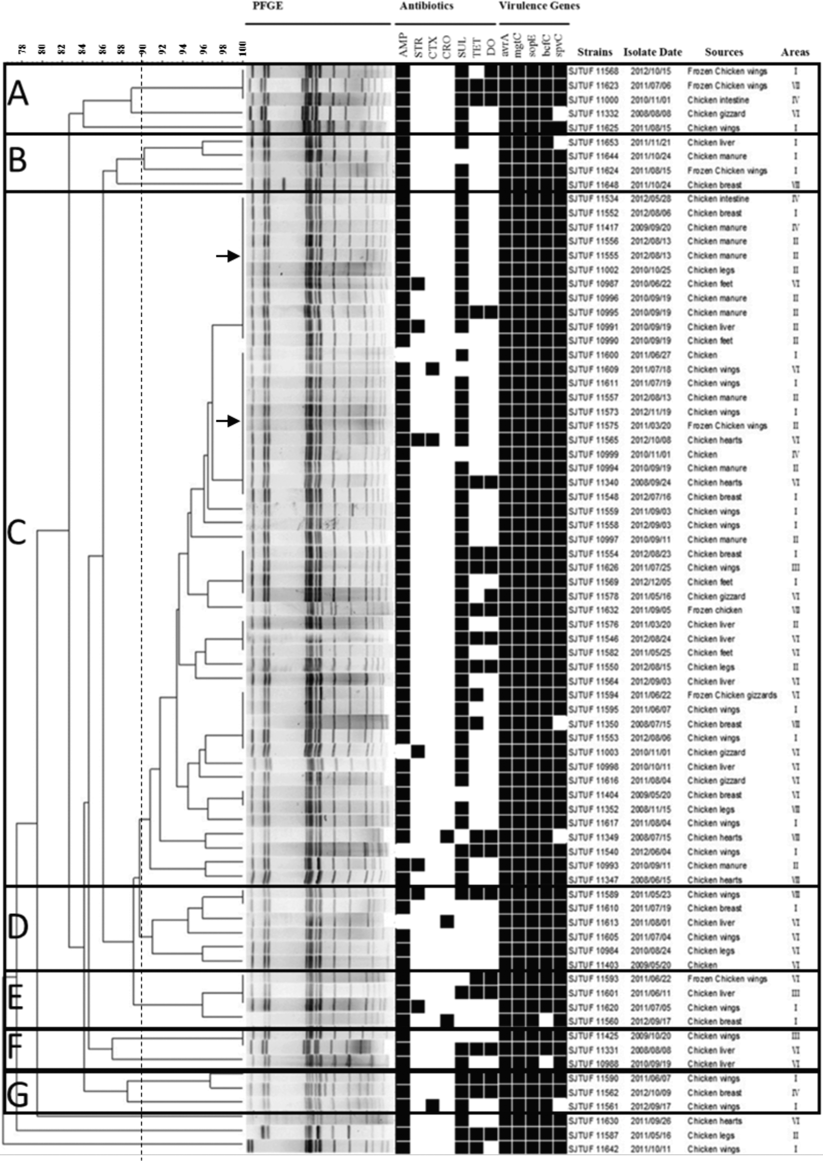
XbaI PFGE patterns, antimicrobial resistance profiles, and virulence gene profiles of isolates identified in this study. A black box indicates resistance to a particular antimicrobial or positive for a particular virulence gene.
Discussion
In this study, we collected 146 Salmonella Enteritidis isolates from retail chicken products in eight districts in Shanghai, China, and determined their AMR, pathogenic potential, and molecular characteristics. Previous studies had indicated that Salmonella Enteritidis was susceptible to most antimicrobial agents (Su et al., 2004; Zhao et al., 2007). However, the increased use of antibiotics in poultry feeds has resulted in an increase in MDR Salmonella Enteritidis strains in China in the last decade (Yang et al., 2010; Wang et al., 2015). Almost 20% (30/146) of our Salmonella Enteritidis isolates were MDR, and these results are overall consistent with other recent reports (Yan et al., 2010; Lu et al., 2011; Wang et al., 2014).
The widespread use of certain antibiotic agents has put a high selective pressure on Salmonella for resistance development; in other words, the development of antibiotic resistance and transference of antibiotic resistance genes might be selected by the widespread use of antibiotic agents, resulting in the emergence and dissemination of antibiotic resistance in Salmonella (Sow et al., 2007). We found that 50.7% of our isolates were resistant to AMP, and similar results have been found in China (61.9%) as well as South Korea (>50%) and Brazil (81.25%) (Hur et al., 2011a; Oliveira et al., 2012; Wang, 2015). This may stem from the widespread and long-term use of AMP in veterinary practices in China (Yang et al., 2010; Wang et al., 2015). Similarly, the higher resistance to TET (17.12%) and DO (15.75%) compared to other antibiotic agents observed in this study was probably due to the incorporation of these two antibiotic agents into livestock and poultry feed as growth promoters (Barbosa and Levy, 2000; Yang et al., 2010). In contrast, the resistance to STR (4.80%) was found to be lower than the above three antibiotic agents. Similarly, low levels of resistances to STR were also observed, particularly among Salmonella Enteritidis and Salmonella Typhimurium isolates recovered from retail chicken meats (Yang et al., 2010).
Importantly, CTX and CRO are third-generation cephalosporins approved for use in livestock and poultry agriculture in China (Liu et al., 2012b), and an increasing rate of cephalosporin-resistant Salmonella of food animal origin can cause important public health implications for the dissemination of Salmonella resistant to cephalosporins through food. Fortunately, cephalosporin-resistant isolates in this study were not MDR strains. However, the low resistance rates to CRO (1.37%) and CTX (2.05%) should be of concern because of the potential for resistance development directly through interference with treatment or indirectly through dissemination of resistance elements to other pathogens. All the Salmonella Enteritidis isolates in this study were susceptible to CIP and OFX, which are frequently used to treat children with Salmonella infections, particularly, invasive infections (White et al., 2001). These data further emphasize the necessity to appropriately manage the use of clinical antibiotics in food production animals.
There was considerable conservation of the virulence gene content among the isolates, and conservation among the SPIs and chromosomal genes was found in this study. This was similar to a previous report of the 100% prevalence of bcfC, mgtC, and sopB genes in Salmonella Typhimurium isolates in Europe (Huehn et al., 2010). In our study, all isolates harbored the two SPI genes (avrA and mgtC), indicating that these virulence genes are widespread and highly conserved among the Salmonella, as previously reported in Salmonella Typhimurium of human clinical isolates (Huehn et al., 2010) and in Salmonella Enteritidis isolated from poultry house and clinical samples (Mezal et al., 2014). In addition, the plasmid-associated gene spvC has been associated with a limited number of serovars, predominately, Salmonella Typhimurium and Salmonella Enteritidis (Chiu and Ou, 1996). It was therefore not surprising that the spvC prevalence in our isolates was 92.4%.
The blaTEM gene, which confers resistance only to penicillins and early generations of cephalosporins, was also found in 75 isolates in this study. However, the resistance spectrum of blaTEM descendants may extend to second, third, and fourth generation cephalosporins (Salverda et al., 2010). In addition, all the blaTEM-positive isolates were negative for the blaSHV gene, and 38.7% (29/75) of the isolates were positive for blaCTX-M. The latter may be located on the same plasmid as blaTEM has been previously reported (Elumalai et al., 2014). Interestingly, none of the three CRO-resistant isolates in the current study harbored blaCMY, indicating the existence of other resistance mechanisms (Chuanchuen et al., 2010).
We observed 100% prevalence rates of sulI, sulII, and sulIII genes in all sulfonamide-resistant isolates and that of the aadA gene in all STR-resistant isolates. Previous reports have shown that the sulfonamide resistance genes are encoded by a 55.0 kb plasmid as well as an aadA gene located on spv-plasmids (Cerro et al., 2003; Taban et al., 2013). The presence of both antimicrobial and virulence genes makes these pathogenic strains potentially hazardous if these genes are expressed.
PFGE analysis revealed a highly similar genetic profile in the 49 Salmonella Enteritidis isolates in cluster C (Fig. 1). The isolates did not cluster based on year of isolation, source, or geographic distribution. Some of the isolates from different origins were grouped together in a cluster and, in some cases, shared identical patterns. For example, 11 isolates from different origins in cluster C showed the same PFGE pattern (Fig. 1 marked with black arrows; Supplementary Table S1), indicating potential cross contamination during processing and cutting or working during retailing and marketing. In addition, some PFGE profiles of Salmonella Enteritidis from clinical cases involving children during the same period were obtained by others in our team (data not shown) that could be compared with these poultry patterns that the same profile(s) might be the prevalent PFGE profile(s). Next step, these PFGE profiles could be compared with globally circulating PFGE patterns by PulseNet. In the current study, PFGE exhibited limited discriminatory power for Salmonella Enteritidis that 63.63% (49/77) of isolates shared the same PFGE cluster, which was consistent with the previous studies (Boxrud et al., 2007; Mezal et al., 2014). Therefore, whole genome sequencing should be carried out on MDR isolates, including those carrying resistance plasmids for more detailed information.
Conclusions
The findings of this study indicated that retail raw chicken products are reservoirs for MDR Salmonella Enteritidis, which is a major food safety concern for public health. Moreover, our study provided baseline antimicrobial-resistant data for food safety risk assessment of Salmonella Enteritidis from retail chicken products. Thus, this study provides information for potentially developing effective intervention strategies and for considering the use of natural biocontrol agents such as bacteriophages to ensure the safety of retailed chicken supplies.
Footnotes
Acknowledgments
This study was supported by the National Natural Science Foundation of China (Grant Nos. 31601562 and 31230058) and Shanghai Agriculture Applied Technology Development Program, China (Grant No. G 20150405).
Disclosure Statement
No competing financial interests exist.
References
Supplementary Material
Please find the following supplemental material available below.
For Open Access articles published under a Creative Commons License, all supplemental material carries the same license as the article it is associated with.
For non-Open Access articles published, all supplemental material carries a non-exclusive license, and permission requests for re-use of supplemental material or any part of supplemental material shall be sent directly to the copyright owner as specified in the copyright notice associated with the article.