




Abstract
Husbandry practices for laying hens in commercial egg production is a topic of interest from a social, economic, and regulatory standpoint. Animal welfare concerns regarding the use of conventional cages have arisen and consumer perceptions of hen welfare have led to a higher demand for cage-free eggs. The aim of this study was to assess the impact of housing systems on prevalence, persistence, and antimicrobial resistance (AMR) of Campylobacter from laying hens and shell eggs. A total of 425 samples were collected over a 10-month period from the North Carolina Layer Performance and Management Test and Campylobacter isolates were identified by serological, biochemical, and molecular tests. Genetic variability was evaluated using pulsed-field gel electrophoresis (PFGE) and AMR testing was performed. Prevalence of Campylobacter spp. ranged from 11.1% in the enrichable cages to 19.7% in the conventional systems. A greater prevalence of Campylobacter was found in the fecal swab samples from free-range birds compared with those of birds housed in the more intensive housing systems (p > 0.05). Overall, 72 isolates were confirmed as Campylobacter spp. by PCR. More than 90% of the isolates (n = 66) were identified as Campylobacter jejuni, followed by Campylobacter coli (n = 6). C. jejuni isolates displayed high levels of resistance to tetracycline (67%). Genetic variability of Campylobacter was high, with more than 20 PFGE patterns identified. Pattern “a” comprised 42% of isolates from all housing systems and was also the most persistent. This study suggests that housing systems of laying hens used for commercial shell egg production may impact the rate of Campylobacter shedding by layers. Isolation rates and tetracycline resistance levels of this pathogen are still of concern, emphasizing the need for well-implemented biosecurity measures on the farm.
Introduction
P
Concerns about biosecurity of extensive housing systems have also been expressed due to increased potential for bacterial contamination (Mollenhorst et al., 2005; De Vylder et al., 2009). This has led to studies evaluating effects of housing systems on hen health and welfare, as well as impact on quality and safety of eggs (Jones et al., 2014, 2016; Karcher et al., 2015). Conflicting findings are common, due to the multifactorial nature of animal welfare, egg safety, and inherent variability of animal husbandry practices (Holt et al., 2011). To assess microbiological impact of different housing systems, researchers have studied the prevalence of Salmonella and Campylobacter in eggs and laying hens, and results are contrasting (De Reu et al., 2005; Cox et al., 2009; Gast et al., 2014; Jones et al., 2014, 2016). Both Salmonella and Campylobacter account for 2.5 million estimated foodborne illnesses annually in the United States (CDC, 2014), and are bacterial pathogens isolated from poultry, including laying hens, rearing environments, and eggshell surfaces (Gast et al., 2014; Jones et al., 2014, 2016). Laying hens commonly shed Campylobacter jejuni in their fecal material, as it is highly prevalent in ceca and lower reproductive tracts increasing the risk of surface contamination of eggs (Stern and Robach, 2003; De Reu et al., 2005; Cox et al., 2009; Jones et al., 2016). However, studies determining sources of contamination and persistence of pathogens in such environments and shell eggs are limited.
The present study further contributes to microbiological comparisons of commercial laying hen housing systems by evaluating the prevalence and persistence of Campylobacter spp. in environmental and eggshell samples obtained from various hen housing systems. Little is known about the antimicrobial resistance (AMR) exhibited by bacteria in these egg production environments. In recent years, international regulatory agencies that track the spread of AMR in the food chain have reported increasing trends of resistance to ciprofloxacin in C. jejuni isolated from poultry production and processing environments (FDA, 2011). Ciprofloxacin is a critically important drug for treatment of severe infections caused by Campylobacter spp. in susceptible patients (WHO, 2017a). Consequently, drug-resistant Campylobacter is now considered a serious threat to public health worldwide (CDC, 2013a; WHO, 2017b). With an estimated annual production of 80 billion eggs in the United States. (USDA, 2017), it is important to develop an extensive study that allows for assessment of drug resistance among pathogenic bacteria that are commonly present in egg production environments. Thus, the objective of the present study was to evaluate the effect of different commercial laying hen housing systems on prevalence, persistence, and AMR of Campylobacter from laying hens and eggshells.
Materials and Methods
Hen management
A flock of Hy-Line Brown layers was hatched and maintained at the North Carolina Department of Agriculture and Consumer Service, Piedmont Research Station, Poultry Unit (Salisbury, NC), in accordance with protocols and regulations established by the North Carolina Layer Performance and Management Test Program (Anderson, 2014, 2015). All animal care and production protocols were approved by the North Carolina State University Institutional Animal Care and Use Committee. Five different layer housing systems were monitored during this study: conventional cage (V), enriched colony cage (E), enrichable cage (H), cage-free barn (C), and free-range housing (R). The specific cage characteristics and hen stocking densities are described by Anderson (2015).
Sampling procedures
Samples were collected during the laying period, starting at 22 weeks of age, every 8 weeks over the course of 10 months, for a total of 425 samples. Sampling consisted of four environmental swab locations and three types of shell pool emulsion distributed among the different housing systems (Table 1). Egg and environmental swab samples were collected according to methods described by Jones et al. (2011). Fecal samples were collected from fecal deposits recovered from clean butcher paper placed under the cages or roosts; overnight fecal droppings were pooled into sterile 50 mL conical tubes for further processing.
Campylobacter spp. isolation and identification
Eggshell pool samples were prepared as described by Musgrove et al. (2005). Fecal deposits were homogenized with sterile cotton-tipped applicators. Environmental and fecal swabs were placed in 10 mL of sterile phosphate-buffered saline at 42°C and blended for 1 min at 230 rpm in a stomacher (Stomacher 400 Circulator, Seward Ltd., London, United Kingdom); Campylobacter recovery and isolation were performed according to Richardson et al. (2009) and Stern et al. (1995). A commercial latex agglutination immunoassay (Microbiology International, Frederick, MD) was used for preliminary identification of Campylobacter spp. and isolates were then stored in 20% glycerol at −80°C for further analysis. Presumptive Campylobacter isolates were grown as microbial lawns on tryptic soy agar plates (TSA; Thermo Fisher Scientific, Asheville, NC) with 5% laked horse blood (Lampire Biological Laboratories, Pipersville, PA) and restreaked onto blood-enriched TSA for recovery of damaged cells. Plates were incubated for 48 h at 42°C under microaerophilic conditions (5% O2, 8% CO2, and 87% N2), using the GasPak™ EZ Campy Container System (BD GasPak supplies, Thermo Fisher Scientific, Asheville, NC), and cryopreserved at −80°C in a 20% glycerol solution. The isolates were later examined using oil immersion phase-contrast microscopy and screened for typical Campylobacter morphology and darting motility (Ng et al., 1985; Jones et al., 1993; On, 1996; Butzler, 2004). Biochemical testing was further performed on the presumptive Campylobacter isolates using the API® Campy biochemical test kit (API™; bioMérieux, VWR, Radnor, PA).
DNA extraction and quantification
The PureLink™ Genomic DNA kit (Thermo Fisher Scientific, Asheville, NC) was used for DNA extraction of pure isolated cultures. A 2 μL sample of pure DNA from each isolate was quantified using an Epoch Microplate Spectrophotometer (BioTek® Instruments, Winooski, VT) by measuring UV absorbance at 260 nm (A260) and purity was assessed using the A260/A280 ratio of absorbance. DNA with a ratio of ∼1.8 was accepted as pure and stored at −20°C until further analysis.
Campylobacter spp. confirmation
Confirmation of Campylobacter spp. was carried out using a TaqMan real-time PCR assay (Lund et al., 2004). An additional 10 percent of the non-Campylobacter isolates were also tested using this PCR assay. The primers and probe for Campylobacter spp. were designed as per Lund et al. (2004) to detect a 16S rRNA gene sequence conserved among species C. jejuni, Campylobacter coli, Campylobacter lari, and Campylobacter upsaliensis. These were primers campF2 (5′-CACGTGCTACAATGGCATAT-3′) and campR2 (5′-GGCTTCATGCTCTCGAGTT-3′), and the TaqMan PCR probe campP2 (5′-6-FAM-CAGAGAACAATCCGAACTGGGACA-BHQ1-3′) (Express PrimeTime®; Integrated DNA Technologies, Inc., Coralville, IA). Template DNA from C. jejuni (ATCC 33560) was used as the positive control, and nuclease-free water was used as the negative control. A variation of the PCR assay developed by Best et al. (2003) was followed for the identification of species C. jejuni and C. coli, targeting genes mapA and ceuE, respectively. The primers selected for the detection of C. jejuni were mapA_F (5′-CTGGTGGTTTTGAAGCAAAGATT-3′) and mapA_R (5′-CAATACCAGTGTCTAAAGTGCGTTTAT-3′), and the TaqMan probe mapA_Probe (5′-6-FAM-TTGAATTCCAACATCGCTAATGTATAAAAGCCCTTT-BHQ1-3′). For identification of C. coli, the primers selected were ceueE_F (5′-AAGCTCTTATTGTTCTAACCAATTCTAACA-3′) and ceueE_R (5′-TCATCCACAGCATTGATTCCTAA- 3′), and TaqMan probe ceueE_Probe (5′-6-FAM-TTGGACCTCAATCTCGCTTTGGAATCATT-BHQ1-3′) (Express PrimeTime; Integrated DNA Technologies, Inc., Coralville, IA). An internal amplification control (IAC) was used based on the methods described by Lund et al. (2004). The IAC consisted of DNA of the bacterium Yersinia ruckeri ATCC 29473. The primers and probe for detection of the Y. ruckeri internal control were located in the 16S rRNA gene sequence; these were primers yersF1 (5′-GGAGGAAGGGTTAAGTG TTA-3′) and yersR1(5′-GAGTTAGCCGGTGCTTCTT-3′), and the TaqMan probe yersP1(5′-HEX-GCGAGTAACGTCAATGTTCAGTGC-BHQ1-3′) (Express PrimeTime; Integrated DNA Technologies, Inc., Coralville, IA). For each assay, a template of DNA from either C. jejuni ATCC 33560 or C. coli ATCC 43481 was used as the positive control.
Campylobacter spp. PCR assay was conducted in a StepOnePlus™ Real-Time PCR System, while the speciation PCR assays were conducted in the QuantStudio™ three Real-Time PCR Systems (Thermo Fisher Scientific, Asheville, NC). Amplification conditions for all assays were identical, consisting of an initial hold stage of 2 min at 50°C, a 10-min polymerase activation stage at 95°C, followed by 40 cycles of denaturation at 95°C for 15 s and annealing/extension at 58°C for 30 s and 72°C for 30 s with a final cycle of 5 min at 72°C. The PCR products were detected based on increases in fluorescence and assigned a threshold cycle value (C T ). A C T value of 40, the total number of cycles, was given for the nontemplate control.
Antimicrobial susceptibility testing
All confirmed Campylobacter were tested for AMR against nine antimicrobial agents, including azithromycin, ciprofloxacin, erythromycin, gentamicin, tetracycline, florfenicol, nalidixic acid, telithromycin and clindamycin, using the microbroth dilution method from Sensititre™ (Trek Diagnostic Systems, Inc., Independence, OH) as recommended by the Clinical and Laboratory Standards Institute (CLSI). A periodic quality control check of the colony count was performed according to the Sensititre protocol. In addition, a plate inoculated with C. jejuni ATCC 33560 was used for quality assurance and incubated with every batch of MIC plates. The MIC plates were read visually according to CLSI guidelines (CLSI, 2016). Resulting MICs were compared to inhibition breakpoints specified by CLSI and NARMS to determine resistance or susceptibility to antimicrobials.
Pulsed-field gel electrophoresis
Genetic characterization of the confirmed Campylobacter isolates was carried out according to the PulseNet protocol for molecular subtyping of C. jejuni by pulsed-field gel electrophoresis (PFGE) (CDC, 2013b). Isolates were cultured microaerobically on blood agar plates at 42°C for 18–20 h. Cell lysis and digestion of DNA were performed following the methods described by PulseNet (CDC, 2013b) using SmaI and KpnI as restriction enzymes (New England Biolabs, Inc., Ipswich, MA). The DNA fragments were separated on a CHEF MapperÒ Pulsed-Field Electrophoresis System (Bio-Rad Laboratories, Inc., Hercules, CA) with ramped pulses of 6.76–35.38 s and 5.2–42.34 s over 19 h, for Campylobacter isolates restricted with SmaI and KpnI, respectively. The resulting macrorestriction patterns were subjected to similarity analysis using the BioNumerics 3.5 software (Applied Maths, Sint-Martens-Latem, Belgium) with the Dice correlation coefficient (tolerance of 1.5%).
Statistical analysis
Campylobacter prevalence was determined based on the number of samples considered positive after PCR confirmation. Prevalence of antimicrobial-resistant phenotypes was calculated based on the number of isolates exhibiting resistance to at least one antimicrobial in proportion to the total number of confirmed Campylobacter. Prevalence results were analyzed using the chi-square goodness-of-fit test with housing system and sample type as the main effect by the PROC FREQ procedure (SAS 9.4). When cell counts were fewer than five in 50% of the cells, Fisher's exact test was performed. In addition, statistical differences in Campylobacter prevalence between sample type within housing systems were determined through pairwise chi-squared analysis. Statistical differences were resolved as p £ 0.05.
Results and Discussion
Campylobacter prevalence
Results from the preliminary identification immunoassay showed that 123 of the 425 samples were presumptively positive for Campylobacter spp. One presumptive Campylobacter isolate per positive sample was recovered for later confirmation. Further analysis identified 73 isolates as presumptive Campylobacter and results from biochemical confirmations by API® Campy test kit showed 91% (n = 66), 7% (n = 5), 1% (n = 1), and 1% (n = 1) of presumptive isolates as C. jejuni, C. coli, C. upsaliensis, and Arcobacter cryoaerophilus, respectively. Confirmation by PCR identified 72/73 isolates as Campylobacter spp. More than 90% (n = 66) of the isolates were identified as C. jejuni and about 8% (n = 6) were identified as C. coli (data not shown).
C. jejuni is the most commonly isolated species from humans and poultry processing environments (Butzler, 2004; CDC, 2013a), while C. coli is more prevalent in swine production systems (Gebreyes et al., 2005; Thakur and Gebreyes, 2005) and turkey meat (FDA, 2011). Numerous studies have reported similar isolation rates of C. jejuni and C. coli from commercial broiler flocks (van Gerwe et al., 2009; Panel, 2010; Wieczorek and Osek, 2015) and our findings are consistent with available literature. C. jejuni was present in all housing environments at similar rates, while C. coli was only detected in fecal swab samples from hens raised in enrichable and enriched caging systems, and those raised in free range huts. Similar observation was reported by Jones et al. (2016), where C. coli was found to be more prevalent in manure scraper blades compared with other environmental sites. No differences (p > 0.05) were observed for the rate of C. jejuni and C. coli isolation among the different housing types.
Lower levels of Campylobacter prevalence were found in our study than those reported by Jones et al., 2016 (46%), and Hariharan et al., 2009 (56%) (Table 2). When comparing housing types, hens raised in enrichable and enriched colony cages had the lowest prevalence of Campylobacter, while birds in conventional battery-style cages had the highest (Table 2). Laying hens housed in less intensive housing systems, that is, cage free and free range, also had higher levels of Campylobacter although differences in Campylobacter prevalence between housing systems were not significant (p > 0.05). Birds raised outdoors are exposed to potential pathogen vectors and are susceptible to stress due to weather variations. Free-range birds can become infected with pathogens that persist in the soil environment of the farm due to the difficulty of disinfection practices in such systems (Holt et al., 2011). Effective farm management practices and biosecurity measures are easier to implement in the more intensive systems (Holt et al., 2011). When analyzing Campylobacter prevalence in fecal swabs across housing systems, results showed that pathogen levels were higher for cage-free and free-range birds compared with birds in the more intensive housing systems (Table 3). All cage production systems featured a dry manure belt removal system, which can favor the growth of Campylobacter (Jones et al., 2016). Similar results were reported by Jones et al. (2014) showing that samples from the aviary system and enriched colony cages had a significantly higher contamination rate than samples from conventional housing. The opposite was seen, however, in another study by Jones et al. (2016) reporting a higher occurrence of C. jejuni in tissues from 77-week-old hens housed in enriched colony cages compared with conventional and aviary hens. Such discrepancies illustrate the challenges of evaluating the microbiological impact of housing systems on egg safety due to the dynamic nature of husbandry practices and confounding factors affecting egg safety (Cox et al., 2009; Holt et al., 2011).
In our study, we observed that eggshells from floor eggs were contaminated with the pathogen, suggesting possible improvement of egg safety by using nest boxes in cage-free and free-range systems. Similar conclusions were drawn by Jones et al. (2014) where aviary floor eggs had the highest levels of total aerobic microorganisms. There was a higher (p £ 0.05) prevalence of Campylobacter in the cage-free and enriched nest box swabs compared with that in the nest box shells of these same systems. The same was observed in the conventional system. While the risk for eggshell contamination with the pathogen seems low, hens should still be managed to prevent the occurrence of Campylobacter in floor eggs.
Antimicrobial resistance
Results from antimicrobial susceptibility testing indicate that C. jejuni was predominantly resistant to tetracycline (Table 4), while C. coli was susceptible to all nine antimicrobials tested. Resistance to tetracycline was high, with more than half (n = 44) of C. jejuni isolates exhibiting MICs ≥2 μg/mL of the antimicrobial based on the current epidemiological cutoff value established by NARMS (FDA, 2014). One of the 72 Campylobacter isolates was simultaneously resistant to three antimicrobials, azithromycin, ciprofloxacin, and nalidixic acid. All C. jejuni and C. coli isolates were susceptible to erythromycin, gentamicin, florfenicol, telithromycin, and clindamycin. Tetracycline-resistant C. jejuni was recovered from all housing systems at similar rates with levels of resistance ranging from 46% in free-range hens to 83% in hens housed in enrichable cages (Table 5). Hens raised in noncage systems had lower levels of tetracycline-resistant C. jejuni than those raised in conventional and enriched housing, however, differences were not significant (p > 0.05). Harisberger et al. (2011) also reported no consistent association of housing system and management practices in egg production, and the prevalence of AMR among Escherichia coli, Enterococcus faecalis, and Enterococcus faecium.
AZI, azithromycin; CIP, ciprofloxacin; NAL, nalidixic acid; TET, tetracycline.
Calculated from total C. jejuni isolates per housing system.
No antibiotics were used on the research farm, yet tetracycline resistance was high. Oxytetracycline, a drug of the tetracycline class, is used in some poultry production systems for treatment of infections by E. coli that develop as sequelae to viral respiratory infections in chickens. Researchers suggest that its use can induce decreased susceptibility to tetracycline due to selective pressure; however, findings are often conflicting as transfer of resistance genes in the environment is a multifactorial issue (Roberts, 1996; Qian et al., 2016). Campylobacter resistance to tetracycline has also been reported from organic production systems, in which no antibiotics are used, suggesting that long-term use of the antimicrobial might have contributed to widespread dissemination of tetracycline-resistant Campylobacter in food animal production systems (Piddock et al., 2000; Luangtongkum et al., 2006).
Higher resistance to tetracycline among C. jejuni isolated from commercial broilers, laying hens, and retail poultry has been reported in recent years worldwide (Cox et al., 2009; Hariharan et al., 2009; Melero et al., 2012; EFSA and ECDC, 2014; FDA, 2014; Guyard-Nicodème et al., 2015). Drugs of the tetracycline class are considered highly important for human medicine as they remain the drug of choice for treatment of specific infections of the urinary tract, skin, and respiratory tract (WHO, 2014). High levels of tetracycline resistance are therefore concerning for both human and veterinary medicine.
In Campylobacter spp., resistance to tetracycline is acquired through a plasmid-mediated gene tetO (Roberts, 1996). This gene can be transferred spontaneously between C. jejuni cells in the gastrointestinal tract of chickens, which might explain the observed levels of resistance (Avrain et al., 2004). Ciprofloxacin resistance in Campylobacter spp. is becoming increasingly common worldwide (EFSA and ECDC, 2014; FDA, 2014), rising steadily in isolates from poultry and humans in the United States since 1997 (FDA, 2014). Contrasting the national trend, only one isolate tested in our study was resistant to ciprofloxacin, a drug of importance for treatment of severe campylobacteriosis in humans (Moore et al., 2005). We report low levels of resistance to nalidixic acid, again differing from the national trend (FDA, 2014, CDC, 2013a), as only one isolate exhibited this phenotype. Concerns with the spread of fluoroquinolone resistance globally have prompted the classification of antibiotic-resistant Campylobacter as one of the leading threats to human health (CDC, 2013a; WHO, 2017). Infections caused by drug-resistant Campylobacter are believed to pose difficulty in providing effective therapy in humans, however, conflicting results have been reported (Wassenaar et al., 1998; Feodoroff et al., 2009).
PFGE genotypes
The PFGE typing of 66 C. jejuni and 6 C. coli isolates yielded 13 SmaI types and 22 KpnI types. This illustrates the higher discriminatory power of enzyme KpnI and importance of using two or more restriction enzymes for typing Campylobacter isolates, an observation previously reported by Gibson et al. (1995). Combination of the macrorestriction patterns resulted in 23 PFGE types (a through w; Table 6) with 5 predominant PFGE types encompassing more than 60% of the C jejuni isolates (Table 7). Pattern “a” was the most common PFGE profile (28 of the 66) and was observed in all housing types despite physical separation of the houses. Dendrograms showing PFGE of Campylobacter isolates using SmaI and KpnI are shown in Figures 1 and 2. The distribution of PFGE patterns of C. jejuni and C. coli isolated across the entire production period can be seen in Table 6. Genetic diversity of isolates increased with the duration of the egg production period, as 63-week-old hens had the highest diversity of C. jejuni and C. coli strains.
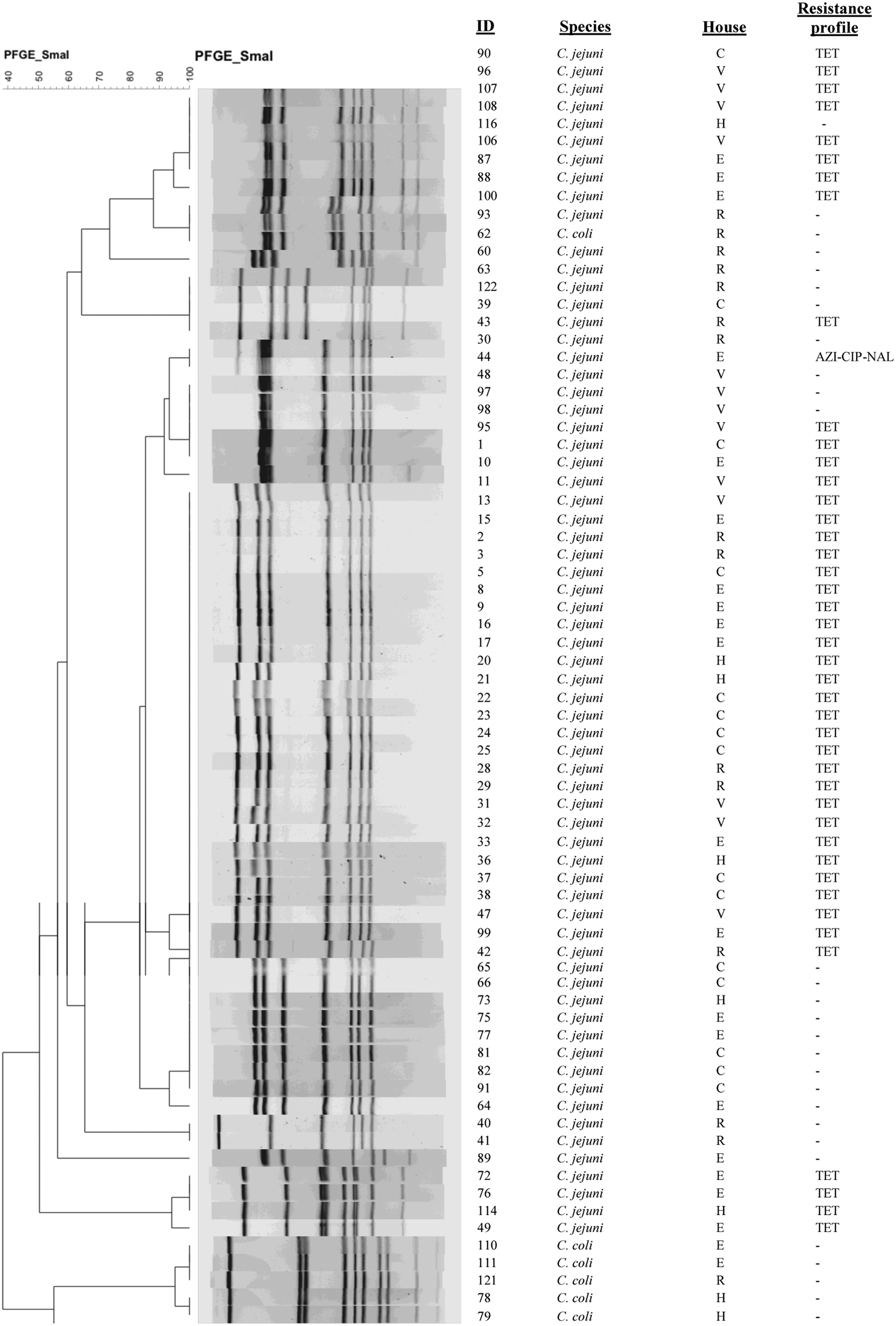
Dendrogram of PFGE Campylobacter isolates restricted with SmaI. PFGE, pulsed-field gel electrophoresis.
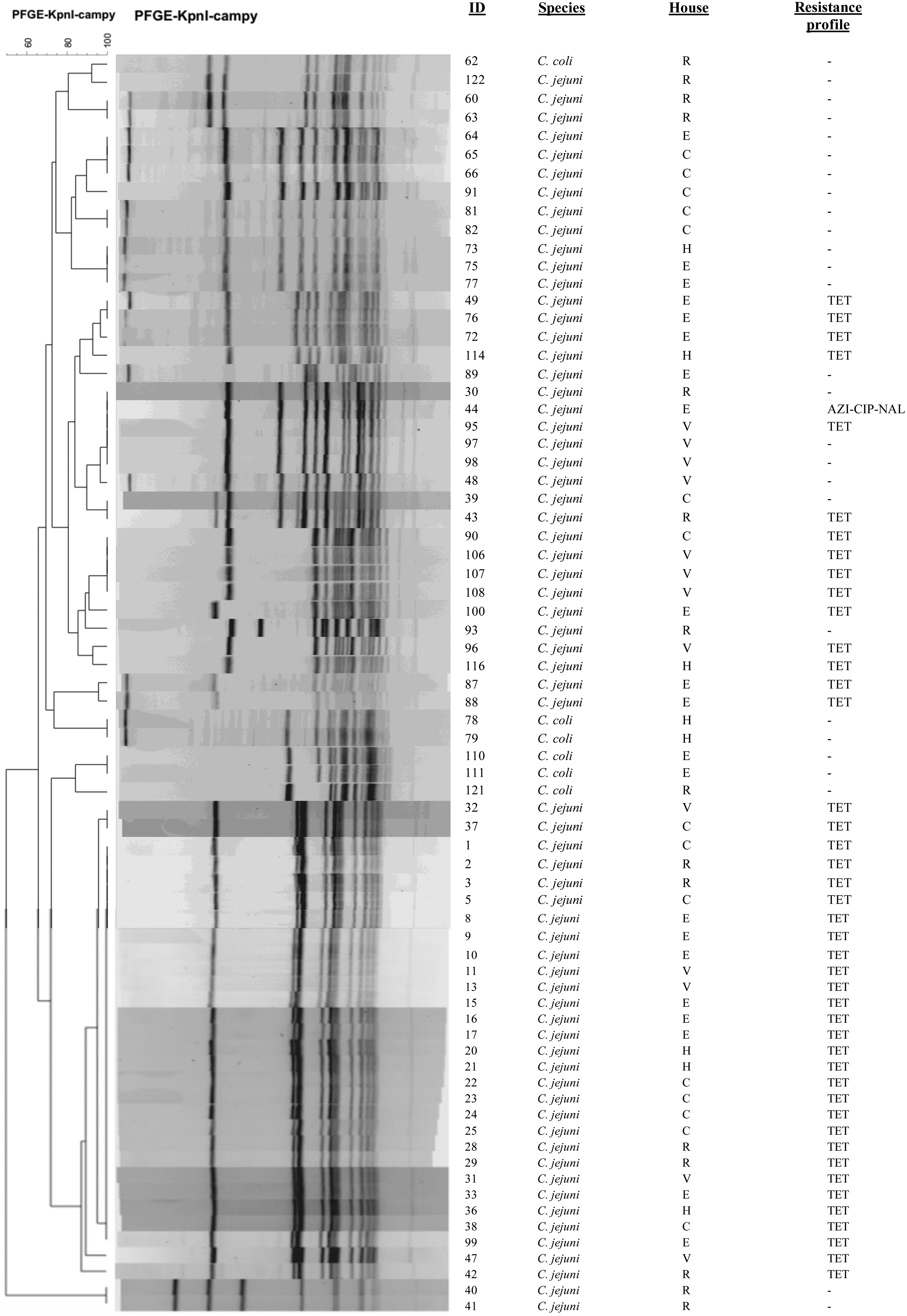
Dendrogram of PFGE Campylobacter isolates restricted with KpnI.
PFGE, pulsed-field gel electrophoresis.
Calculated from total C. jejuni-confirmed isolates.
C, cage-free housing; E, enriched environmental housing; H, enrichable cage; R, free-range housing; V, conventional cage.
At the farm level, cross-contamination between flocks is likely as Campylobacter can easily spread to surrounding environments and persist for several weeks in high numbers (Sahin et al., 2002; Johnsen et al., 2006). Colonization of breeder hens by multiple strains is likely (Jacobs-Reitsma, 1995; Jorgensen et al., 2011). Farm management practices and age of birds are factors that may contribute to the genetic diversity displayed by Campylobacter (Newell et al., 2001; Newell and Fearnley, 2003; Johnsen et al., 2006); yet, the dynamics of Campylobacter strains in the intestinal tract of birds remains poorly understood. Observed differences in genetic profiling of Campylobacter evaluated in this study might be attributed to the inherent genomic instability of C. jejuni strains during poultry colonization (Wassennaar et al., 1998; Parkhill et al.; 2000, Ridley et al., 2008).
All 28 isolates belonging to PFGE pattern “a” exhibited resistance to tetracycline. The wide spread of this resistant clone among young hens thus influences the high levels of resistance to tetracycline reported in the present study. Similar correlation was observed for most PFGE patterns comprising more than one bacterial isolate and is in agreement with reports from other studies (Praakle-Amin et al., 2007, Zhao et al., 2010). More accurate correlation between resistance genotypes and phenotypes can be achieved through whole genome sequencing by identification of corresponding AMR genes (Zhao et al., 2016).
Conclusions
This study suggests that there is no consistent association between laying hen housing and rate of Campylobacter colonization in layers. Features such as nest boxes can reduce the incidence of floor eggs, which have a greater potential of being contaminated with Campylobacter. Campylobacter has shown the ability to thrive in poultry production environments even when biosecurity protocols are followed, suggesting a need for more stringent actions to reduce the incidence of this pathogen at the farm. Overall, tetracycline resistance was highly prevalent among C. jejuni isolates recovered from all housing types, yet no correlation with housing characteristics was observed. Most tetracycline-resistant isolates shared a common PFGE pattern; further research is warranted to determine the presence and characterization of resistant genes. Finally, genetic diversity assessed by PFGE genotyping appeared high. A trend regarding hen age was observed, with older hens having a wider variety of Campylobacter strains in their environment. Reasons for this, however, remain poorly understood, and thus, further research is necessary for characterizing the genes involved in colonization and virulence mechanisms of Campylobacter.
Footnotes
Disclosure Statement
No competing financial interests exist.