
Abstract
Antimicrobial resistance genes play an important role in mediating resistance to sulfonamide in Gram-negative bacteria. While PCR is the current method to detect sulfonamide resistance genes (sul1, sul2, sul3), it is time-consuming and costly and there is an urgent need to develop a more convenient, simpler and rapid test for the sul. In this study, we describe a multiplex loop-mediated isothermal amplification (m-LAMP) assay we developed for the rapid and simultaneous detection of three sul. This m-LAMP assay successfully detected seven reference strains with different sul genotypes, but was negative for nine sul-negative reference strains. The m-LAMP products were verified by HinfI restriction enzyme digestion and the detection limit of the test was 0.5 pg genomic DNA per reaction. Testing 307 sulfonamide-resistant Enterobacteriaceae clinical isolates with the m-LAMP revealed all were positive for the sul with sul2 (79.5%) and sul1 (64.5%) being most prevalent, and sul3 the least (12.1%). Of the Enterobacteriaceae isolates tested, the Salmonella Indiana, a newly emerging serovar resistant to numerous antimicrobials, were most commonly positive with 33% having sul3.
Introduction
S
With the long-term and extensive use of sulfonamides, resistance has developed in many bacteria (Kozak et al., 2009). This sulfonamide resistance (SR) in Gram-negative bacteria has been shown to be mediated mainly by mutations in the floP (dihydrofolate synthase) (Sköld, 2001; Perreten and Boerlin, 2003) with three drug-resistant variants of the gene described to date, sul1, sul2, and sul3. The sul1 and sul2 were reported first (Rådström and Swedberg, 1988; Sundström et al., 1988) and are most common in SR Gram-negative strains. The sul1 is usually located in the 3′-terminal conserved regions of the class I integron, and sul2 in the nonconjugative plasmid or mobile multidrug-resistant plasmid (Carattoli, 2001; Byrne-Bailey et al., 2009). The sul3 was identified in 2003 (Perreten and Boerlin, 2003) and is mainly distributed in various large plasmids. The discovery of the SR genes has enabled the more comprehensive investigation of the molecular mechanisms of SR.
PCR and multiplex PCR are commonly used for the detection of sul genes (Antunes et al., 2005; Hammerum et al., 2006; Phuong Hoa et al., 2008; Kozak et al., 2009) but these methods are generally time-consuming and costly, making them unsuitable for the rapid screening for multiple sul in large numbers of samples (Kokkinos et al., 2014). For this, there is an urgent need for a more simple and convenient test.
Loop-mediated isothermal amplification (LAMP) is a novel detection technology in which four specific primers can rapidly (1 h) recognize six regions of the target DNA under isothermal conditions (60°C–65°C) (Notomi et al., 2000; Nagamine et al., 2001, 2002). The LAMP amplification products can be determined visually by the turbidity of the reaction fluid or color changes that occur when products combine with fluorescence dyes (Boehme et al., 2007). The LAMP assay stands out as a highly sensitive and specific diagnostic test that is particularly useful in developing countries as it requires sophisticated equipment or skilled personnel and is cost effective (Notomi et al., 2000). Currently, LAMP technology is mainly used for detecting infectious organisms and there are only few reports of its use to detect drug-resistance genes (Li et al., 2017).
In this study, we describe the development of a multiplex LAMP (m-LAMP) for the rapid, specific, and sensitive simultaneous detection of the three sul and its use for the rapid screening of clinical isolates for sul.
Materials and Methods
Bacterial strains
Seven sul-positive reference strains and nine sul-negative reference stains were used as controls in this study (Table 1). A total of 307 SR Enterobacteriaceae isolates (113 Salmonella spp., 168 Escherichia coli, 20 Klebsiella pneumoniae, and 6 Proteus mirabilis) made from clinically ill poultry in previous studies were screened for sul with the m-LAMP we developed. The Salmonella spp. isolates were serotyped using standard agglutination tests with O and H antisera (S&A Reagent Laboratory LMT, Bangkok, Thailand).
DNA extraction
The purified bacteria were inoculated into Luria-Bertani (LB) broth (Tianhe, Hangzhou, China) for overnight incubation at 37°C. Then, a rapid DNA isolation kit (Sangon, Shanghai, China) was used to extract bacterial DNA according to the manufacturer's instructions.
Design of the m-LAMP primers
The nucleotide sequences of the three sul were obtained from GenBank, and online software Primer Explorer version 4 (
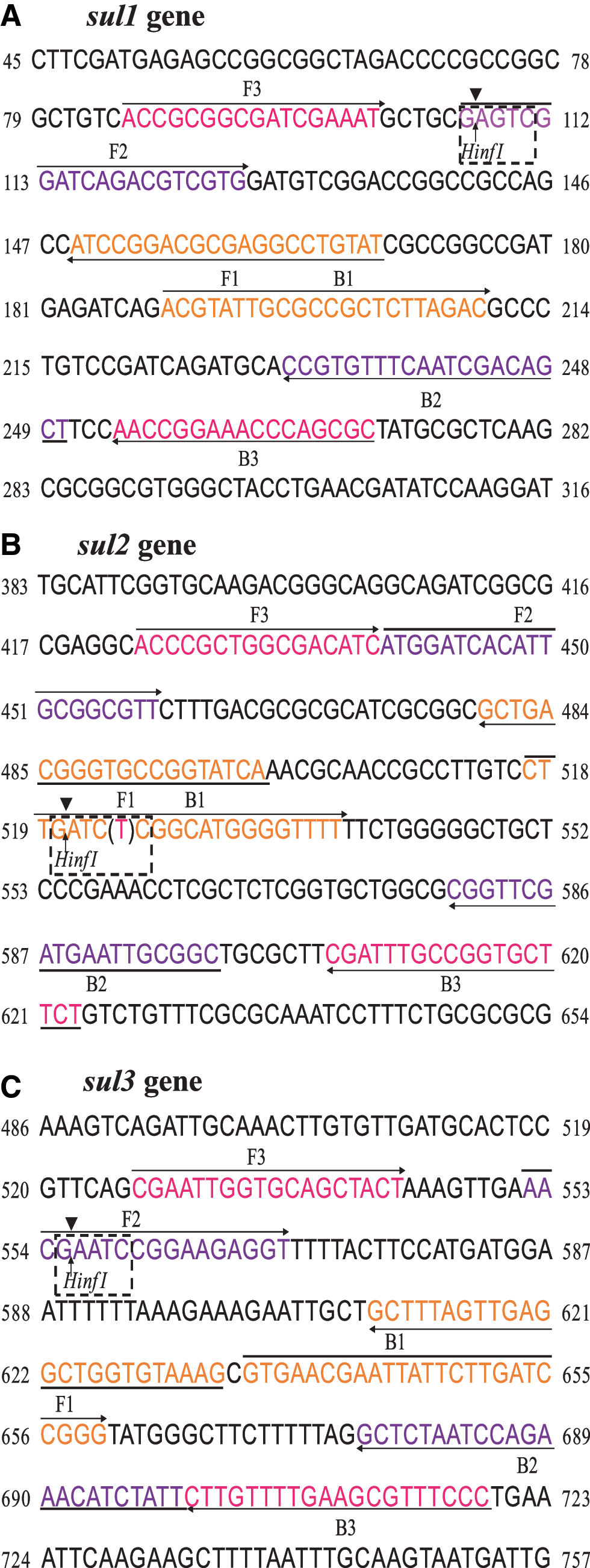
Locations of the m-LAMP primers and HinfI restriction sites in the sulfonamide resistance genes. Outer primers (F3 and B3) and the inner primers (FIP and BIP) were used in the m-LAMP. The dashed box shows the restriction site of HinfI.
m-LAMP reaction
The LAMP reaction was carried out in a final volume of 25 μL reaction mixture containing 1 μL of primers [FIP and BIP (40 μM), F3 and B3 (5 μM), 2.5 μL buffer [200 mM Tris–HCl pH8.8, 100 mM KCl, 100 mM (NH4)2SO4, 40 mM MgSO4, 8 M Betaine, 1% Triton X-100], 1 μL Bst DNA polymerase (8 U), 3.5 μL dNTPs (10 mM), 2 μL DNA template, and 4 μL ddH2O. The mixture was incubated for 50 min at 64°C in the Loopamp real-time turbidity system (LA-500, Beijing Lanpu Bio-tech, Beijing, China).
Specificity of the m-LAMP assay
The specificity of the m-LAMP assay was established by testing seven reference strains with known sul genotypes (sul1, sul2, sul3, sul1+sul2, sul1+sul3, sul2+sul3, and sul1+sul2+sul3) and nine sul-free reference strains. The m-LAMP products (5 μL) were incubated at 37°C for 1 h with 1 μL HinfI, 2 μL CutSmart buffer, and 12 μL ddH2O, before being electrophoresed through 2% agarose gels and visualized under ultraviolet light.
Sensitivity of the m-LAMP assay
Serial 10-fold dilutions of the genomic DNA templates of the seven sul–positive reference strains were made (5 × 10⁴ pg to 5 × 10⁻² pg per reaction) to determine the detection limit for the m-LAMP.
Detection of sul genes in clinical isolates
The 307 SR clinical isolates were screened for sul with the m-LAMP assay and amplification products identified by color change when they combined with SYBR GREEN I (1 μL per reaction) that could be observed with the naked eye (green indicated positive, orange negative). Products were digested with HinfI to determine which of the three sul were present.
Statistical analysis
The prevalences of the different sul were compared with Chi-squared Test. A p value below 0.05 was considered to indicate significant difference.
Results
Specificity of the multiplex LAMP assay
The Primer-BLAST program showed the m-LAMP primers selected for the study had 100% identity with the sul1, sul2, and sul3. The m-LAMP method successfully amplified all the reference strains known to have sul but were negative for the sul-free reference strains. The presence of sul1, sul2, and sul3 in the samples was reliably demonstrated with HinfI restriction enzyme digestion of the LAMP amplification products (Fig. 2).
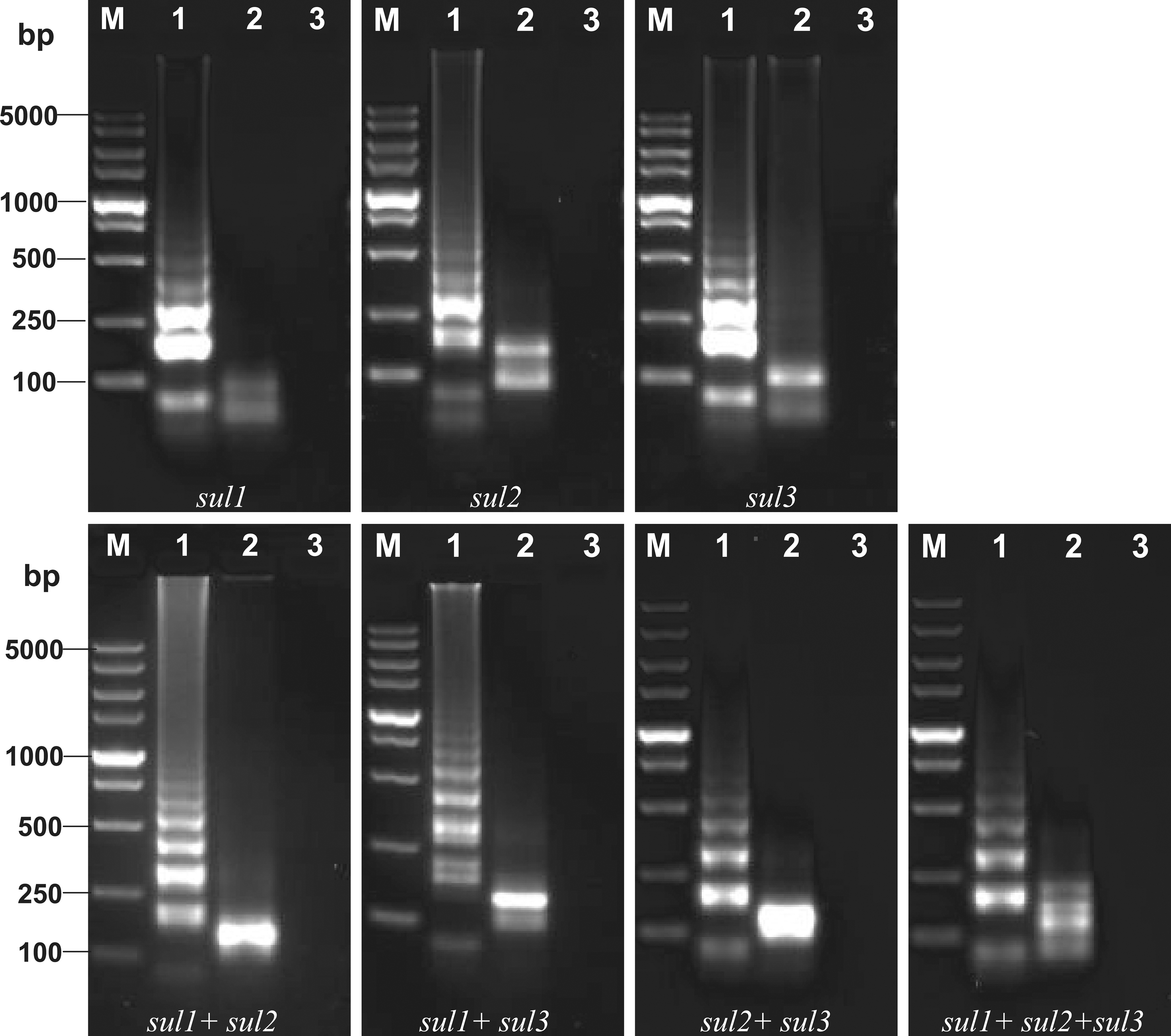
Specificity of the m-LAMP amplification products of different sulfonamide resistance genotypes digested by HinfI restriction enzyme. Lane M, DL-5000 DNA Marker. Lane 1–3, LAMP amplification products, the digested products and negative control.
Sensitivity of the multiplex LAMP assay
The m-LAMP assay was used to amplify 10-fold serial dilutions of S. Albany D234 (sul1), S. Indiana B354 (sul2), S. Indiana B142 (sul3), S. Pullorum C1891 (sul1+sul2), K. pneumoniae KP156 (sul1+sul3), S. Pullorum C1451 (sul2+sul3), and S. Indiana B157 (sul1+sul2+sul3) and the detection limit determined to be 0.5 pg genomic DNA per reaction (Fig. 3).
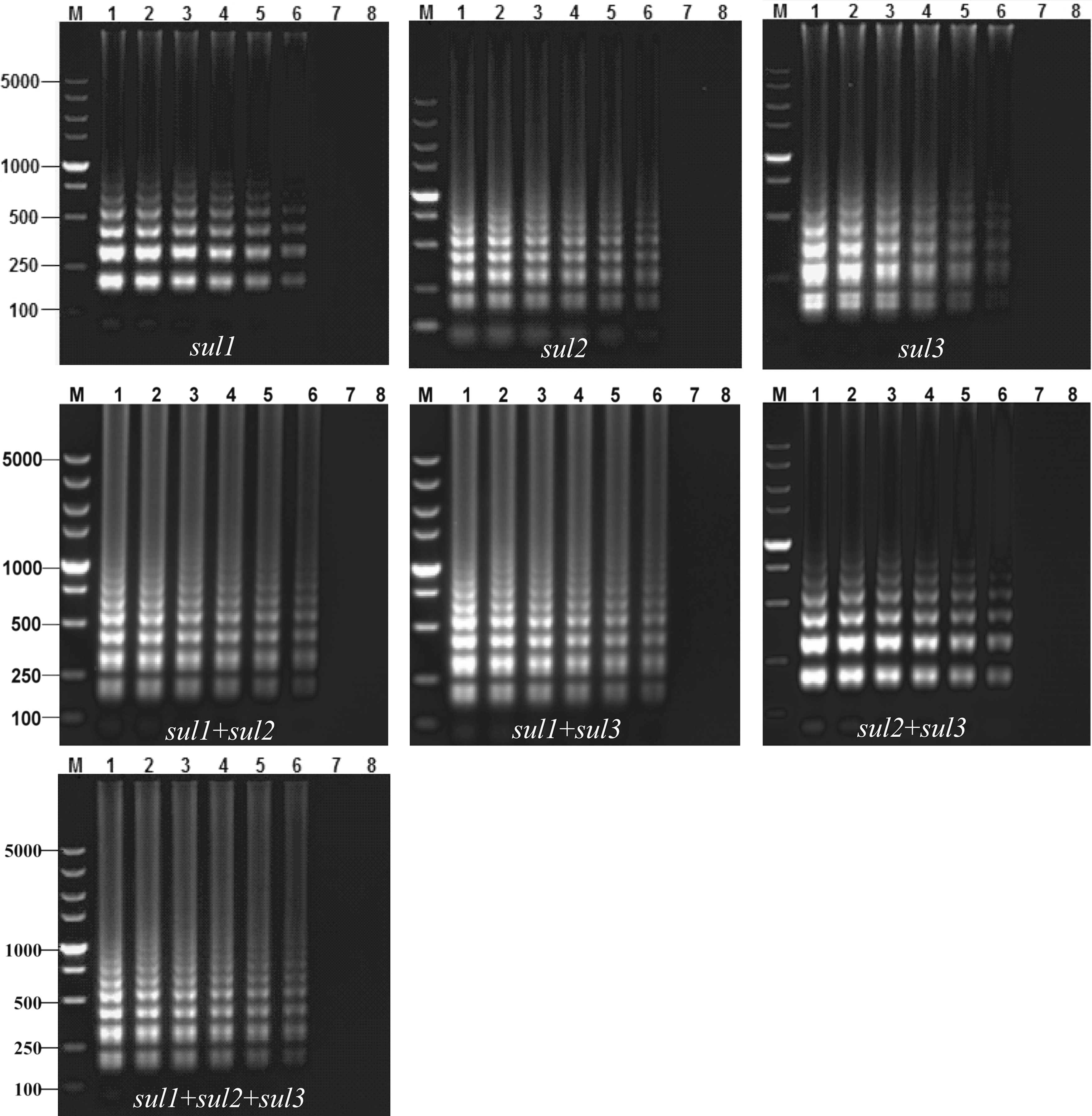
Sensitivity of the m-LAMP in detecting sulfonamide resistance genes. Lane M, DL-5000 DNA Marker. Lane 1–7, 5 × 10⁴, 5 × 10³, 5 × 10², 5 × 101, 5 × 100, 5 × 10⁻¹, 5 × 10⁻² pg per reaction, Lane 8, negative control.
Prevalence of SR genes in clinical isolates
The m-LAMP showed all the clinical SR isolates contained at least one of the sul. The prevalence of the sul3 (37/307, 12.1%) was significantly lower than that of sul1 (198/307, 64.5%), and sul2 (244/307, 79.5%) (p < 0.01) (Table 3). Half the isolates (155/307; 50%) carried a single sul, while 132 (43%) carried two of the sul, and 20 strains (7%) carried all three sul. The most common resistance patterns were the combination of sul1and sul2 (n = 125, 40.7%) followed by the presence of only sul2 (n = 97, 31.6%) (p < 0.01) (Table 4). Of the SR Enterobacteriaceae isolates, the Salmonella spp. had six resistance genotypes (patterns?) none of which contained sul3, the E. coli strains had five and the Klebsiella spp. and Proteus spp. three (Table 4).
The positivity in sul3 (37/307, 12.1%) was significantly lower than that of sul1 (198/307, 64.5%), and sul2 (244/307, 79.5%) (p < 0.01).
The dual positive for sul1and sul2 (n = 125, 40.7%) and positive for sul2 (n = 97, 31.6%) were more prevalent than other sul genotypes.
There were differences in the prevalence of the different sul among the 113 Salmonella serovars: 95.6% (43/45) of Salmonella Indiana isolates carried two or three sul (Table 4) while, in strong contrast, 91.2% (62/68) of the other Salmonella serovars carried only a single sul (Table 4). In addition, the Salmonella Indiana isolates showed more diverse sul genotypes than the other Salmonella serovars (Table 4).
Discussion
There are now many studies showing widespread resistance to sulfonamides among bacteria due to the long-term and often injudicious use of these antimicrobials. The resistance in the Gram-negative bacteria is mainly mediated by sul (sul1, sul2, sul3) (Sköld, 2001; Perreten and Boerlin, 2003; Kozak et al., 2009; Gong et al., 2013) and simple, rapid, and cost-effective tests to these genes is important to guide antimicrobial therapy and to monitor and investigate mechanisms of resistance.
In this study, we successfully established a m-LAMP detection method for the rapid detection of the three sul. The specificity of the m-LAMP method with 16 control reference strains was 100% with the HinfI digestion of the m-LAMP products enabling the rapid identification of the resistance genotype. The sensitivity of the m-LAMP method was 0.5 pg from genomic DNA per reaction system, which is similar to that of other LAMP assays, including 2.6 pg for Bordetella pertussis, 1 pg for cfr gene, 0.26 pg for Staphylococcus aureus, and 0.23 pg for K. pneumoniae and Enterobacter sakazakii (Liu et al., 2012, 2015; Qi et al., 2012; Dong et al., 2015; Sheet et al., 2016). It is of note the detection limit of LAMP assays are usually 10-fold higher than those of PCRs (Liu et al., 2012, 2015; Qi et al., 2012; Dong et al., 2015).
When we used the m-LAMP we developed to screen for sul in 307 SR Enterobacteriaceae isolates, we found all to be positive (especially for sul1 and sul2). This is consistent with the sul being responsible for SR in SR Gram-negative bacteria and is in agreement with other studies (Gomes-Neves et al., 2014; Lopes et al., 2015; Yahiaoui et al., 2015; García-Fierro et al., 2016). We detected the sul 2 most commonly and the sul3 least commonly, which is similar to some previous studies (Aslam et al., 2009; Mąka et al., 2015; Yahiaoui et al., 2015; Manyahi et al., 2017) but not others (Gomes-Neves et al., 2014; Lopes et al., 2015; García-Fierro et al., 2016; Ben Salem et al., 2017; El-Sharkawy et al., 2017). The reason for the differences is unclear but might be due to regional differences or the different species examined the studies.
Although our study and reports from other groups have generally shown a low prevalence of sul3 in Enterobacteriaceae isolates, we found that Salmonella Indiana isolates had a significantly higher sul3 prevalence than the other species we studied. Salmonella Indiana is a newly emerging Salmonella serovar with high levels of Antimicrobial resistance in China (Gong et al., 2017). The finding that the majority of our Salmonella Indiana isolates had multiple sul genotypes suggests that Salmonella Indiana may enhance its drug-resistance by acquiring various resistance-related mobile components (Gong et al., 2016).
Overall, genotyping of the SR Enterobacteriaceae isolates showed that most carried one or two of the sul (n = 287, 93.5%), mainly sul2 or sul1-sul2, which were the most prevalent genotypes. Similar results have been reported previously which appears to indicate that sul1-sul2 or sul2 may be the major sul genotypes in SR Enterobacteriaceae bacteria (Zhang et al., 2012; Yahiaoui et al., 2015; Manyahi et al., 2017).
Conclusions
The m-LAMP we developed proved to be a rapid, simple, specific, and sensitive test for the three known SR genes. The test can be used for the detection and genotyping of sul in clinical specimens, which will enable the more judicious use of antimicrobials in patients. Further, the m-LAMP could serve as an important tool in studies of the mechanisms and evolution of SR in bacteria.
Footnotes
Acknowledgments
This project was supported by grants from the National Natural Foundation of China (31772758 and 31402200), and the Scientific and Technological Project of Yangzhou (YZ2016039, YZ2017062, and YZ2017171).
Disclosure Statement
No competing financial interests exist.