
Abstract
Although Mycobacterium bovis is the major etiological agent of tuberculosis in bovines, it can infect other mammalians. Previously reported cases of tuberculosis caused by M. bovis in cats from the Autonomous City of Buenos Aires (CABA) led to the conclusion that the main source of infection for these felines was the ingestion of raw bovine lungs. Thus, for this study, we collected samples of bovine viscera from butchers' shops of the Greater Buenos Aires (GBA) and the CABA to assess presence and viability of these mycobacteria in bovine lungs (including the lymph nodes) and livers. We analyzed 216 different samples and obtained 5 isolates of M. bovis (4 from lungs and 1 from liver) by culture analysis. We also confirmed the presence of different isolates by polymerase chain reaction, spoligotyping, and MIRU-VNTR assays. The results obtained in this work emphasizes the need of social education for food hygiene, and to change the habit of feeding pets with raw viscera, which carries the risk of epizootic and zoonotic transmission. Moreover, control and eradication programs of bovine tuberculosis should be strengthened and improved.
Introduction
B
Cats are more susceptible to mycobacterial infections such as M. bovis (Underwood et al., 1999; Gunn-Moore, 2014). They are infected by the ingestion of raw lungs and milk without a thermal treatment (Fernández and Morici, 1999; Underwood et al., 1999). Although it has been suggested that the risk of cat to human transmission of M. bovis may be very low, a report in England revealed the first documented case (
Additionally, the zoonotic risk increases when domestic animals are infected, because of the close relationship between humans and pets. Because M. bovis infection might cause lesions that are imperceptible on macroscopic evaluation, some infected organs may be sold despite having viable bacilli.
The aim of this study was to assess the food contamination risk because of the presence of viable M. bovis in viscera from different markets of the Autonomous City of Buenos Aires (CABA) and the Greater Buenos Aires (GBA). The selected samples were bovine lungs (including the lymph nodes) and livers and the presence of the bacilli were confirmed by molecular tools.
Materials and Methods
Samples
Six establishments from the (GBA) and one from the CABA were selected by convenience. The visits to each establishment were made on different days.
A total of 216 complete organs, including 210 lungs (with their respective mediastinal lymph nodes) and six livers were studied (Table 1). One hundred thirty-seven lungs were collected from a slaughterhouse after passing food inspection (Table 1). The remaining 73 lungs and all the livers were bought in different butchers' shops.
Samples Information
The results of culture and molecular typing are also shown. Each establishment were identified with a number (1–6).
CABA, Autonomous City of Buenos Aires; GBA, Greater Buenos Aires; GL, granulomatous lesion.
All the viscera were inspected and palpated to find any distortion of the tissue that may indicate granulomatous lesions (GL). Transversal cuts were made 1 cm apart on the complete organ and a sample was taken when GL were found. If GL were not found in the organ, normal tissue and mediastinal lymph nodes were also sampled. A detailed inspection of the complete organs was performed individually to prevent cross contamination during manipulation in the laboratory.
Culture
The sampled tissue was cut into small pieces with scissors and put into a sterile bag with 20 mL of double-distilled sterile water. Mechanical maceration was performed using a Masticator (IUL Instruments, Spain). Petroff's decontamination method with NaOH 4% was performed to culture the samples in Stonebrink media (CEPANZO, 1988) in triplicate. Cultures were held at 37°C for 60 d and evaluated on a weekly basis.
Detection by polymerase chain reaction
Colonies obtained by culture were collected with a sterile disposable loop and suspended in 200 μL of double-distilled sterile water in a 2 mL-tube with a screw cap. The tube was incubated at 95°C for 30 min to lyse the bacilli and release the DNA according to Supply et al. (2000) with modifications. After that, it was centrifuged at 12,000 rpm for 10 min. The supernatant was collected in a sterile tube and 10 μL were used as polymerase chain reaction (PCR) template. For the identification of mycobacteria of MTC, the IS6110 target sequence was amplified by using Touch-Down cycling, previously described by Zumárraga et al. (2005).
Additionally, to confirm that the M. bovis strains isolated were wild types, we analyzed the presence of esxA and esxB genes, which codify the proteins Esat-6 and Cfp-10 deleted in vaccinal M. bovis Bacillus Calmette-Guérin (BCG) strains, respectively. For the amplification of esxA, the esxAfLM (5′GAGAGATCTCATGACAGAGCAGCAGTGGAATTTC3′) and esxArLM primers (5′GTTGGATCCTGCGAACATCCCAGTGACG3′) were used. This pair of primers amplifies a 288 bp fragment. For the amplification of esxB, the esxBf (5′TGACAACAGACTTCCCGG3′) and esxBr (5′CGATACCCGCGAAATTC3′) primers were used. These primers amplify a 450 bp fragment. For both genes, the reaction was carried out at 95°C for 2 min (1 cycle), followed by 30 cycles of 1 min at 95°C, 1 min at 55°C, and 30 s at 72°C, and a final elongation at 72°C for 5 min (Encinas, et al., 2018).
Molecular typing
Molecular typing by spoligotyping was conducted as previously described by Kamerbeek et al. (1997). Materials such as primers and membrane with bound probes were provided by Mapmygenome (Hyderabad, India). The spoligotypes were identified at the M. bovis Spoligotype Database (
Results
By evaluating 216 samples, we obtained five IS6110 PCR positive isolates from five different organs from GBA. One of them came from a liver and the remaining four positive isolates belonged to lungs and lymph nodes. Four of the positive samples were taken from butchers' shops (three lungs and one liver) and one from a slaughterhouse (lung). The liver presented a GL but this lesion was imperceptible until the organ was sliced (Fig. 1).
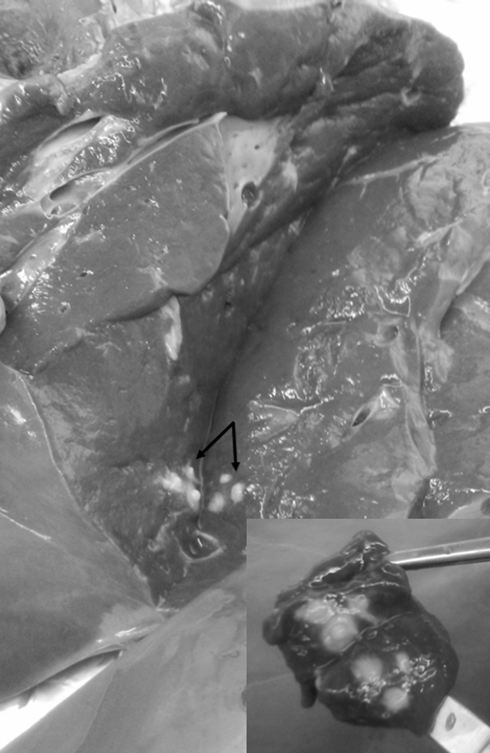
Granulomatous lesions detected in a liver. Multifocal nodular granulomatous lesion with liquefactive necrosis. The organ did not evidence enlargement or abnormalities detectable during inspection.
From all the organs collected in the slaughterhouse, 29 showed different lesions such as congestion, lymphadenitis, bronchopneumonia foci lesions, and fibrosis, but only 15 exhibited GL and small nodular lesions. However, we could only obtain a M. bovis isolate from one of these organs. The remaining four organs sold in butchers' shops, from which the other four isolates were obtained, did not show any GL that may indicate tuberculosis.
Spoligotyping confirmed that all the isolates were M. bovis and identified three different spoligotypes: SB0120 (n = 3), SB0130 (n = 1), and SB0140 (n = 1). MIRU-VNTR detected also three different patterns: 232314251212; 232214253322; and 233324253322. Each MIRU-VNTR pattern was related to each different spoligotype.
Considering that spoligotype SB0120 is present in both wild-type and vaccinal BCG strains, a deeper analysis based on MIRU-VNTR was performed confirming that the three M. bovis isolates were wild-type strains (Table 1). Additionally, these data were confirmed with a specific PCR for the esxA and esxB genes (absent in all BCG strains) (Fig. 2).

PCR results of IS6110
Discussion
The results presented in this study confirm the presence of viable M. bovis strains in bovine organs sold in butchers' shops, evidencing escapes from the routine slaughterhouse inspections mainly due to the inherent characteristics of the disease. In early infections, the incipient development of the lesions can go undetected during the manual and visual inspection, even if the inspection at the slaughterhouse is exhaustive. Accordingly, in this study 1.8% (4/216) of the organs without lesions were infected with viable M. bovis.
These results match those obtained by de Kantor et al. (1987), where the authors describe that 2.8% (5/178) of the inspected organs that were not condemned after slaughterhouse inspection were later confirmed as M. bovis by culture. Although our study is preliminary and limited to a restricted area of Buenos Aires province, the prevalence rate described above suggest an underestimation of the disease.
With regard to the molecular epidemiology of the M. bovis isolates, spoligotype SB0140 is most frequently obtained from isolated studies in Argentina (46%), followed by SB0130 (12%), whereas SB0120 represents only 4.9% (Zumárraga et al., 2013). In the case of statistics for the GBA, these spoligotypes represent 40%, 8%, and 4%, respectively, from the total of isolates studied between 1994 and 2016 in Buenos Aires province (n = 390) (unpublished data). In the present study, three samples presented SB0120 spoligotype, which is identical to the M. bovis BCG vaccine strain. However, using MIRU-VNTRs, these strains could be differentiated from BCG. Additionally, the presence of esxA and esxB genes was assayed, both of which are deleted in M. bovis BCG strains, confirming that these isolates correspond to wild-type strains. In cats, the spoligotype most frequently obtained in Argentina is SB0140, as it is in bovines.
Between 1998 and 2006, 19 isolates of M. bovis from cats living in the CABA were typed by spoligotyping (Zumárraga et al., 2009). In one of the reported cases, the owner of a cat with tuberculosis suffered from septic arthritis of the glenohumeral joint, caused by M. bovis, and she lived with 20 other cats that she fed daily with bovine lungs bought in a butcher's shop in CABA (Colmegna et al., 2004). This suggests a possible epizootic transmission between cats and bovines when cats are fed raw offal. However, more appropriate techniques such as MIRU-VNTR should be considered to establish those epidemiological links.
Recently, a rare and noteworthy pulmonary TB disease due to M. bovis has been reported in a patient who worked as a butcher in a slaughterhouse, who developed a cutaneous granulomatous inflammatory reaction on the dorsal side of his hand (Mertoglu et al., 2018). Thus, the manipulation of bovine organs such as lungs and livers by humans, represents an additional risk factor as they might contain viable mycobacteria, with the associated health concern for zoonotic transmission.
In this study, lungs and livers were assayed to detect viable zoonotic M. bovis strains taking into account that livers are consumed by people in Argentina and lungs are mainly used for feeding pets. In the first case, the risk to acquire an infection by consumption of an infected liver is low because the organ is previously cooked, whereas lungs dispensed to pets are generally given raw. However, the manipulation of raw organs represents a possible source of M. bovis infection for both, consumers and pet owners.
Food inspection is still a reliable and economical technique to identify tuberculosis and serves as a protection before consumption by humans and pets. Our findings lead to reinforce social education strategies for food hygiene practices to avoid the risk of epizootic and zoonotic transmission of bovine tuberculosis. Moreover, the results raise the need to perform a more exhaustive manual and visual inspection in slaughterhouses with complementary methods to detect potential sources of infection.
Footnotes
Acknowledgments
We thank Dr. Julia Sabio-García and Pablo Gabriel Crescentini for their critical reading of this article. S. Barandiaran, M.E. Eirin, and M.J. Zumárraga are career members of CONICET, Argentina. This work was supported by the National Agency of Research Promotion of Argentina Grant PICT 2012-0368.
Disclosure Statement
No competing financial interests exist.