




Abstract
The study aimed to monitor the fecal shedding of cefotaxime-resistant Escherichia coli (CREC) in a cohort of healthy calves on a dairy farm with documented antimicrobial usage and to characterize selected AmpC beta-lactamase-producing E. coli isolates. Fecal samples from 13 suckling calves (1–63 d of age; 113 samples in total) were repeatedly collected and cultivated on MacConkey agar with cefotaxime (2 mg/L). Resistant colonies were counted, and one colony obtained from the highest dilution of each fecal sample was identified by matrix-assisted laser desorption–ionization time-of-flight mass spectrometry. Susceptibility to antimicrobials and production of AmpC and extended-spectrum beta-lactamase (ESBL) were tested. No ESBL-producing E. coli was found, but representative AmpC-positive E. coli isolates were subjected to further typing and whole-genome sequencing (WGS) for the analysis of clonal relationships, resistance genes, virulence factors, and plasmid replicons. High amounts of CREC were detected in the feces of all 13 calves during the study. The number of CREC colonies varied from 1.0 log10 to 8.0 log10 colony-forming unit per gram. Drops in CREC density or its discontinued shedding were recorded at the end of the study period. A total of 82 (94%, n = 87) CREC isolates were confirmed as AmpC producers and all but one showed resistance to multiple antimicrobials. Twenty-nine selected AmpC-positive E. coli isolates belonged to 12 and 13 unique rep-PCR fingerprints and pulsed-field gel electrophoresis types, respectively, highlighting the variation in E. coli genotypes in individual calves. WGS of 10 selected isolates showed diverse antimicrobial resistance and virulence gene content and the presence of a bla CMY-2 gene carried by an IncK2 plasmid. Clinically important multiresistant E. coli isolates belonging to emerging extraintestinal pathogenic E. coli ST69 and ST648 lineages were found. Our findings reinforce the urgency of efforts to prevent the spread of ESBL-/AmpC-producing bacteria in dairy cow farms.
Introduction
In the past decade, resistance to extended-spectrum cephalosporins (ESCs) has increased because of the dissemination of plasmid-mediated extended-spectrum beta-lactamases (ESBLs) and/or AmpC type (AmpC) beta-lactamases in Enterobacteriaceae (Collignon et al., 2016). AmpC beta-lactamases fall into six phylogenetic groups with CMY-2 being the most commonly found enzyme (Philippon et al., 2002).
AmpC-producing Escherichia coli isolates have been frequently detected in healthy animal carriers and also among dairy and feedlot cattle, especially in animals with a history of ESCs treatment (Singer et al., 2008; Gonggrijp et al., 2016; Hutchinson et al., 2017). CMY-2-producing E. coli was also found in the milk of a cow with recurrent subclinical mastitis (Endimiani et al., 2012). In this context, cattle are considered to be the primary reservoirs for several zoonotic agents causing serious foodborne infections in humans, and bla CMY-2 may be potentially disseminated to these pathogens by a horizontal transfer of plasmids.
Although many genes of several pathotypes of E. coli have already been shown to contribute to virulence, little is known regarding the occurrence of virulence-associated determinants among ESBL-/AmpC-producing E. coli isolates of cattle origin, except for Shiga toxin–producing E. coli (Iweriebor et al., 2015; Dong et al., 2017). Factors contributing to the horizontal transfer of ESBL/AmpC genes into pathogenic E. coli strains on dairy farms also remain unclear.
The purpose of this study was to determine the fecal shedding density of cefotaxime-resistant E. coli (CREC) in calves on a dairy farm with documented antimicrobial usage for treatment of bovine mastitis in cows and to characterize CREC isolates using whole-genome sequencing (WGS)-assisted characterization of sequence types (STs), antimicrobial resistance genes, plasmids, and virulence factors.
Materials and Methods
Farm description, antimicrobial usage, and sampling
The parent herd at the investigated farm consisted of 620 Holstein dairy cows with an average yield ∼12,000 L per year. Cows were housed free in boxes and a standard hygiene and mastitis prevention program was carried out. The annual usage of antimicrobial substances in 2014 was collected by interviewing a field veterinarian and a farm owner and included lincomycin, 2767 defined daily doses (DDDs) per adult cow; oxytetracycline, 1259 DDDs; amoxicillin–clavulanic acid 672 DDDs; marbofloxacin, 632; cefquinome 71 DDDs; and cefoperazone 64 DDDs. Cows suffering from clinical or subclinical mastitis underwent antimicrobial therapy. Some pregnant cows were treated with intramammary injections of antimicrobial drugs 60 d before calving. During the investigated period, the monitored calves were not treated with antibiotics.
The newborn calves were placed immediately after birth into individual boxes and fed with 4 L of unpasteurized colostrum within 2 h. Individual calves were not in contact with each other or their mother cows. Further feeding consisted of two daily doses of pasteurized colostrum and milk; the quality of colostrum was regularly checked.
During February–April 2014, 13 calves at 1–63 d of age were periodically investigated using fecal sampling; 113 samples were obtained. In addition, 17 farm environment samples (water supply, n = 1; in-line milk filters, n = 3; surface smears of calf hutches, n = 13), 5 feed samples (colostrum and milk), and fecal samples from 13 mother cows of the examined calves before calving were taken. During the investigated period, all calves were clinically healthy. Experiments performed in this study were approved by the Ethics Committee of the University of Veterinary and Pharmaceutical Sciences Brno under project IGA no. 102/2014/FVL.
Bacteriological methods and susceptibility tests
Fecal samples were inoculated directly on MacConkey agar (Oxoid, United Kingdom) with 2 mg/L cefotaxime (CZ, MCACEF; Sigma) and incubated 24 h at 37°C. Fecal samples showing no growth after the direct inoculation, environmental samples, and feed were cultured as described previously with an additional overnight pre-enrichment step in MacConkey broth (Oxoid) in a ratio of 1:10. Fecal samples showing the growth of cefotaxime-resistant colonies were homogenized in phosphate-buffered saline in a ratio of 1:10 and 10-fold dilutions were plated on MCACEF and incubated overnight at 37°C. Presumptive lactose-fermenting colonies were identified using matrix-assisted laser desorption–ionization time-of-flight mass spectrometry (MALDI biotyper; Bruker Daltonics). Isolates confirmed as E. coli were examined for ESBL/AmpC phenotype and susceptibility to 12 antimicrobials using the disk diffusion method (Jamborova et al., 2015). In selected isolates, minimum inhibitory concentrations of nalidixic acid and ciprofloxacin were determined by the broth microdilution method (Clinical and Laboratory Standards Institute, 2013). AmpC-producing isolates were tested by PCR targeting genes of bla CMY-2 group (Jamborova et al., 2015).
Analysis of clonal relationship
rep-PCR and pulsed-field gel electrophoresis (PFGE) were used to analyze clonal relationships of 29 representative AmpC-producing E. coli isolates from different samples. rep-PCR including the image analysis was performed as previously described (Svec et al., 2008) with the following modifications. A full loop of individual colonies cultivated on blood agar were transferred into clean tubes, resuspended in 180 μL solution (20 mM Tris-HCl, 2 mM ethylenediaminetetraacetic acid, 1% Triton X-100), supplemented with lysozyme (20 mg/mL), and incubated for 60 min at 37°C. The genomic DNA was purified using a NucleoSpin Tissue kit (Macherey Nagel, Inc., France) and 200 ng of DNA was used in each reaction. PFGE using XbaI enzyme was utilized as a control method in parallel to rep-PCR. The final image data were processed using the BioNumerics v. 6.6 software and the Unweighted Pair Group Method with Arithmetic mean clustering analysis with the Dice correlation coefficient. The minimum level of similarity between the PFGE patterns was defined to be 95% and, according to these criteria, the isolates were assigned to one cluster.
WGS and data analysis
Genomic DNA was purified from 10 representative isolates using the Easy-DNA extraction kit (Invitrogen, Carlsbad, CA). Fragment libraries were constructed using the Nextera Kit (Illumina, Little Chesterford, United Kingdom) followed by 2251 bp paired-end sequencing (MiSeq; Illumina) according to the manufacturer's instructions.
Initial pair-end reads were quality trimmed using Trimmomatic v0.36 (Bolger et al., 2014), assembled by SPAdes v3.12.0 (Bankevich et al., 2012), and deposited in the NCBI Short Read Archive (Bioproject number PRJNA487519). Assembled data were analyzed using the CGE tools (
Comparison of bla CMY-2-carrying plasmids
To obtain plasmid DNA, reads were quality trimmed using Trimmomatic v0.36 and mapped to the reference E. coli K-12 substr. MG1655 genome (GenBank accession number U00096.3) using BWA-MEM algorithm from BWA v0.7.17 (Li, 2013). Consequently, all the unmapped pair-end reads were extracted and assembled by de novo assembler SPAdes v3.12.0. Plasmid contigs of nine isolates were mapped to the IncK2 reference pDV45 plasmid originating from E. coli DV45 strain from poultry meat (KR905384.1; Seiffert et al., 2017) and visualized through BRIG v0.95 (Alikhan et al., 2011).
Results
Isolation of cefotaxime-resistant E. coli
Fecal samples (n = 126) from 13 calves and their respective 13 mother cows and environmental samples (n = 17) were collected. A total of 87 CREC isolates were recovered from all 13 calves (n = 83), 2 mother cows (n = 2), and 2 samples of in-line milk filters (n = 2; Table 1). The remaining fecal samples from 11 mother cows, environmental samples, and calf feed (colostrum or milk) were negative. Most CREC (94%, n = 87) isolates produced AmpC and were found to be positive for bla CMY-2 group gene by using PCR.
Fecal Shedding and Antimicrobial Resistance Phenotypes of Cefotaxime-Resistant Escherichia coli in Individual Calves, Positive Mother Cows, and In-Line Milk Filters
Isolates analyzed by rep-PCR and PFGE are given in bold.
Grey boxes represent resistance to the tested antimicrobial agent.
The isolates subjected to WGS.
CREC, cefotaxime-resistant Escherichia coli; <1, CREC cultivated after enrichment; AmpC, production of AmpC type beta-lactamase; AMC, amoxicillin–clavulanic acid (20/10 μg); AMP, ampicillin (10 μg); KF, cephalothin (30 μg); CAZ, ceftazidime (30 μg); C, chloramphenicol (30 μg); CIP, ciprofloxacin (5 μg); CN, gentamicin (10 μg); NA, nalidixic acid (30 μg); S, streptomycin (10 μg); SXT, trimethoprim–sulfamethoxazole (1.25/23.75 μg); S3, sulfonamides compounds (300 μg); TE, tetracycline (30 μg); PFGE, pulsed-field gel electrophoresis; WGS, whole-genome sequencing.
Excretion of cefotaxime-resistant E. coli in calf feces
CREC colonies were detected in the feces of all 13 calves from 1 to 63 d of age (Table 1). All calves were positive for CREC on the first or second day after birth. Fecal CREC counts fluctuated from 2.0 log10 to 8.0 log10 colony-forming unit (CFU)/g (median = 6.0 log10 CFU/g) and noticeable drop in CREC density or its discontinued shedding were recorded at the end of the study. From the total of 83 fecal samples from calves positive for resistant E. coli, 52% and 34% showed a very high (≥6.0 log10 CFU/g) and high (3.0 log10 < 6.0 log10 CFU/g) density of CREC colonies.
Antibiotic resistance profiles of CREC isolates
All CREC isolates (n = 87) from calves, mother cows, and in-line milk filters showed resistance to multiple antibiotics, except for one isolate from in-line milk filter FO1 (Table 1). Apart from resistance to beta-lactam antibiotics, resistance to tetracycline (90%) was the most common phenotype followed by resistance to streptomycin (84%), sulfonamides (78%), chloramphenicol (66%), and nalidixic acid (66%). Similar antibiotic resistance profiles among CREC isolates from seven calves (calves A, D, H, J, K, L, and Q) were observed, whereas isolates from the other six calves displayed significant differences in the profiles.
Clonal relationship of AmpC-producing isolates
PFGE and rep-PCR were used to examine the clonality of 29 representative CMY-2-producing E. coli isolates from 12 calves (24 isolates), 2 mother cows (2 isolates), and 2 in-line milk filters (2 isolates) and one AmpC-negative isolate from a calf (Fig. 1). The PFGE analysis provided 13 different pulsotypes and in all but one of the isolates these results were compliant with the rep-PCR fingerprinting. Ten (34%) isolates originating from six different calves and a mother cow showed >97% similarity of PFGE patterns and were assigned to cluster A. A high level of similarity among isolates from a mother cow (isolate QK1) and her calf (QT isolates) was found, whereas the isolates from calf M and its mother cow (isolate MK1) had different profiles. Both isolates from in-line milk filters showed PFGE and rep-PCR patterns distinct from the cattle isolates.
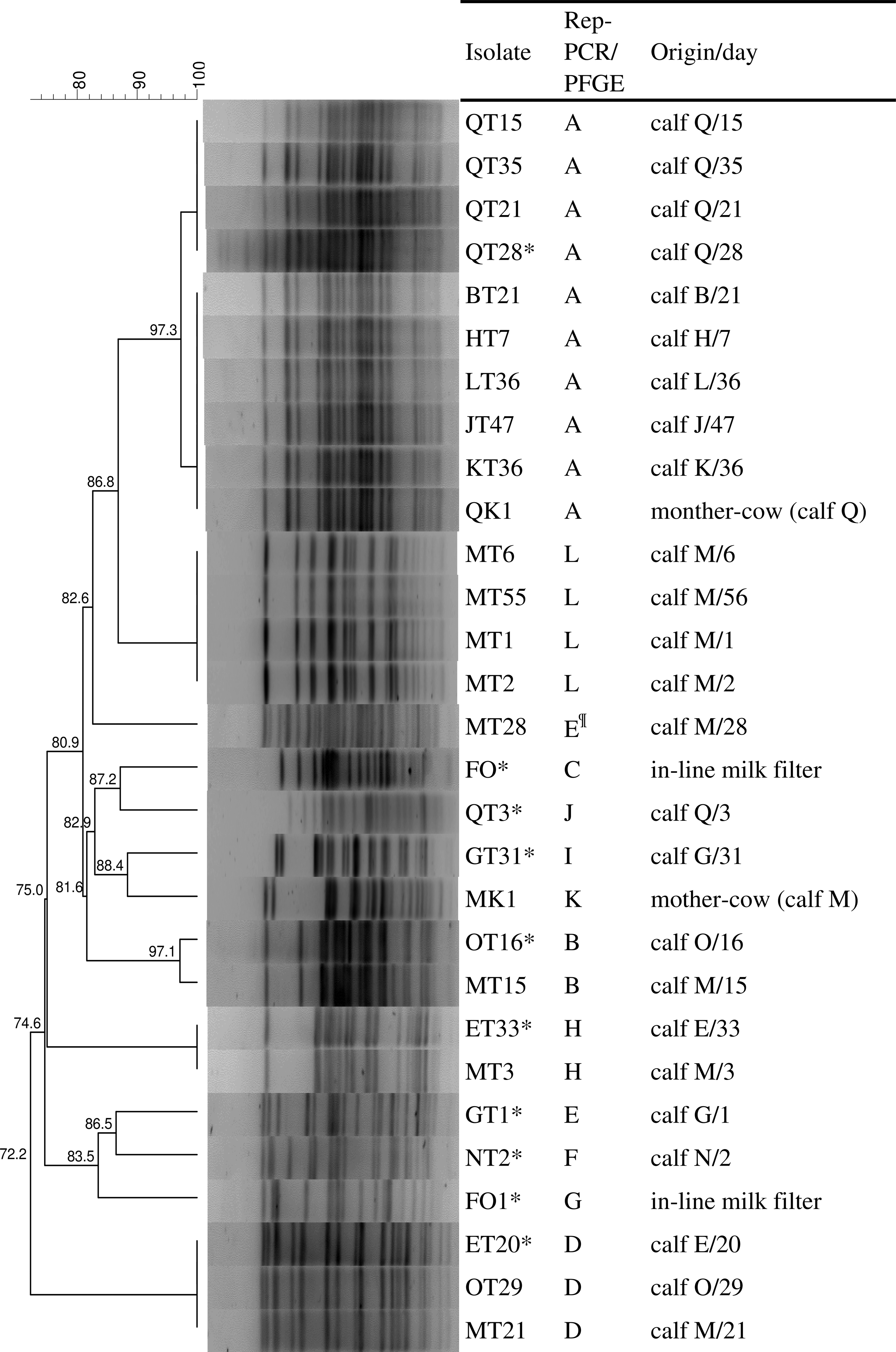
PFGE analysis of clonal relationship of selected cefotaxime-resistant Escherichia coli isolates. *Isolates subjected to WGS; ¶PFGE analysis unlike rep-PCR indicated the presence of another unique restriction profile. PFGE, pulsed-field gel electrophoresis; WGS, whole-genome sequencing.
Multilocus sequence typing, serotype, and phylogenetic groups
WGS was applied to nine CMY-2-producing isolates and one AmpC-negative isolate originating from eight individual calves and two in-line milk filters and showing different PFGE profiles (Table 2). All isolates were confirmed as E. coli and based on multilocus sequence typing they were assigned to nine different STs. Five phylogenetic groups were distinguished among investigated isolates including A (three isolates), B1 (one isolate), B2 (one isolate), C (three isolates), and D (two isolates). Both O and H antigens were predicted for eight isolates, whereas only H antigen types were found in two isolates.
Characteristics of Ten Escherichia coli Isolates from Calves and In-Line Mil Filter Subjected to Whole-Genome Sequencing
The gray box indicates the presence of the gene or other marker. Amino acid substitutions in quinolone resistance determining region (QRDR) are given in the gray box. MIC to quinolones are given in the box and resistance (R) values, ≥4 mg/L for ciprofloxacin and ≥32 mg/L for nalidixic acid, are highlighted in gray.
According to wzx gene the isolate was assigned to O17/O77 serotype while based on wzy gene to O17/O44 serotype.
MLST, multilocus sequence typing; MIC, minimum inhibitory concentration.
Detection of antibiotic resistance genes
Antibiotic resistance gene content of 10 isolates subjected to WGS corresponded to resistance phenotypes obtained by phenotypic testing. The presence of the bla CMY-2 gene was confirmed in all nine AmpC producers. Various genes encoding narrow-spectrum beta-lactamases and determinants for resistance to other antimicrobials were detected (Table 2). Various substitutions in quinolone resistance-determining region of GyrA, ParC, and ParE were found. Five isolates contained class 1 integron with three types of gene cassette arrangements (Table 2). A single isolate contained two different class 1 integrons, whereas a class 2 integron was found in the genome of one isolate.
Presence of virulence factors and plasmid replicons
All isolates carried at least one gene encoding fimbriae and other adhesion factors with predominance of prfB for P-related fimbriae regulation and lpfA for long polar fimbriae. Bacteriocin-encoding or serum resistance genes were detected in eight and eight isolates, respectively. Four isolates carried factors (air and eilA) associated with enteroaggregative E. coli pathotype, whereas three were positive for the astA gene responsible for production of enteroaggregative E. coli heat-stable enterotoxin 1. The highest number of virulence factors was found in two isolates, BT21 and GT31. The later isolate was equipped with a set of type III secretion system genes and their effectors as well as the eae gene for intimin.
Eight different plasmid types were found with a predominance of K, FIB, and FII replicons. In two isolates, the K replicon type together with the bla CMY-2 gene was identified within the same contig of the draft genomes. Further mapping analysis of plasmid contigs showed that all AmpC-producing isolates carried IncK2 plasmid with bla CMY-2 similar to the reference pDV45 (Fig. 2).

Circular visualization of bla
CMY-2-carrying IncK2 plasmids of Escherichia coli isolates from calves and in-line milk filters. Plasmid contigs are mapped against plasmid pDV45 (KR905384.1) as a reference sequence using Blast Ring Image Generator (BRIG). Color images available online at
Discussion
This study investigated the fecal shedding of cefotaxime-resistant E. coli on a dairy farm with well-documented antimicrobial therapies. Cefotaxime-resistant isolates were found in all studied calves but only two (15%) mother cows, and a resistance phenotype was associated with CMY-2 beta-lactamase. On the contrary, in the European Union, CTX-M-positive E. coli was more frequently isolated in calves, whereas the finding of CMY-2-producing isolates was rather sporadic (Hordijk et al., 2013; Brunton et al., 2014).
Antimicrobial treatment of food-producing animals with ESCs may create favorable conditions for the selection and persistence of ESBL/AmpC-producing Enterobacteriaceae in their intestine (Landers et al., 2012). Most CMY-2-positive E. coli isolated in this study showed multiresistance phenotype associated with diverse antimicrobial resistance genes as demonstrated by WGS. Considering that the annual consumption of cephalosporins was the lowest while other antimicrobials such as tetracyclines were used more frequently, it is likely that co-selection by other antimicrobials was involved in the maintenance of CMY-2 producers on the farm.
All studied calves carried CMY-2-producing E. coli in their gut despite not having been treated with antibiotics. It has been demonstrated that antimicrobial drug residues in waste milk commonly fed to dairy calves may contribute to the selection and shedding of resistant fecal E. coli in calves from birth to weaning (Thames et al., 2012; Brunton et al., 2014). In our study, calves were fed with colostrum and milk; therefore, antibiotic residues present in milk may result in the selection and persistence of cefotaxime-resistant E. coli in calf intestine. In addition, dairy colostrum or waste milk fed to calves has been suggested to be potential sources of multiresistant bacteria (Randall et al., 2014; Awosile et al., 2017). In our study, no cefotaxime-resistant bacteria were isolated from pasteurized colostrum or milk. However, unpasteurized colostrum used for the first feeding was not tested; therefore, we cannot exclude the feed being a source of resistant isolates for calves.
All examined cohort calves became colonized by CMY-2-producing E. coli shortly after the birth and fecal shedding of these bacteria was observed during the whole study period. As two mother cows were found to carry CMY-2-producing isolates, it is likely that the initial colonization of calves occurred during delivery or by indirect transmission during housing and postnatal care. Drops in CREC density or discontinued shedding were recorded at the end of the study as demonstrated previously (Hoyle et al., 2004; Watson et al., 2012; Hordijk et al., 2013; Horton et al. 2016).
The high number of CMY-2-producing E. coli detected in calf fecal samples in our study may be partly related to colonization factors and growth within the calf intestine. Dynamics of CREC shedding is likely associated with several factors such as diverse microbiome and epithelial structure of the calf gastrointestinal tract pre- and postweaning, which are changing with calf age (Meale et al., 2017; Mir et al., 2018). These changes in calf intestine can explain noticeable drop in density shedding at final part of the investigated period. WGS analysis of selected E. coli isolates showed that genes for fimbriae were possessed by a large majority of the isolates. One isolate was equipped with genes encoding intimin that mediates the tight binding of entheropathogenic E. coli to the intestinal epithelia (Watson et al., 2017). Moreover, three isolates belonged to extraintestinal pathogenic E. coli (ExPEC) lineages ST648 and ST69 that may be better equipped to colonize the calf intestine. ST648 is an emerging high-risk clone associated with CTX-M enzymes reported in various habitats (Ewers et al., 2014; Helldal et al., 2017). ST69 is another globally distributed ExPEC responsible for community-onset and hospital-acquired antibiotic-resistant infections in humans (Riley, 2014; Helldal et al., 2017). One isolate belonged to commensal E. coli ST10, a widely reported lineage among human and food-animal sources and frequently associated with resistance to cephalosporins (Manges et al., 2015).
PFGE analysis of isolates obtained from individual calves showed both different and identical E. coli genotypes. Given that we used 10-fold diluted fecal samples for isolation of AmpC-producing E. coli, it enabled the selection of the most abundant clones for its subsequent characterization. The observed variety of E. coli genotypes in individual calves implied temporal heterogeneity in the colonization of calf intestine. This may indicate the presence of various sources of possible intestinal colonization of newborn calves and/or the importance of the horizontal transfer of the bla CMY-2 gene within the calf microbiome. WGS of nine representative E. coli isolates of diverse PFGE profiles confirmed that the bla CMY-2 gene was carried by IncK2 plasmids, suggesting its role in the dissemination of the gene among different E. coli clones in animals on the studied farm.
Conclusion
This study describes the occurrence and persistence of cefotaxime-resistant E. coli isolates in calf feces from different sampling visits on a Czech dairy farm. To the best of our knowledge, this is the first study investigating quantitatively the shedding density of CMY-2-producing E. coli from individual calves over a prolonged period of up to 63 d after birth. Despite the limited number of isolates subjected to further detailed analysis, WGS gave us an actual overview of genes associated with antibiotic resistance and virulence and also mobile genetic elements of cefotaxime-resistant E. coli circulating in suckling calves on the studied farm. Our findings reinforce the urgency needed to prevent the spread of ESBL-/AmpC-producing bacteria on dairy cow farms. Analysis of their bacterial genomes can help to understand the patterns of their selection and circulation in this environment, and predict the risks associated with the occurrence of the highly resistant E. coli pathotypes.
Footnotes
Acknowledgments
The study was supported by the projects IGA no. 102/2014/FVL to J.S., MSM/EE no. EE2.3.30.0053. (CZ.1.07/2.3.00/30.0053) to I.M., GACR (18-23532S) to M.D. and CEITEC 2020 (LQ1601). The authors thank Tomas Haloun and Katerina Sedlarova for their help in the field and laboratory. Their special thanks go to Adam Valcek for his help with plasmid data analysis.
Disclosure Statement
No competing financial interests exist.