
Abstract
Escherichia coli causes a significant number of clinical mastitis cases in dairy cattle worldwide. The antimicrobial susceptibility of E. coli is important for both human and animal health. Surveillance reports recorded that the efficacy of most antibiotics is substantially preserved but detection of E. coli from clinical mastitis cases producing extended-spectrum beta-lactamases and plasmid-encoded AmpC beta-lactamases has been reported. These resistance determinants have frequently been associated with multidrug resistance. The aim of this study was to determine if a MacConkey agar medium supplemented with 8 mg/L of ceftiofur (MC-CEF) could be a useful tool to identify cephalosporin-resistant and multidrug-resistant (MDR) E. coli among bovine mastitis isolates. During the period 2010–2011, 773 E. coli were isolated from bovine clinical mastitis milk samples collected in 80 dairy farms in Northern Italy. A total of 105 E. coli were selected and assigned either to group randomly selected E. coli (RSEC; n = 53), based on a random selection among the whole collection of 773 E. coli, or to group ceftiofur-resistant E. coli (CEFREC; n = 52). CEFREC isolates were identified by spreading the 773 E. coli isolates on MC-CEF. Minimum inhibitory concentration (MIC) was used to test the phenotypic antimicrobial susceptibility to 16 antibiotics. The MIC results confirmed the ceftiofur resistance in 73.1% (38/52) of CEFREC isolates, whereas all RSEC isolates were susceptible to ceftiofur. The comparison of MIC values for each antibiotic tested between the two groups revealed significantly higher frequencies of resistance to antimicrobials other than ceftiofur in the CEFREC group. Resistance profiles highlighted a significantly higher frequency of MDR isolates among CEFREC (73.1%) than RSEC (17%) E. coli. The results showed that MC-CEF may be a useful selective medium to identify cephalosporin-resistant and MDR E. coli on dairy farms, without performing MIC on all the isolates.
Introduction
Mastitis is the costliest disease on dairy farms (Halasa et al., 2007) and Gram-negative bacteria are regarded as important causative agents of environmentally associated mastitis. High proportions of clinical mastitis are caused by coliforms and in particular by Escherichia coli (Blum et al., 2014; Hertl et al., 2014; Bradley et al., 2015). In a recent field trial, E. coli is ranked as the second most frequently isolated pathogen in clinical samples (14.5%) after Streptococcus uberis (Addis et al., 2017). Several reports confirm that E. coli is susceptible to antibiotics commonly used for treatment of coliform mastitis in dairy cattle (Srinivasan et al., 2007; Bengtsson et al., 2009; Thomas et al., 2015). Several specific surveys have reported resistance among E. coli isolated from mastitis because of the production of extended-spectrum beta-lactamases (ESβL) (Locatelli et al., 2009, 2010; Dahmen et al., 2013). A recent study found that ESβL and plasmid-encoded AmpC-producing E. coli could be frequently isolated in samples from the environment of dairy farms, which represents the main source of E. coli that causes mastitis (Gonggrijp et al., 2016).
An increasing trend has been recorded for the prevalence of E. coli showing important co-resistance phenotypes in both human and veterinary medicine (Tadesse et al., 2012; Walther et al., 2017). The resistance of a bacterial isolate to antibiotics belonging to at least three different classes is defined as multidrug resistance (Magiorakos et al., 2012). Multidrug-resistant (MDR) E. coli spread is a public health concern (Walther et al., 2017), representing a zoonotic risk for farm workers and contact people. The horizontal transfer of resistance genes from commensal to pathogenic bacteria through mobile elements is possible in farm settings and through the food chain (Persoons et al., 2011).
A rapid and efficient phenotypic test able to detect isolates potentially resistant to therapy in animal and public health settings could be an important tool in surveillance programs. The use of a second cephalosporin is recommended but for most diagnostic laboratories it could be too costly. A single molecule testing would be the best solution (Aarestrup et al., 2010). Ceftiofur is a third generation cephalosporin and is a Veterinary Critically Important Antimicrobial, according to the World Health Organization (WHO, 2017). It is the main registered antibiotic for dairy cows considered useful in detecting E. coli producing ESβL or AmpC, using both the minimum inhibitory concentration (MIC) and disk diffusion methods (Aarestrup et al., 2010; Persoons et al., 2011). ESβL-producing E. coli frequently exhibit resistances that classify them as MDR E. coli (Walther et al., 2017).
Ceftiofur has also been added in concentration related to MIC cutoff value MC-CEF is the culture media obtained by the addition of ceftiofur to MacConkey (MC-CEF) to detect ceftiofur-resistant Gram-negative bacteria in feces (Donaldson et al., 2006; Persoons et al., 2011). Plating the E. coli isolates on MC-CEF after their primary isolation from milk could be applied to rapidly detect resistant isolates and the farms where resistance phenomena occur to prevent the transmission to consumers of raw milk and raw milk products or through direct contact.
The aim of this study was to determine if MC-CEF could be a useful tool to identify (1) cephalosporin-resistant E. coli and (2) MDR E. coli among bovine mastitis isolates. For this purpose, we compared the resistance profiles of E. coli identified using this selective medium, based on resistance to ceftiofur, to the resistance profiles of randomly selected E. coli (RSEC). An association between cephalosporin resistance and multidrug resistance was also investigated.
Materials and Methods
In 2010–2011, 773 E. coli were isolated from bovine clinical mastitis samples, routinely collected on 80 dairy farms in Northern Italy and sent to the laboratory of the Department of Veterinary Medicine (University of Milan). The farms had from 50 to 600 lactating cows with an average farm population of 177 cows. Milk samples were collected by farm personnel trained to detect mastitis cases of varying degrees of severity. After disinfection of teat ends and discarding the first streams of foremilk, milk was collected in 10 mL sterile vials, labeled with cow number and quarter, frozen at −20°C, and sent frozen to the laboratory. Milk sample analysis and pathogen identification were carried out following the National Mastitis Council (2017) guidelines.
Each sample was thawed at room temperature and 100 μL were spread onto 5% defibrinated sheep blood agar plates. After overnight incubation at 37°C, E. coli isolates were identified using colony morphology, Gram-staining characteristics, and biochemical reactions on MacConkey and Eosin Methylene Blue agar (Oxoid, Basingstoke, United Kingdom). All E. coli isolates were frozen at −80°C in nutrient broth with 15% glycerol for further analysis. If multiple samples from the same cow were submitted, only the first E. coli isolated was considered and stored.
Of 773 E. coli isolates, based on two different inclusion criteria, two groups were created using a total of 105 isolates. The resistance to ceftiofur, meant as the ability to grow on MC-CEF plates, was the first inclusion criterion. The plates of MC-CEF were manufactured in our laboratory supplementing MacConkey agar (Oxoid) with 8 mg/L of ceftiofur (Sigma-Aldrich, St. Louis, MO) (Donaldson et al., 2006). All 773 isolates were cultured on MC-CEF and the 52 (6.7%) E. coli isolates that grew were assigned to ceftiofur-resistant E. coli (CEFREC) group. The second group was named RSEC (n = 53) and was generated using a systematic random selection by SPSS 22.0 (IBM; SPSS, Inc., Chicago, IL) starting from a randomized spreadsheet containing all 773 E. coli isolates from all farms. MICs of the 16 antibiotics listed in Tables 1 and 2 were determined for each E. coli isolate in both the CEFREC and RSEC groups, using a broth dilution test according to the procedure described in the CLSI guidelines Vet01A4 (CLSI, 2013a). The antibiotics listed in Tables 1 and 2 were selected to test antimicrobial resistance to the different classes of antimicrobials and to determine the presence of MDR isolates. Antimicrobials not approved for treatment of dairy cattle (cefpodoxime, cefoxitin, and imipenem) were also tested to detect isolates potentially producing ESβL, AmpC, or carbapenemases.
Overall Resistance to Each Antimicrobial of Escherichia coli of 105 Included Isolates and the Distribution of Resistant Isolates in the Ceftiofur-Resistant E. coli and Randomly Selected E. coli Groups, with Statistical Results
The antimicrobial categories reported and used to evaluate multidrug resistance are those proposed by Magiorakos et al. (2012).
Statistics not applicable.
CEFREC, ceftiofur-resistant E. coli; RSEC, randomly selected E. coli.
Percentages of Resistant Escherichia coli in Ceftiofur-Resistant E. coli and Randomly Selected E. coli Groups
Breakpoint interpretation was based on CLSI guidelines VET01-S2 (2013b) and M100-S24 (2014).
CEFREC, ceftiofur-resistant E. coli; I, Intermediate; R, Resistant; RSEC, randomly selected E. coli; S, Susceptible.
Picking isolates from a fresh, pure culture on blood agar plates, a commercially available microdilution MIC system (Sensititre compan1F; Trek Diagnostics System, East Grinstead, United Kingdom) was used according with the manufacturer's instructions. The manufacturer certified that the method conformed to CLSI guidelines and was for veterinary use only. The Sensititre plate reading was performed manually by the same laboratory technician, blind to the isolates status, who recorded the last concentration of antibiotic without turbidity or deposit of cells at the bottom of the well. CLSI resistance breakpoints were used (CLSI, 2013b). Quality control tests were performed according to CLSI guidelines VET01A4 (CLSI, 2013a) on a weekly basis with Staphylococcus aureus ATCC 29213 and E. coli ATCC 25922 as reference strains. All quality controls performed were within acceptable MIC limits required by VET01-S2 CLSI guidelines (CLSI, 2013b).
A χ 2 test was conducted using commercial statistical software (IBM SPSS Statistics 25.0; IBM, Armonk) to compare the resistance frequencies for each antimicrobial in the CEFREC and RSEC groups. Values of p < 0.05 were considered significant. Sensitivity, specificity, positive predictive value (PPV), negative predictive value (NPV), and accuracy for MC-CEF were calculated.
Results
The resistance to ceftiofur was confirmed by MIC in 73.1% of E. coli included in the CEFREC group (38/52). No E. coli isolate from the RSEC group was found to be ceftiofur resistant on the MIC test nor grew on MC-CEF. Table 1 gives the total number of E. coli resistant to each antimicrobial agent and the percentages out of the 105 isolates included in the study. For most antimicrobials considered, the distribution of resistant isolates between the two groups was significantly different, with p values ranging between 0.001 and 0.061 (Table 1). Cefoxitin was the only antimicrobial with a p value leaning toward the significance (p = 0.061), having only four resistant isolates included in the CEFREC group. E. coli selected by ceftiofur resistance was more frequently resistant to one or more other compounds than the isolates of the randomly selected group (Fig. 1). All the isolates, regardless of the group, were susceptible to amikacin and imipenem, for which statistics were not applicable.
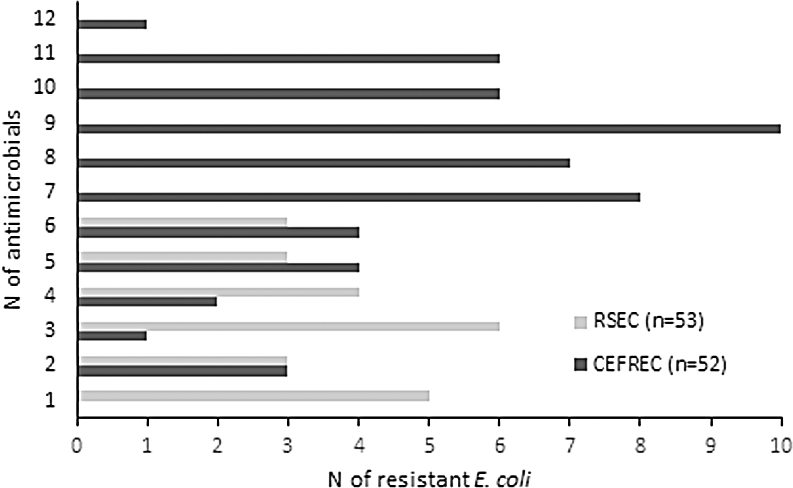
Comparison between the resistance profiles of the RSEC group, (pale gray bars) and Escherichia coli selected using the ceftiofur-supplemented medium (CEFREC group, dark gray bars). The graph shows the number of isolates that were resistant to up to 12 antimicrobials, regardless of the antimicrobial class. CEFREC, ceftiofur-resistant E. coli; RSEC, randomly selected E. coli.
Table 2 shows the percentages of E. coli resistant to each antimicrobial in the two groups, of the E. coli isolates included (i.e., n = 52 in CEFREC and n = 53 in RSEC groups). Figure 1 represents and graphically compares the extent of co-resistance to different antibiotics in the E. coli isolates belonging to the two groups. In all, 100% of the CEFREC E. coli (52/52) were resistant to at least two antibiotics and 94.2% (49/52) to at least three antibiotics (Fig. 1). The widest resistance profiles, including between 9 and 12 antibiotics, were found in 23 E. coli (44.2%) with 1 E. coli resistant to 12 antibiotics. Of the 52 E. coli isolates, 42 were resistant to at least 1 cephalosporin (80.8%).
Despite the inclusion in the CEFREC group, 14 E. coli were susceptible to ceftiofur on the MIC test. These ceftiofur-susceptible E. coli isolates displayed resistance to at least 1 beta-lactam antibiotic: 13 were resistant to ampicillin and ticarcillin, 6 to ticarcillin/clavulanic acid, 3 to cefpodoxime, 2 to cefoxitin or amoxicillin/clavulanic acid, and 1 to cefazolin. Most of the CEFREC isolates (13/14, 92.9%) susceptible to ceftiofur and resistant to 1 beta-lactam antibiotic were resistant to doxycycline. In 4 isolates (28.6%), the resistance to doxycycline was associated with resistance to fluoroquinolones, in 7 (50%) to trimethoprim/sulfamethoxazole, in 3 (21.4%) to fluoroquinolones, trimethoprim/sulfamethoxazole, and chloramphenicol concomitantly and in 2 (14.3%) gentamicin was the fourth antibiotic included in the resistance profile. Figure 2 represents and compares the extent of co-resistance to at least three classes of antibiotics in the E. coli isolates belonging to the CEFREC and RSEC groups.
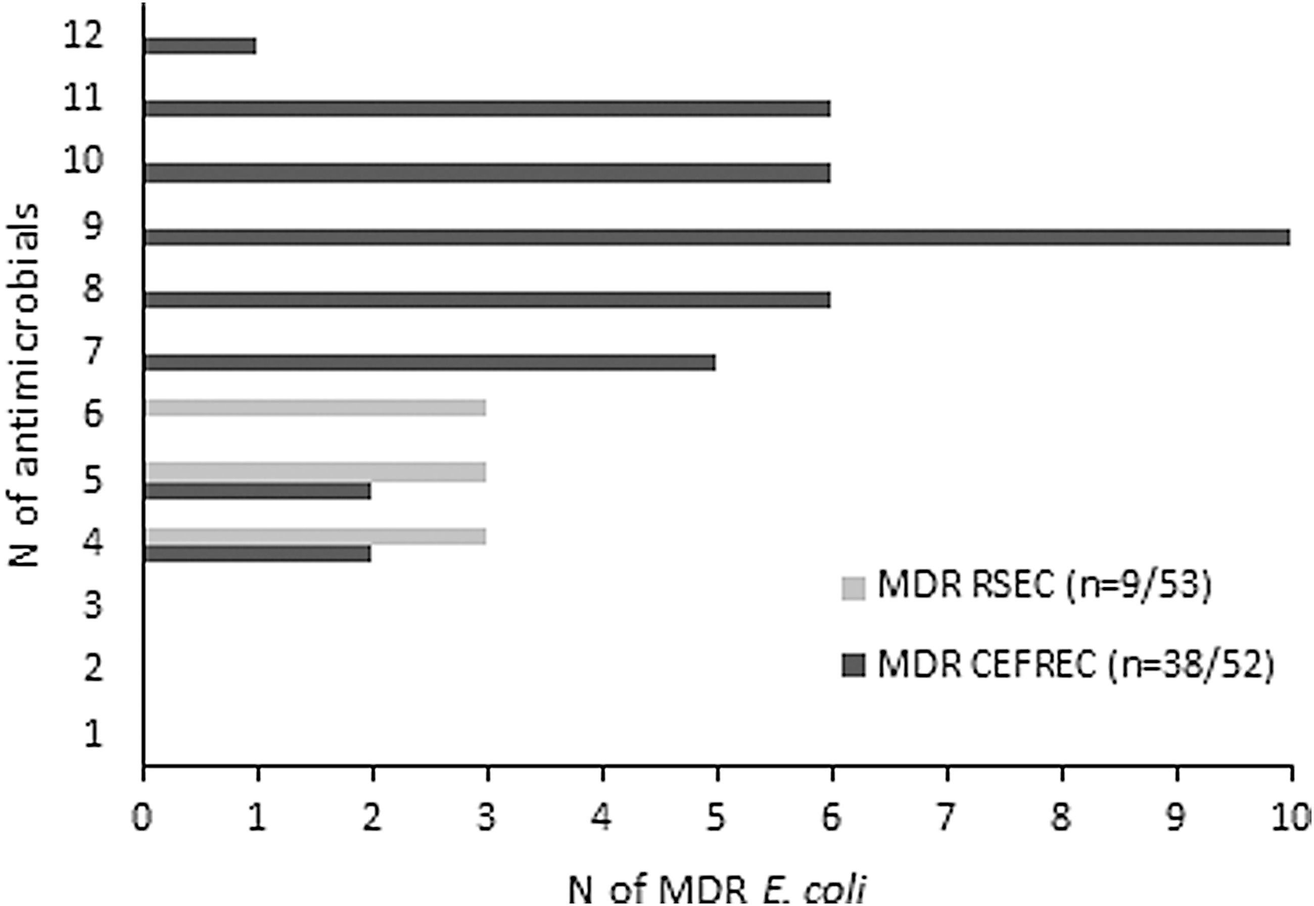
Comparison between MDR E. coli in the randomly selected group (RSEC, pale gray bars) and the group of isolates selected using the ceftiofur-supplemented medium (CEFREC, dark gray bars). The graph shows the number of isolates resistant to up to 12 antimicrobials belonging to more than three antimicrobial classes. MDR, multidrug resistant.
Thirty-eight of the 52 E. coli isolates (73.1%) met the definition provided by Magiorakos et al. (2012) (Fig. 2). According to this definition, 14 CEFREC E. coli isolates should not be considered MDR. Despite their inclusion in the resistant group, 6 E. coli were resistant only to agents belonging to beta-lactams, 7 were resistant to beta-lactams and doxycycline, and 1 was resistant to beta-lactams and trimethoprim/sulfamethoxazole.
Twenty-nine of 53 RSEC E. coli (54.7%) were susceptible to all tested antibiotics and 5 isolates (9.4%) were resistant to 1 antibiotic (3 to doxycycline and 2 to ticarcillin); 19 and 16 isolates were resistant to at least 2 and 3 antibiotics, respectively (35.8% and 30.2% vs. 100% and 94.2% in the CEFREC group, respectively). The widest resistance profile was limited to only 3 RSEC isolates and included a maximum of 6 antibiotics (vs. 12 antibiotics for the CEFREC group): mostly beta-lactam antibiotics together with doxycycline and trimethoprim/sulfamethoxazole.
Among the isolates selected in the RSEC group, there was no resistance toward cefoxitin, cefpodoxime, gentamicin, marbofloxacin, ceftiofur, amikacin, and imipenem. Only one isolate was resistant to enrofloxacin. In the RSEC group, 35.8% and 37.7% of the isolates were resistant, respectively, to ampicillin and ticarcillin (vs. 98.1% in the CEFREC group), 28.3% were resistant to doxycycline and 13.2% to trimethoprim/sulfamethoxazole. Of the 53 RSEC isolates, only 3 were resistant to at least 1 cephalosporin (5.7%), a frequency significantly lower than that in the CEFREC group (p = 0.001). Very low levels of resistance were observed toward the other antibiotics tested (Tables 1 and 2). In this group, only 9 of 53 E. coli (17%) exhibited resistance to 3 and 4 antimicrobial classes, coinciding with the definition of MDR E. coli (Fig. 2).
Finally, a significantly higher number of MDR E. coli (p = 0.001) were identified in the CEFREC group than in the RSEC group. Sensitivity of MC-CEF in detection of MDR isolates was 81%, specificity 76%, PPV 73%, NPV 83%, and accuracy 80%. A restricted group of farms (n = 18; 22.5%) of the 80 dairy herds screened yielded MDR E. coli of both groups. Within the CEFREC group, 11 farms contributed with 1 MDR isolate, 3 farms with 2 isolates, 2 farms with 4 isolates, and 2 farms contributed with 7 and 6 isolates, respectively. In the RSEC group, MDR E. coli isolates (n = 9), each originated from a different farm.
Discussion
The results of this study showed the utility of MC-CEF as a tool for the identification of MDR E. coli isolated from cases of bovine clinical mastitis as the resistant levels recorded were higher than those of the RSEC group. The comparison between two selection methods highlighted a significantly higher detection of cephalosporin-resistant E. coli (p = 0.001) and MDR E. coli (p = 0.001) using the tested MC-CEF as a selective medium. This confirmed the efficacy of the veterinary cephalosporin ceftiofur as a molecule useful in identifying MDR Gram-negative bacteria (Aarestrup et al., 2010). In the CEFREC group, 14 E. coli isolates resulted as ceftiofur susceptible, although they grew on MC-CEF. Two of 14 were resistant to cefoxitin and for 2 others the MIC results were at the limit of susceptibility (2 μg/mL). Although 10 of 14 ceftiofur-susceptible E. coli were susceptible to all tested cephalosporins, all were resistant to at least 1 non-cephalosporin beta-lactam antibiotic. The growth of cefiofur-susceptible E. coli isolates on MC-CEF may be the result of cross-resistance with other beta-lactam antibiotics (Persoons et al., 2011). Cephalosporin-resistant E. coli are frequently but not necessarily MDR E. coli and that may be consistent with a specificity of 76%; likewise MDR E. coli could be not resistant to cephalosporin. That could justify the discrepancy between the sensitivity of MC-CEF (81%) and the percentage of ceftiofur-resistant CEFREC isolates (73.1%).
This study represents one of the few reports describing 47 MDR E. coli from clinical mastitis in a defined time and geographical area (Ali et al., 2016; Keane, 2016). Overall, 80.8% of the isolates selected by MC-CEF were resistant to at least one of the tested cephalosporins. This method allowed detecting significantly higher frequencies of multiple resistances, providing information about MDR E. coli presence on a farm. A random selection of E. coli to be tested is able to detect MDR isolates, but to a lower extent and with less information about resistance to cephalosporins (only 5.7% of RSEC E. coli are resistant to at least one cephalosporin).
The evidence that few farms (22.5%) among those screened yielded MDR E. coli may suggest that several factors could be involved in the on-farm development of resistance (Persoons et al., 2011). The screening of mastitis isolates by MC-CEF media could be a suitable tool for laboratories interested in detecting farms at risk and monitoring the trend of resistance over time. This is important for future perspective as, once established, resistant strains could survive, potentially spread and persist in a farm environment becoming a threat to animal and possibly human health (Szmolka and Nagy, 2013; Gonggrijp et al., 2016). ESβL-producing E. coli could belong to different genetic types (Odenthal et al., 2016). Clonal spread could not be excluded in this study as on seven dairy farms more than one MDR E. coli was detected. Genotyping is necessary to demonstrate the genetic similarity but the purpose of this study was limited to testing the ability of MC-CEF medium to identify MDR E. coli in different and independent samples.
The co-selection of additional resistance determinants with the ESβL genes could explain the significantly higher frequencies of MDR isolates among CEFREC rather than RSEC E. coli. A very low level of resistance to fluoroquinolones was confirmed in the RSEC group, whereas almost all the isolates in the CEFREC group were found resistant.
The World Health Organization includes both fluoroquinolones and cephalosporins of third and fourth generation in the list of Critically Important Antimicrobials to prevent their effectiveness in human medicine. The aim should be to implement risk assessment and risk management strategies for containing antimicrobial resistance because of nonhuman antimicrobial use (WHO, 2017). Animals and food contaminated with animal commensal bacteria as E. coli could be a source of resistant determinants for human pathogens (WHO, 2017). An evidence-based approach should also be applied to the treatment of E. coli clinical mastitis. Finally, the use of antibiotics should be reserved for cases of particular severity and in consideration of the relationship between beneficial effect and possible risk of increased resistance toward cephalosporins and other critically important antibiotics (Greko et al., 2009; Suojala et al., 2013).
Conclusions
This study demonstrates that screening E. coli isolates responsible for bovine clinical mastitis on MC-CEF medium allowed to detect resistance profiles including cephalosporins and more than two other antimicrobial classes, without testing the MIC of all isolates. Comparing the MIC results between the two selection groups, screening by MC-CEF demonstrated a significantly higher probability to identify MDR E. coli than with a random selection. A relationship between resistance to cephalosporins and multidrug resistance is possible and requires further detailed molecular studies. Further studies should be performed to evaluate the ability of an easy screening process like that proposed to identify dairy farms where resistance issues are present to avoid further spread of resistant strains.
Footnotes
Acknowledgments
This work is a part of the project “Survey on the risk factors related to the spreading of antimicrobial resistance in production animals” that Università degli Studi di Milano funded as a postdoctoral grant. The authors thank Professor Giovanni Re (Department of Veterinary Science, Università degli Studi di Torino) for the critical revision of the article.
Disclosure Statement
No competing financial interests exist.