
Abstract
The study was conducted to describe the dynamics of ST398 methicillin-resistant Staphylococcus aureus (MRSA) on a dairy herd in northeastern Italy. MRSA was first identified in this herd of 120 cows in 2016, after which the herd was sampled once every 3 months for 1 year (April 2016–May 2017). Samples collected included nasal swabs and milk samples from cows and nasal swabs from farmworkers. In addition, pen fencing and teat milk liners were swabbed and air samples from cow pens and the milking parlor were collected. All samples were tested for MRSA using a selective medium; positive isolates were confirmed by mecA PCR. A representative set of MRSA isolates was genotyped using spa typing and multilocus sequence typing. Overall, 34 (mean 23%, range 16–30%) milking cows were found harboring MRSA in the mammary gland and only 6 recovered from infection or colonization. The mean incidence rate was 14% (range 8–20%), mean cure rate was 23% (range 13–43%), and estimated basic reproduction number (R0) was 1.08. The average of positive quarters found was 35.1% and most of the positive quarters (82.4%) developed subclinical mastitis. The mean duration of MRSA colonization in quarters during the study was 247 days, but quarters affected by subclinical mastitis harbored MRSA for a longer time than healthy ones (285 days vs. 131 days). After the second sampling, the farmer segregated MRSA-positive cows from the uninfected cows and milked them last. Despite segregation, 25 newly infected or colonized cows were detected. MRSA isolates from cows, environment, and two farmworkers belonged to the same sequence type (ST398) and spa type (t034). This study highlights the ability of ST398 MRSA to cause a persistent infection of the mammary gland and to survive in the farm environment.
Introduction
Methicillin-resistant Staphylococcus aureus (MRSA) causes a wide range of severe human infections such as endocarditis, osteomyelitis, and sepsis (Forbes et al., 2008). Several studies demonstrated MRSA contamination in pork, beef, veal, poultry meat, and milk (Normanno et al., 2007; de Boer et al., 2009; Agersø et al., 2012; Haran et al., 2012), but few foodborne human infections have been described (Kluytmans et al., 1995; de Jonge et al., 2010). MRSA has been detected in milk and nasal cavities of dairy cows in several countries (Paterson et al., 2012; Luini et al., 2015; Locatelli et al., 2017). Data about MRSA diffusion in dairy herds are controversial. Some reports refer to a low prevalence, ranging from 0.2% up to 4% (Kwon et al., 2005; Huber et al., 2010; Cortimiglia et al., 2016; Locatelli et al., 2016; Parisi et al., 2016), while higher prevalence, from 8.2% to 17.2%, was detected by Türkyılmaz et al. (2010) and Luini et al. (2015).
Since its emergence in swine, the spread of clone ST398 MRSA, also known as livestock-associated MRSA, raised concerns of risk for direct or foodborne transmission to humans (Lee, 2003). Zoonotic transmission of ST398 MRSA strains from livestock to humans with consequent infection or colonization has been reported (Soavi et al., 2010; Lozano et al., 2011; Wendlandt et al., 2013).
In cows, ST398 MRSA has the ability to colonize the udder and to induce a chronic subclinical intramammary infection (Feßler et al., 2010; Vanderhaeghen et al., 2010; Luini et al., 2015). MRSA mastitis has epidemiological behavior similar to methicillin-susceptible Staphylococcus aureus (MSSA), but MRSA responds poorly to antibiotic treatment as it displays multiple resistance and poses a greater risk to humans if transmission occurs (Vanderhaeghen et al., 2010). Little information concerning spreading and persistence of MRSA in dairy herds is available for risk management of this issue and our study aims to fill this knowledge gap. During a 1-year longitudinal study, cows, farmworkers and the environment of a dairy farm were assessed for ST398 MRSA with the aim to investigate its within-farm diffusion.
Materials and Methods
Herd
After the first discovery of MRSA in clinical samples, a dairy herd in northeastern Italy was investigated to assess MRSA diffusion and persistence over a 1-year period, from April 2016 to May 2017.
The herd averaged 73 lactating cows and had not introduced new cows for 5 years. Three family member workers attended the animals. Cows were housed in free-stall barns and milked twice a day in a herringbone parlor with a back flushing system in place. Cows were dried off for an average of 55 days, using blanket antibiotic therapy (amoxicillin plus clavulanic acid). After MRSA detection, infected cows were segregated and milked last. Some chronically infected cows were culled and others with more than 250 days of lactation were dried off without any antibiotic treatment.
Study design
The study planned to describe whether MRSA passed from cow to cow and whether the farm environment and facilities were contaminated and might contaminate people attending the animals. With respect to the scope, MRSA presence was assessed in all lactating cows, farm environment, and facilities in five sampling sessions every 3 months. An individual composite milk sample (ICMS) was collected from each lactating cow to assess the presence of MRSA in the udder. On MRSA-positive cows, the following investigations were scheduled: MRSA detection from single quarters and nostrils and somatic cell count (SCC) enumeration at the quarter level to distinguish between intramammary infection and colonization. Individual milk quarters and nasal swabs from nostrils were collected beginning with the next scheduled herd sampling.
MRSA contamination of the herd environment was assessed by sampling dust and air in pens and the milking parlor during sampling sessions 2 to 5.
Exposure to the MRSA-contaminated environment was evaluated by testing the MRSA nasal carriage in farmworkers (including veterinarian) and contamination of cow udder skin and milking teat liners. Farmworkers provided their nasal swab at the second sampling. Contamination of the udder skin was assessed at the last sampling session by swabbing all MRSA-positive cows and 13 randomly selected MRSA-negative cows. Teat liners used to milk MRSA-positive cows were tested once at the first sampling session.
Sample collection
All milk samples (ICMS and quarter samples) were collected according to the National Mastitis Council procedure (NMC, 2016).
Nasal swabs were obtained using a sterile swab after cleaning and disinfecting the nose with a gauze soaked in 1% Virkon™ solution (Rely+On Virkon; Antec International Limited, Sudbury, United Kingdom).
Liners of clusters used for milking positive cows were sampled by swabbing the inside with a sterile swab immediately after milking of each positive cow, of the next cow milked on the same cluster, and at end of milking before cleaning.
Dust samples from the surfaces of tubular pen fences (50 cm2) were collected by streaking with a sterile swab dipped in sterilized water.
All swabs were kept in a commercial Amies transport medium (Amies CLR; Meus s.r.l., Piove di Sacco, Italy).
Air was absorbed (50 L per plate) by a portable air sampler (SAS Super 100®; PBI International, Italy) on a CHROMagar MRSA II™ plate (Becton Dickinson GmbH, Heidelberg, Germany).
MRSA isolation and identification
Milk samples were inoculated (1/9 mL) in Mueller-Hinton broth (MHB; Biolife Italiana, Milan, Italy) mixed with 6.5% of NaCl. After 16–20 h of incubation at 37°C, 1 mL of MHB-NaCl was subcultivated in tryptone soy broth (Biolife Italiana) plus cefoxitin (3.5 mg/L) and aztreonam (75 mg/L) (Van Loo et al., 2007; EFSA, 2009a) and incubated for 16–20 h at 37°C. After incubation, 10 μL of broth culture was streaked onto a CHROMagar MRSA II plate (Becton Dickinson GmbH) and incubated at 37°C for 24–48 h (EFSA, 2009b). Putative MRSA colonies (up to five from the suspected plate) were collected from chromogenic MRSA plates and subcultured on Columbia agar with 5% sheep blood added (Biolife Italiana). These isolates were tested for susceptibility to cefoxitin by disk diffusion test (EUCAST, 2013, 2017). Resistant isolates were tested for the bacterial 16S rRNA (16S) genus-specific gene, the species-specific nuc gene, and mecA by PCR (Louie et al., 2002). The S. aureus isolate, which was mecA positive, was classified as MRSA.
Swabs were inoculated in MHB-NaCl, incubated at 37°C for 16–20 h, and then processed as the milk samples. CHROMagar MRSA II plates inoculated with collected air samples were directly incubated and processed as other samples.
MRSA genotyping
Genotypic characterization of MRSA isolates was performed by spa typing and multilocus sequence typing (MLST), as previously described (Shopsin et al., 1999; Enright et al., 2000). To classify and identify spa types, the sequences were analyzed with the Ridom StaphType software program (version 1.4; Ridom, GmbH, Wurzburg, Germany [
Somatic cell count
The SCC of milk quarters was measured by the DeLaval cell counter (DeLaval International AB, Tumba, Sweden) according to manufacturer's instructions.
Data analysis
The epidemiologic parameters governing infection or colonization of cows by MRSA were calculated as follows. The incidence rate was calculated assuming the population at risk being the cows found negative at the previous test, along with fresh cows (cows in first 90 days of lactation) and heifers. The cure rate was calculated for cows found to be infected or colonized at the previous control according to Dietz (1993). The basic reproductive number (R0) of MRSA infection or colonization was estimated from the attack rate under the assumption of free movement and the hypothesis of no change or intervention during epidemics. The estimated attack rate used in this calculus was the mean incidence rate of the last three samplings. This analysis was performed in R (R Core Team, 2017) with the Boelle and Obadia (2015) package.
The association between MRSA detection and the SCC of the mammary quarters was estimated by linear regression using a mixed effect model that accounted for data hierarchy. For this purpose, SCC data were converted to linear scores (LSs) (Shook and Seaman, 1983; Noordhuizen et al., 1987).
The duration of infection or colonization in quarters was estimated using the Kaplan–Meier survival curve, accounting for censored observations. According to Leelahapongsathon et al. (2016), the duration of MRSA infection or colonization was calculated at the quarter level as the interval from first detection (midpoint between the last negative and first positive milk sample) to clearance (midpoint between the last positive and first negative sample). In the case of a negative result in between two or more positive results, the quarter was considered persistently infected, and the last positive result was used for calculation. For MRSA detected at first sampling or at first sampling after calving, the infection or colonization date was assumed to be the date of sampling. We further verified whether MRSA was detected for a longer time in infected quarters compared with only colonized quarters using the somatic cell count LSs ≤4 and >4 to classify quarter colonization and quarter infection, respectively (NMC, 2001). Moreover, we assessed the possibility of longer persistence of MRSA in cows with more than one quarter with MRSA. Differences were evaluated by log-rank test (p < 0.05) after Cox regression. SPSS 22.0 software (IBM; SPSS, Inc., Chicago IL) was used for survival analysis.
Results
Within-herd MRSA diffusion
We collected 293 ICMSs, 220 quarter milk samples, 58 nasal swabs, and 28 udder skin samples from cows.
Results of samples collected in five sampling sessions are outlined in Table 1, which provides crude data and statistics on MRSA detection in single cows. The mean percentage of MRSA-positive cows was 23% (range 16–30%), the mean incidence rate was 14% (range 8–20%), and the mean cure rate was 23% (range 13–43%). Details on cows found with MRSA are provided in Table 2. During the whole monitored period, 34 cows were found harboring MRSA in milk at least once, 6 recovered from intramammary infection or contamination, and 8 were culled before the end of the study. Of those positive cows, 7 were in first lactation, 8 in second, and 19 had more than 2 lactations; the mean lactation number of positive cows was 2.6. Intermittent shedding of MRSA was demonstrated in four cows, and eight cows remained MRSA positive after dry off. After the second sampling, those cows found with MRSA were segregated and milked last, but 9 newly MRSA-positive cows were detected at the third, 5 at the fourth, and 11 at the fifth sampling sessions.
Results of Individual Composite Milk Samples Collected from the Methicillin-Resistant Staphylococcus aureus-Infected Dairy Herd from April 2016 to May 2017
Data are reported for each sampling and, in the last column, as the mean of all the sampling performed. Data are expressed as number of cows except for the rates that are expressed as percentages.
Results of Individual Composite Milk Samples, Milk Quarter Samples, and Nasal Swabs for Each Cow Detected as Methicillin-Resistant Staphylococcus aureus Positive in Milk During the Study (April 2016–May 2017)
The results of ICMSs are shown for each sampling with nasal swab results summarized in a single column. The lactation status of the cow or heifer, dry off, and culling are described for each sampling.
Sampling not performed.
Persistently colonized.
ICMSs, individual composite milk samples.
Among 34 cows, 22 cows were tested at the quarter level (Table 3). The mean percentage of positive quarters was 35.1% and the mean infected/colonized quarters per cow was 1.9. Only five quarters were recovered from the infection during the study. On average, 82.4% of positive quarters had an LS >4, the mean score of positive quarters was 5.6, while the score of negative quarters was 2.5. Detection of MRSA was associated with high LS (p < 0.01).
Results of the Quarter Milk Samples and Nasal Swabs Collected from All Cows Found to Be Methicillin-Resistant Staphylococcus aureus Positive in Individual Milk Sampling
All newly infected cows were sampled beginning with the following sampling after a positive result for the remainder of the study, unless culled. Data are reported for each sampling and, in the last column, as the mean of all the sampling performed. Data are expressed as number of quarters or of cows, except for rates that are expressed as percentages. Quarters or cows were defined as chronic when MRSA positive, both at the present and previous samplings, and as cured when MRSA positive at the previous sampling and negative at the present.
Mean adjusted according to the mixed effect model.
LS, linear score; MRSA, methicillin-resistant Staphylococcus aureus.
Overall, 10 cows showed MRSA contamination in the nose: the mean positive rate was 3 (20.1%) ranging from 1 (8%) to 4 (27%). The number of newly infected cows was on average three for each sampling. Two cows were found positive at two subsequent samplings.
Udder skin swabs were collected from 15 cows with IMIC and 13 negative cows. Positive samples were 21 (75%) and negative 7 (25%), 12 positive samples (80%) belonging to cows with IMIC and 9 (69.2%) to negative cows. The trend and positive rates of milk samples and nasal swabs for each sampling are displayed in Figure 1.

Percentage of positive samples detected at each sampling in milk (individual and quarter samples) and nasal swabs from June 2016 to May 2017. The solid black line shows the trend of milk quarter samples, the dashed black line shows the trend of individual composite milk samples, the dashed gray line shows the trend of nasal swab samples.
Environmental and human samples
MRSA was assessed in 8 dust swab samples from pens, 12 air samples from pens and the milking parlor, and 21 swabs from the liners. All environmental samples collected in the pens were MRSA positive (Table 4) and MRSA contamination of air was found in two of four sampling sessions.
Results of Environmental Samples Collected During the Time of Observation
Samples contaminated by MRSA are reported as positive.
Sampling not performed.
MRSA, methicillin-resistant Staphylococcus aureus.
Among the liner swabs collected after milking the positive cows, five of eight were MRSA contaminated. The contamination disappeared after milking the next cow and this was probably due to the previous swab wiping and milk flow. However, two liners sampled at the end of milking were still MRSA contaminated.
Five people were tested for MRSA carriage in the nose: two of them, both farmworkers, were MRSA positive.
MRSA genotyping
Seventeen MRSA isolates were spa typed and sorted as follows: two from farmworkers (second sampling), five from pool milk samples (first and fifth samplings), seven from quarter milk samples (fourth and fifth samplings), one from udder skin (fifth sampling), and two from environmental air (fifth sampling). All typed isolates were spa type t034. Among those 17 isolates, 4 (2 from milk and 2 from farmworkers) were typed by MLST: all the isolates were ST398.
Data analysis
The R0 estimated with 14% mean incidence and a mean population size of 59 cows was 1.08 (confidence interval [CI]: 1.03–1.14).
The duration of IMIC was estimated for 25 quarters and 11 cows, 8 with multiple infected quarters. The presence of multiple infected quarters in some cows does not significantly affect the duration of MRSA infection (p > 0.05). The censored cases were 18. The mean duration of IMIC in quarters was 247 days (CI: 209–285) and ranged from 49 to 309 days. The 17 infected quarters harbored MRSA for 285 days (CI: 253–316), a significantly longer time (p < 0.05) compared with the mean duration of IMIC, 131 days (CI: 99–163), in the 8 colonized quarters (SCC LS ≤4). Survival curves of MRSA-infected and colonized cows are shown in Figure 2.
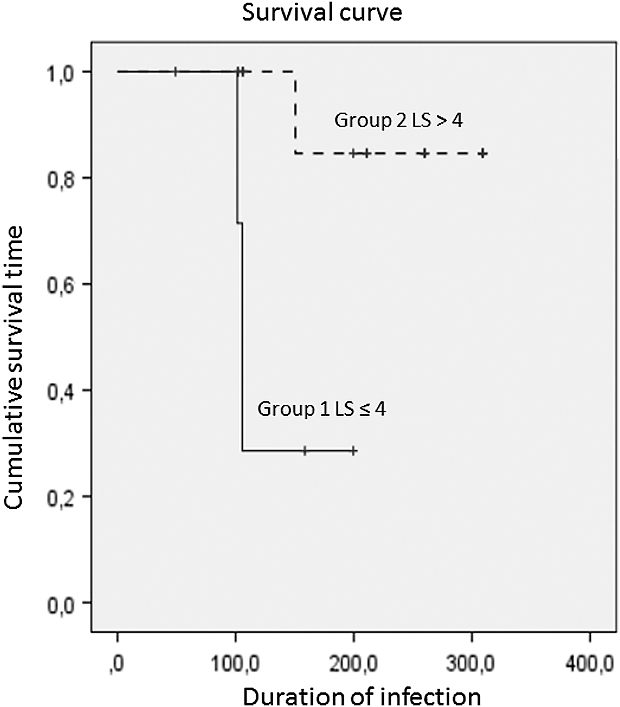
Kaplan–Meier survival curve of duration of methicillin-resistant Staphylococcus aureus infection in quarters with LS >4 (group 2, dashed line) and in quarters with LS ≤4 (full line) observed from July 2016 to May 2017. Lines are significantly different (log-rank test, p = 0.01); + indicates censoring of observation. LS, linear score.
Discussion
The mean within-herd MRSA prevalence we detected was higher (23%) than the ones reported by other authors (Kwon et al., 2005; Huber et al., 2010; Türkyılmaz et al., 2010; Luini et al., 2015), but those studies were performed by testing only animals affected by clinical or subclinical mastitis. The high incidence rate (14%) and poor cure rate (23%) of IMIC cows we detected are consistent with S. aureus infection patterns (Vanderhaeghen et al., 2010). Yet, in MRSA, the cure rate is worsened either by the ineffectiveness of antimicrobial treatment, due to the resistance of MRSA to all β-lactam antimicrobial agents (CLSI, 2013), or by the need to avoid development of further antimicrobial resistances. MRSA was detected in 82.4% quarters with subclinical mastitis where it persisted longer compared with MRSA-colonized quarters. This result is consistent with MSSA infection in cows, mostly producing chronic mastitis (Barkema et al., 2006), and previous studies on ST398 MRSA mastitis (Feßler et al., 2010; Vanderhaeghen et al., 2010; Luini et al., 2015). Udder or quarter colonization occurs when MRSA multiplies without causing disease. Colonization has a shorter excretion period compared with MRSA udder infection, yet the former is not fully predicting the latter, so a different management of colonized or infected MRSA cows is not feasible.
In this study, 20% of the cow nasal mucosa samples were colonized by MRSA, 2 animals having persistent colonization. MRSA colonization of cow nasal mucosa is seldom reported in cows (Feßler et al., 2012), and there was no previous information on persistence.
Environment contamination by MRSA, typically ST398, is well known in pig (EFSA, 2010) and rabbit farming (Agnoletti et al., 2014), demonstrating the ability of this microorganism to survive in the farm environment. In this study, MRSA environmental contamination was found in dust from pens and air in spring and summer samples. This was most likely due to mechanical airborne dissemination of microbial cells by fans that were switched on during the hot season in pens and the milking parlor. Contamination of teat liners, detected after milking cows with IMIC, may occur after MRSA excretion from the mammary gland or by the contaminated udder skin, the latter presumably causing contamination of two liners detected at the end of milking. Indeed, the same MRSA genotype detected in the environment, mammary gland, and udder skin allows for many possible cross-contamination pathways that have to be accounted for in management of MRSA infection in dairy cows. Those findings are consistent with the result of the estimated R0, slightly higher than 1 (1.08), which describes a slow, yet feasible, animal-to-animal transmission presumably favored by the environment and fomite contamination with MRSA.
Two farmworkers were detected with ST398 MRSA in the nose mucosa, which is consistent with heavy environmental contamination. This outcome was predictable because persons attending to MRSA-contaminated herds are more likely to be MRSA colonized (Verkade and Kluytmans, 2014).
We conclude that management of MRSA infection in dairy cows should consider the importance of environmental contamination on diffusion and maintenance of the microorganism differently from MSSA control, which is solely based on milking hygiene and segregation of infected cows (Barkema et al., 2006).
Conclusions
This study highlights the ability of ST398 MRSA to slowly diffuse within a dairy cow herd, to cause a long persistent infection (247 days) of the mammary gland, to contaminate liners, to colonize the udder skin and nasal mucosa of cows, to survive the farm environment, and to colonize humans closely attending the cows. The spread of MRSA between cows and the environment, despite animal segregation, provides evidence of the importance of controlling environmental contamination for risk management of MRSA infection in dairy herds, together with culling cows with udder infection.
Footnotes
Acknowledgments
This research was funded by the Italian Ministry of Health (RC IZSVE 12/2014). Authors thank Serena Genovese, Tiziana Ferro, Elena Tonon, Katia Capello, Dr. Luciano Mondin, Dr. Belinda Gross, and Dr. Frank Welcome for their cooperation.
Disclosure Statement
No competing financial interests exist.