
Abstract
Liver samples from finisher pigs were collected at the slaughterhouses for the analysis of zearalenone (ZEA), alfa-/beta-zearalenone (α-ZE, β-ZE), zearalanone (ZA), alfa-/beta-ZA (α-ZA, β-ZA), aflatoxin B1 (AFB1) and aflatoxin M1, fumonisin B1 (FB1), ochratoxin A (OTA) and ochratoxin B, deoxynivalenol and deepoxi-deoxynivalenol (DOM-1). For the analysis liquid chromatography-triple quadrupole coupled with mass spectrometry was applied. Liver samples with detected FB1 were further histopathologically evaluated after hematoxylin and eosin staining. Various levels of liver mycotoxins were detected in all farms. Pig livers with 2.91–8.30 μg/kg of FB1 were detected in three farms, estimate of 850–2400 μg/kg of FB1 intake, whereas 0.54 μg/kg of OTA was detected in one farm, estimate of 75 μg/kg of OTA intake. Moreover, pig livers with 0.30 μg/kg of ZEA, 1.87 μg/kg of α-ZE, and 0.63 μg/kg of β-ZE were detected in one farm, estimate with of 300 μg/kg of ZEA intake. The histopathological analysis revealed that the lesions' grading and necrosis grading were analogously increased when FB1 concentration increased from 2.91 to 4.36–8.30 μg/kg. The severity of megalocytosis was analogously increased with FB1 detection levels and particularly in levels of 4.36–8.3 μg/kg. However, the increased FB1 detection levels did not show analogous behavior with the severity of hepatic cell vacuolization. Results showed that FB1 remained the most critical risk factor in the Greek pig industry, whereas ZEA and AFB1 were also prevalent. The OTA contamination in pig farms raised a high risk for animal and human health.
Introduction
Contamination of food and animal feed with various mycotoxins remains a high-risk agro-economic problem (Hazel and Patel, 2004; Maresca and Fantini, 2010; Bryden, 2012). Food and feed mycotoxin-contamination results in significant economic losses for grain producers, and when the contaminated food is consumed it reduces the growth and health in a wide range of animal species (Chaytor et al., 2011). Mycotoxins are secondary metabolites of fungi (e.g., genus Aspergillus, Fusarium, and Penicillium), which have toxic properties and are commonly found in cereal grains (Binder et al., 2007). Some major mycotoxins include aflatoxins, ochratoxins, trichothecenes, zearalenone (ZEA), fumonisins, and ergot alkaloids (Richard, 2007; Marasas et al., 2008; Stein and Bulboacă, 2017).
Mycotoxins' economic impact on livestock includes decreased productivity and increased health care and veterinary care costs (Hussein and Brasel, 2001; Zain, 2011). Traditionally, mycotoxin detection relies on laboratory examination, which is time-consuming and laborious. Thus, it is necessary to develop a more precise and fast detection methodology, such as using biomarkers for the detection of mycotoxins. The chosen biomarker should be specific for each mycotoxin and target species, reflect the real exposure load, and be easily detectable with sensitive analytical methods validated for the matrix used (EFSA, 2010; Yang et al., 2015; Vidal et al., 2018; Lauwers et al., 2019). EFSA has proposed different biomarkers of exposure to aflatoxin B1 (AFB1), deoxynivalenol (DOA), ZEA, ochratoxin A (OTA), and fumonisins (EFSA, 2010).
Co-occurrence of mycotoxins under field conditions is common (Smith et al., 2016), and studies have investigated their effects on pigs by utilizing purified mycotoxins or cultured feed material (Bracarense et al., 2012; Mateos et al., 2018; Schertz et al., 2018). Some studies have documented the occurrence and toxicity of mycotoxins and the risk they represent in pig production (Broekaert et al., 2015; Pierron et al., 2016). There are limited data on dietary exposure of pigs with naturally mycotoxin-contaminated feed and its effects on liver function (Andretta et al., 2012; Pierron et al., 2016), especially on the occurrence of mycotoxins in the Greek livestock sector. Although some results of mycotoxins contamination in feed materials have been published (Griessler et al., 2010; Streit et al., 2012).
The aim of this study was the investigation of mycotoxin biomarkers traced in livers of pigs reared under field conditions in Greece, as well as the evaluation of the associated histopathological lesions.
Materials and Methods
Farms/sampling
During the herd health management routine, applied from the faculty of veterinary medicine, University of Thessaly, liver samples from finisher pigs were collected at slaughterhouse from eight commercial farrow-to-finish pig farms. The farms' capacity ranged between 350 and 500 sows, and they were located in different regions of Greece (two farms in Central Greece, two in South Greece, two in West Greece, and two in North Greece).
All farms vaccinated their sows against Aujeszky's disease virus, parvovirus, atrophic rhinitis, erysipelas, Porcine Reproductive and Respiratory Virus (PRRSV), Escherichia coli, and Clostridium infections. Weaners were vaccinated against Porcine Circovirus type 2 (PCV2) and Mycoplasma hyopneumoniae. Control of endo/ectoparasites was currently obtained by sows' treatment with ivermectin. Based on the serological routine monitoring program, no viremia of PRRSV and PCV2 were detected in sows, weaning, and finishing pigs of these farms. The feed provided was home-mixed corn/barley/wheat–soybean-based meal. All farms used toxin binders in the feed of sows during gestation and lactation period and in the feed of weaning pigs. However, the use of toxin binders in the feed of growing/finishing pigs was intermittent (3–4 times per year). The farms of this study used different commercial products of toxin binders, but for the same periods.
Compliance with ethical standards
All procedures during this clinical study were performed according to the Code of Practice for the Conduct of Clinical trials for Veterinary Medical Products, and the Guide for the Care, and Use of Agricultural Animals in Research and Teaching, whereas animals were maintained in compliance with national and European animal welfare requirements. All procedures regarding animal care and handling were approved by the Ethical Committee of the Faculty of Veterinary Medicine, School of Health Sciences (University of Thessaly, approval number 92).
Sampling/preparation of samples
At the slaughterhouse, liver samples (50 g) were collected from four animals from each farm. All samples were frozen and sent to the laboratory within 24 h, using dry ice and freezing gels. Simultaneously liver samples from each animal were fixed in 10% neutral buffered formalin.
Four liver samples from each farm were homogenized by cutting into pieces and crushing into the good mass before lyophilization. To process samples, all materials were properly washed with ethanol and water. Initially, 20.0 ± 0.1 g of homogenate liver was kept in 100 mL polypropylene jars, and then the jars were frozen at −20°C for 24 h. All jars were immediately placed into the lyophilizer. All samples were lyophilized at −50°C and 4 × 10⁻⁴ mbar for 17 h, and then the jars were removed from the lyophilizer and were closed tightly, sealing the lid with parafilm. Finally, all samples were stored in a dry environment at 25°C, and remained stable for 10 d.
Analyzed parameters and detection methods in liver samples
The parameters analyzed for all liver samples, as shown in Table 1, are the following: (1) ZEA and its metabolites: alfa-zearalenone (α-ZE), beta-zearalenone (β-ZE), zearalanone (ZA), alfa-zearalanone (α-ZA), and betazearalanone (β-ZA), (2) AFB1 and its metabolite aflatoxin M1 (3) fumonisin B1 (FB1), (4) OTA and its metabolite ochratoxin B, and (5) DON and its metabolite deepoxi-deoxynivalenol (DOM-1).
Analyzed Parameters (Mycotoxins and Mycotoxin Biomarkers, Estimated Intake) in Liver Samples of Each Farm
Detected-D (μg/kg): detected below the limit of quantification, therefore concentrations in liver and estimated intake cannot be indicated with an adequate precision
EI: Estimated intake (μg/kg) according to detection in liver according to detection in liver.
ND, not detected; ZEA, zearalenone; α-ZE, alfa-zearalenone; β-ZE, beta-zearalenone; α-ZA, zearalanone; β-ZA, beta-zearalanone; AFB1, aflatoxin B1; AFM1, aflatoxin M1; FB1, fumonisin B1; OTA, ochratoxin A; OTB, ochratoxin A; DON, deoxynivalenol; DOM-1, deepoxi-deoxynivalenol
The analytical methods adopted were (i) liquid chromatography coupled to a mass spectrometry detector tandem triple quadrupole type (QqQ) for the determination of parameters 1, 2, 3, and 5 mentioned earlier and (ii) liquid chromatography coupled to a fluorescence detector (FLD) for the determination of parameter 4 mentioned earlier (Table 1). Quantification methods were used for the determination of mycotoxins and mycotoxin biomarkers. The FLD method set detection and quantification limits, and the QqQ method set quantification limits. The detection limit was the lowest concentration of mycotoxin or metabolite detected but not quantified with accuracy and precision. The quantification limit was the lowest concentration of mycotoxin or metabolite with accuracy and precision. If neither mycotoxin nor metabolites were detected, it was recorded as “Not Detected’’ and the detection limit value was assigned. If the analytical results were below the quantification limit, but over the detection limit, they were recorded as “Detected’’, and the quantification limit value was assigned. The estimated mycotoxin intake was calculated according to the quantification or detection limits, without taking into consideration for this estimation the periodical use of toxin binders (Table 1), based on the maximum limit levels recommended by the technical literature and by the European Union (EU) legislation (Gimeno et al., 2007, 2011; The European Commission, 2011, 2016)
Histopathological analysis
Based on the analysis results for mycotoxins and mycotoxin biomarkers in liver samples, positive FB1 samples were further histopathologically examined. The samples were collected, fixed in 10% neutral buffered formalin, and embedded in paraffin by a routine procedure. Dewaxed 3–6 μm-thick sections were obtained and stained with hematoxylin and eosin to be evaluated.
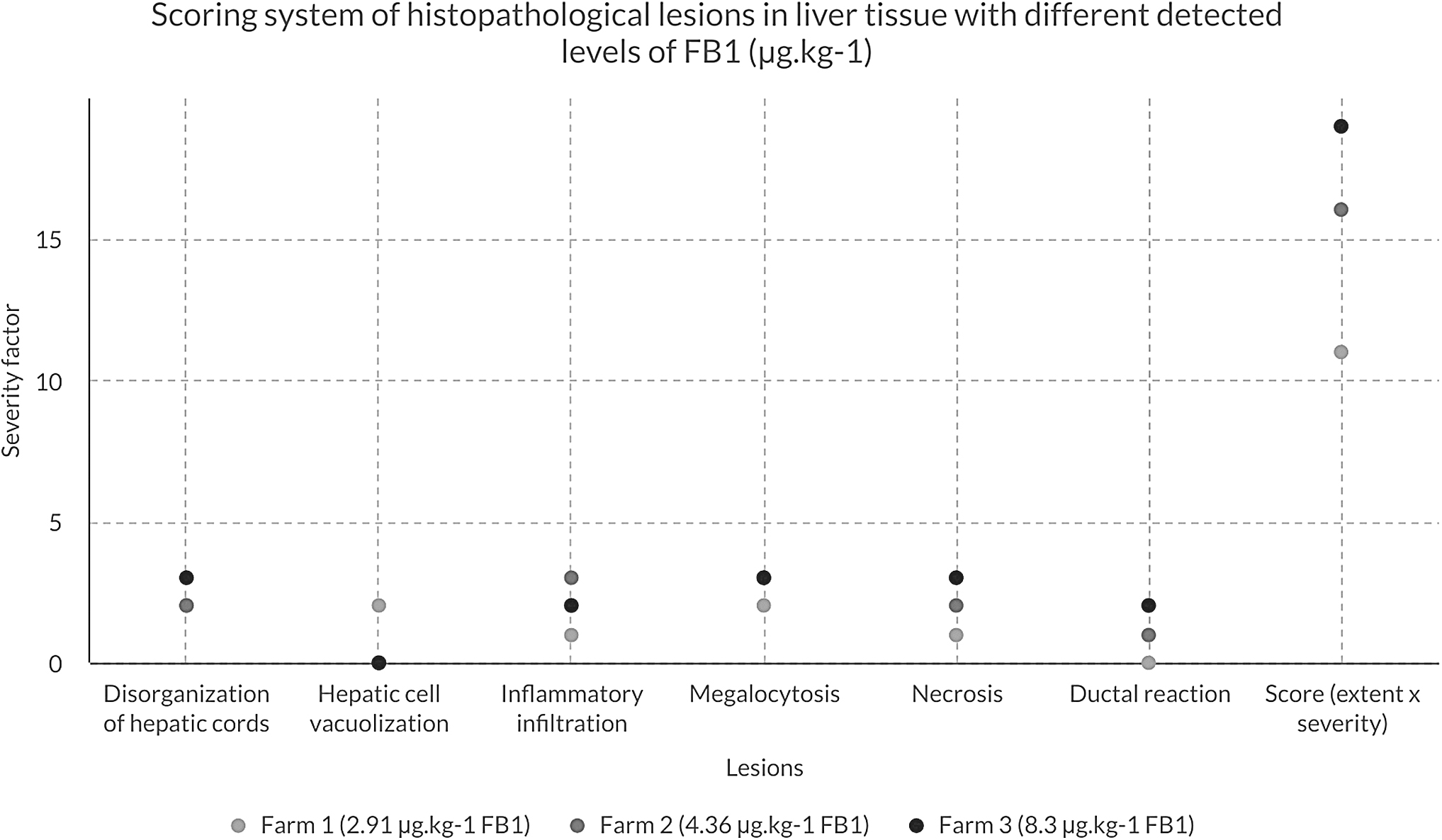
The histopathological evaluation was performed, following a modified grading system based on previous studies that have described similar systems (Grenier et al., 2011, 2012; Bracarense et al., 2012). More specifically, various types of lesions were assessed according to Table 2. The lesions' score was calculated by considering the degree of severity (severity factor) and the extent of each lesion. Each lesion's extent was evaluated by scoring 0–3 intensity or frequency of each lesion. Finally, on each lesion, the score of the extent was multiplied by the severity factor. Each group included four liver samples from four animals per farm, and for each sample, three slides were examined.
Scoring System of Histopathological Lesions in Liver Tissue with Different Detected Levels of Fumonisin B1 (μg/kg)
Severity factor: 1 = mild, 2 = moderate, 3 = high.
FB1, fumonisin B1.
Results
Exposure biomarkers analysis in livers
Various levels of liver mycotoxins were detected from all farms. The exposure of biomarker levels in the pool of livers of each farm and the estimated feed intake are shown in Table 1.
Pig livers with 2.91–8.30 μg/kg FB1 were detected in three farms, estimating with 850–2400 μg/kg of FB1 intake. Moreover, liver samples with 0.54 μg/kg of OTA were detected in one farm, estimating of 75 μg/kg of OTA intake. Finally, pig livers with 0.30 μg/kg of ZEA, 1.87 μg/kg of α-ZE, and 0.63 μg/kg of β-ZE were detected in one farm, estimating with 300 μg/kg of ZEA intake (Gimeno et al., 2007, 2011; The European Commission, 2011, 2016).
Histopathological results
Pig liver from three sampled farms had detected FB1, with different histopathological aspects.
Liver samples from farm 1 (FB1 2.91 μg/kg)
Swollen hepatocytes with abundant granular eosinophilic cytoplasm (megalocytosis) were seen to an intermediate extent. In the periphery, there was multifocal vacuolization of bundles of hepatic cells (Fig. 1).
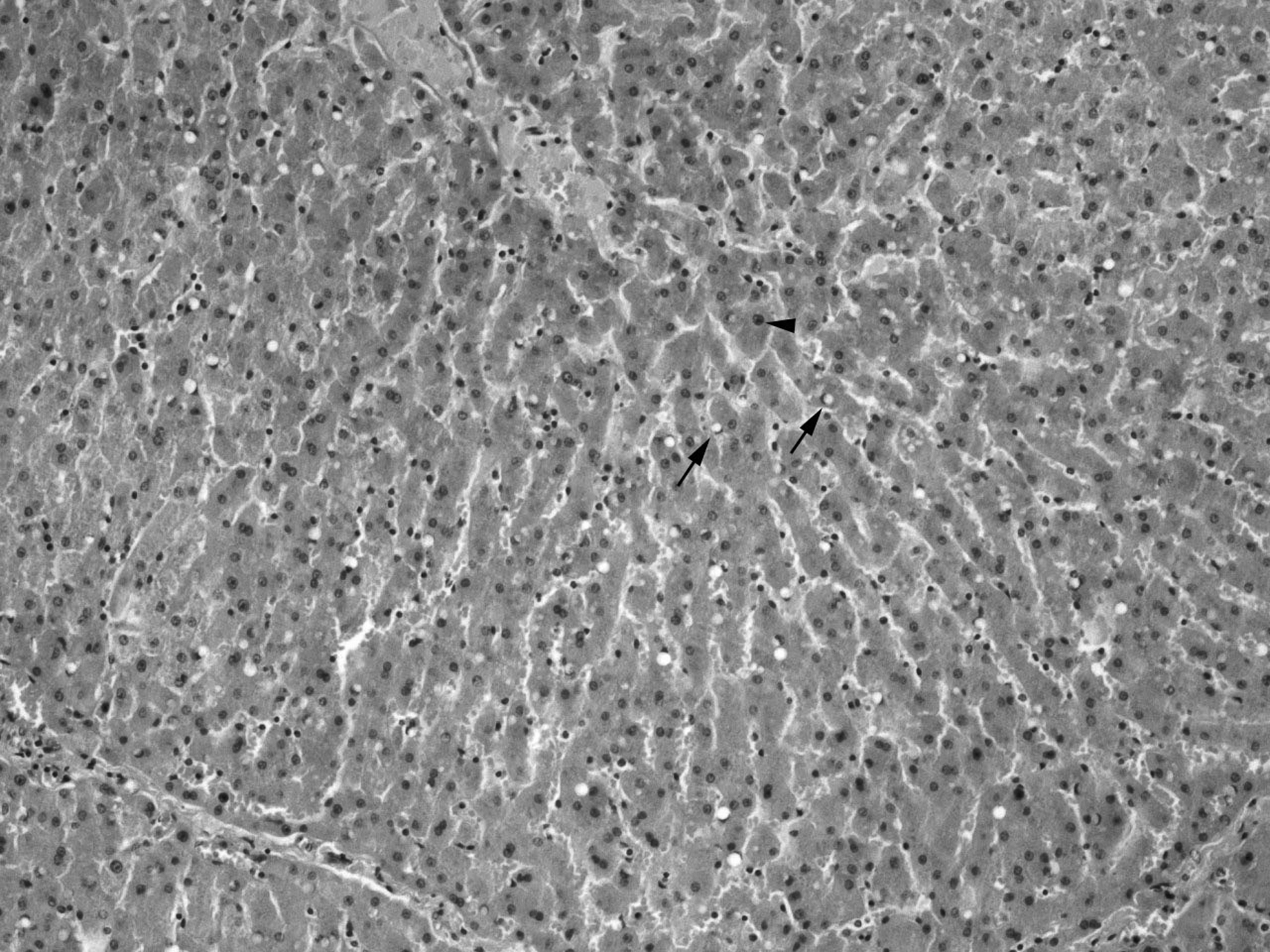
Megalocytosis (arrowhead) and vacuolization (arrows) in liver with detection of FB1 2.91 μg/kg, H&E, × 10. FB1, fumonisin B1; H&E, hematoxylin and eosin.
Liver samples from farm 2 (FB1 4.36 μg/kg)
Severe diffuse megalocytosis scattered with small angular hepatocytes and Kupffer cells were detected. Centrilobular necrosis and disorganization of the hepatic cords were also noticed. Ductal reaction, accompanied by local portal and stromal neutrophilic and eosinophilic inflammatory reaction and fibrosis, was found (Fig. 2).
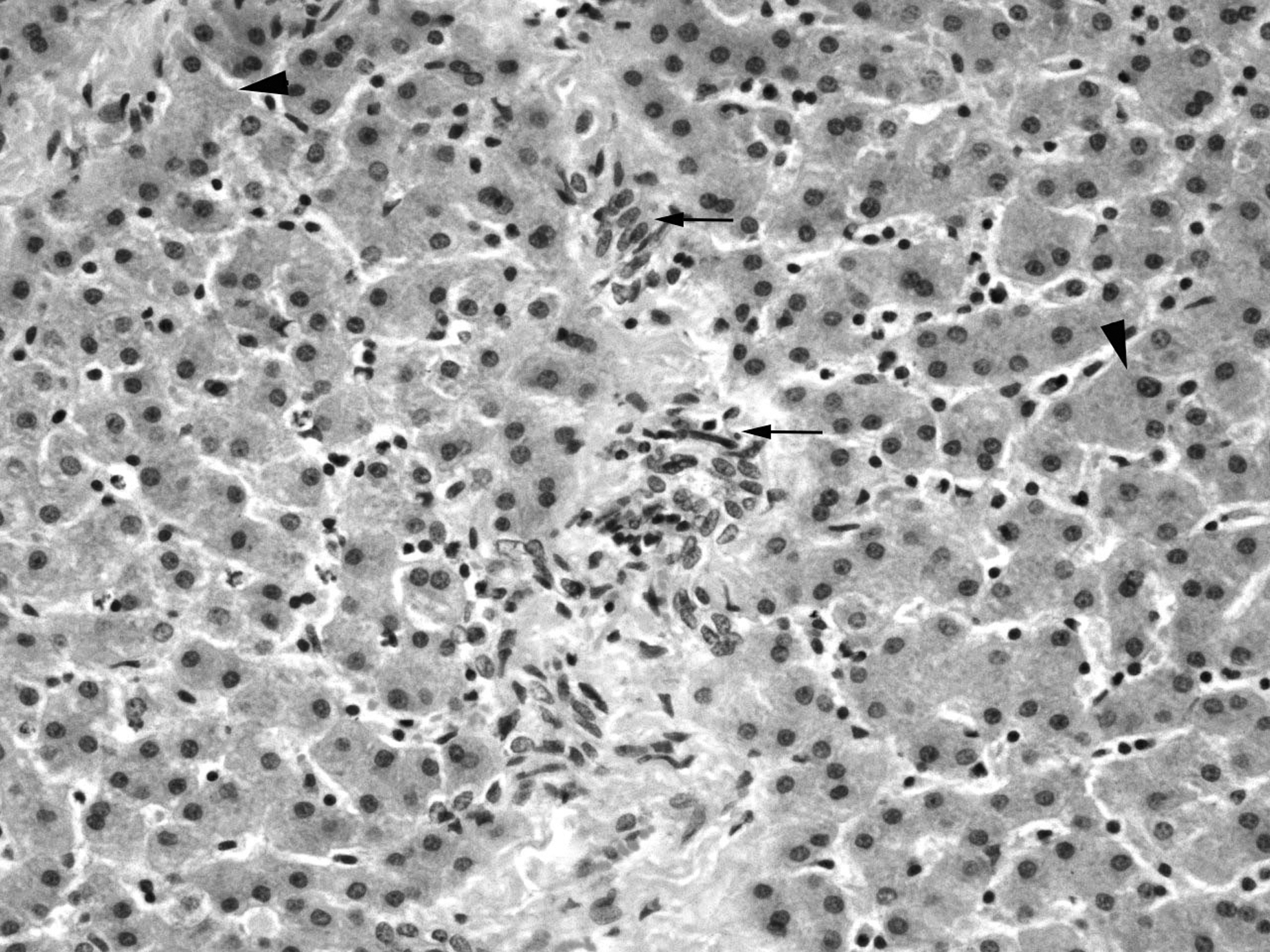
Irregular ductular structure lined by flattened basophilic cells in the lobular parenchyma (arrows) and megalocytosis (arrowhead) in liver with detection of FB1 4.36 μg/kg, H&E, × 20. FB1, fumonisin B1; H&E, hematoxylin and eosin.
Liver samples from farm 3 (FB1 8.30 μg/kg)
Severe diffuse megalocytosis scattered with small angular hepatocytes and Kupffer cells were seen. Massive necrosis of the lobules without zonal distribution, leading to restriction or loss of the intervening stroma and multifocal absence of the typical histological lobular organization wass observed. Ductular reaction prominent in the hepatic parenchyma and to a milder extent in the portal tract connective tissue interface was also revealed (Fig. 3).
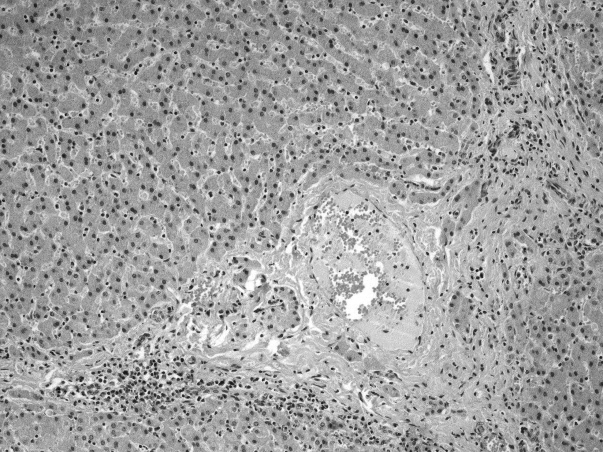
Portal necrosis and stromal inflammatory infiltration in liver with detection of FB1 8.3 μg/kg, H&E, × 10. FB1, fumonisin B1; H&E, hematoxylin and eosin.
Table 2 shows the lesions in each farm's liver samples, using a modified scoring system. The lesions' grading was analogously increased with FB1 detection levels of 2.91, 4.36, and 8.30 μg/kg, respectively (Fig. 4).
Histopathological grading of liver lesions due to different detected levels of FB1. FB1, fumonisin B1; H&E, hematoxylin and eosin.
Gross pathological observations
The liver samples from farm 1 and farm 2 (detection of 2.91 and 4.36 μg/kg of FB1, respectively) had a yellowish to dark color and brittle characteristic to the touch, whereas from farm 3 (high detection of 8.30 μg/kg of FB1) had dark color and hard touch.
Discussion
Pigs, as monogastric animals, are particularly vulnerable to mycotoxins because of the lack of a rumen with a microbiota able to degrade mycotoxins before their intestinal absorption (Zain, 2011; Pierron et al., 2016; Dang et al., 2017). The most notorious mycotoxins on swine health are FBs, which are poorly absorbed from the gastrointestinal tract and the absorbed fraction remains mostly in the liver and kidneys (Prelusky et al., 1994, 1996; Pierron et al., 2016). Previous studies have determined the effects of FB on pigs, using purified FB toxin or cultured feed material in the experimental trial (Bracarense et al., 2012; Mateos et al., 2018; Schertz et al., 2018). In contrast to the aforementioned studies, our results are based on natural dietary exposure of pigs and for this reason offer important information for clinical practice. Similar to our study lesions in liver (megalocytosis, nuclear, and cytoplasmic vacuolization of hepatocytes) were recently reported using naturally contaminated feed with low doses of FB (Terciolo et al., 2019). Our study, which is also based on naturally contaminated dietary exposure of pigs with FB, indicated that necrosis grading, which is a hallmark lesion of FB toxic effect, was analogously increased with FB1 detection levels (2.91, 4.36, and 8.3, respectively). Respectively, the severity of megalocytosis was increased in accordance with the level of FB1 detection and particularly in levels of 4.36 and 8.3 μg/kg. The presence and severity of hepatic cell vacuolization was not increased according to the elevation of FB1 levels. On the contrary, it was detected only in the farm with the lowest levels of FB1, indicating that hepatic cell vacuolization could possibly be an “early” lesion of fumonisin intoxication. In our histopathological results in the applied scoring system, we added the ductal reaction evaluation as a marker of mature hepatocytes' inability to replicate. Similar results were reported in other studies, but most of them included experimental contamination and did no report lesions under field conditions and at different FB1 detection levels (Haschek et al., 1992; Casteel et al., 1993; Colvin et al., 1993; Motelin et al., 1994; Gumprecht et al., 1998; Grenier et al., 2013). Since pig is an excellent model for human diseases, the mechanism for FB toxicosis in swine must be characterized to permit assessment of its potential toxicity in humans (Yoshisawa et al., 1994; Hendricks, 1999; Haschek et al., 2001). Moreover, in our study, the contamination of feed with ZEA and AFB1 seems to be also prevalent, but the mycotoxin biomarkers analysis did not reveal their importance as risk factors. However, the detection of OTA in one farm raises a high risk for animal and human health.
Fusarium mycotoxins have attracted increasing attention due to the risk of contamination by mycotoxins in the food chain. EU has published regulations and recommendations for several mycotoxins in pig feed (EFSA, 2010; The European Commission, 2011, 2016). Among various mycotoxins, FB1 greatly impacts on pig health and growth performance (Pierron et al., 2016). In our study, the detected FB1 levels exceeded the maximum limit levels recommended by the technical literature and EU legislation (The European Commission, 2011, 2016). Therefore, based on our histopathological findings, FB1 could be a high-risk mycotoxin for the Greek pig industry, with an important negative impact on health and growth performance. To our knowledge, there are limited data on the dietary exposure of pigs with naturally mycotoxin-contaminated feed in Greece. Our study is the first report on the detection of mycotoxins and mycotoxin biomarkers in Greek pig industry. In contrast to our study, previous surveys about mycotoxin occurrence in the Greek livestock sector reported only the incidence of mycotoxins in feed materials (Griessler et al., 2010; Streit et al., 2012). Feed official control system in Greece reported high compliance with EU regulation 1881/2006 in the majority of samples tested in 2010 (Streit et al., 2012). Furthermore, our results for the estimated intake of mycotoxins comply partially with the results of Griessler et al. (2010) who evaluated the occurrence of mycotoxins in feed materials and compounded feed samples originating from Southern European countries, including Greece. In this study, 85 feed samples from Greece were analyzed (including samples from Cyprus) and the occurrence of mycotoxins was 63%, 33%, 40%, and 75% for DON, ZEA, OTA, and FB, respectively.
The patterns of mold growth and mycotoxin production are strongly influenced by environmental conditions and ecophysiological factors, such as temperature and water activity (Northolt and Bullerman, 1982). Recent studies reported that climate change factors would substantially affect patterns of growth and mycotoxin production (Miraglia et al., 2009; Tirado et al., 2010; Paterson and Lima, 2010, 2011; BEST, 2011). Especially in Europe, mycotoxins' occurrence patterns are expected to alter due to the increase in average environmental temperatures (Miraglia et al., 2009; Goertz et al., 2010). For this reason, the risk of mycotoxins is increasing year by year as a result of climate change to warmer and drought-like environmental conditions (Streit et al., 2012). Previous studies reported that these FB1 have become dominant in Europe due to rising environmental temperatures (Stroia et al., 2010; Goertz et al., 2010; Griessler et al., 2010). The Mediterranean zones, including Greece, have been identified as a climate-change hot spot, resulting in negative effects on the agriculture sector (Bank of Greece, 2011). Therefore, further studies are required to investigate the possible effects of mycotoxins on livestock production in relation to climate change.
Conclusions
FB1 has an essential impact on pigs' health status in Greece, as severe lesions and probably negative effects on health and growth performance accompany its detection in the liver at high levels. Exposure biomarkers of pigs are related to their negative effects, as the severity of the lesions increased in a dose-dependent manner.
Footnotes
Disclosure Statement
No competing financial interests exist.
Funding Information
The author(s) received no financial support for the research, authorship, and/or publication of this article.