
Abstract
A flow cytometry (FCM)-based method was developed for the faster detection of Staphylococcus aureus in milk and milk powder. Viable S. aureus cells were recognized by highly selective, fluorescently labeled antibodies and Propidium Iodide, and then analyzed by FCM. Using a 5-h pre-enrichment period, the method could detect low numbers of S. aureus cells in 6 h, with a limit of detection of 7.50 cells/mL in milk and 8.30 cells/g in milk powder. The established method was compared with the plate-based method using 75 ultra-high-temperature-treated milk samples, 25 pasteurized milk samples, 66 raw milk samples, and 123 milk powder samples. The two methods yielded similar results for the detection of the pathogen in all sample types. The FCM-based method allows effective and faster monitoring of S. aureus contamination and can be applied to the rapid detection of microorganisms in milk and dairy products.
Introduction
S
European Commission regulations (EC, 2007) state that quantitative determination of S. aureus in milk and dairy products is with a limit of detection (LOD) of 10–100 colony-forming units (CFU)/mL or CFU/g. Notably, one of the main consumers of milk powders is infant, which may be at greater risk of disease from products with a low level of S. aureus contamination because of its lower immunity. In consequence, for a more accurate and effective detection, faster methods that can efficiently detect low numbers of viable S. aureus cells are urgently needed (Templier and Roupioz, 2017).
The conventional culture and plate-based method is the gold standard and the most common method used for S. aureus detection in standardized laboratories given its high sensitivity and ideal specificity (Banada et al., 2009), nevertheless, it is excessively time-consuming, with a long time-to-results (TTR) (it can take up to a few days to obtain a result), and is labor intensive. In the past few decades, numerous efforts have been made to develop rapid methods for S. aureus detection, including immunoassay methods (Yan et al., 2017; Alamer et al., 2018), PCR-based assays (Liu et al., 2019a; Moezi et al., 2019), biosensors (Ghali et al., 2016; Templier and Roupioz, 2017; Zheng et al., 2019), and flow cytometry (FCM) (Bassoe, 1984; Meng et al., 2017; Shan et al., 2019). Unfortunately, immunoassay methods suffer from poor sensitivity and/or lack quantitative results (Schaumburg et al., 2019), while biosensor-based methods have difficulty in distinguishing between viable and nonviable bacterial cells. In addition, there are certain limitations in simplicity and rapidity in the PCR-based methods (Wang et al., 2018).
FCM is a good option as it allows rapid and automatic quantification of viable cells among nonviable cells (Ananta et al., 2005). Previous studies have reported FCM-based methods for the rapid detection of single bacterial cells of species such as Escherichia coli (Liu et al., 2019b) and Salmonella (Wang et al., 2020) in dairy food matrices, while that of viable S. aureus cells in low numbers in dairy, although of great importance, has not been explored.
Therefore, in this study, we combined FCM with a pre-enrichment procedure to achieve faster detection of viable S. aureus cells in low numbers in milk and milk powder.
Materials and Methods
Bacterial strains and culture conditions
The information of all bacterial strains used in this study is listed in Table 1. Moreover, S. aureus ATCC 6538P was the target strain. Cells of all strains were grown at 37°C in nutrient broth (Oxoid, Basingstoke, UK), centrifuged at 10,000 × g for 5 min, and then resuspended in phosphate-buffered saline (PBS; pH 7.2).
Bacterial Strains Tested for the Reactivity with Anti-Staphylococcus aureus Antibody Using Flow Cytometry
ATCC, American Type Culture Collection; BNCC, BeNa Culture Collection; CICC, China Center of Industrial Culture Collection; FITC, Fluorescein isothiocyanate; GDMCC, Guangdong Microbial Culture Collection Center; N, negative, means that the strain could not be recognized by the FITC-labeled antibody; P, positive, means that the strain could be recognized by the FITC-labeled antibody.
Dual staining of S. aureus and FCM
The FCM used was an A50-Micro model (Apogee, Hemel Hempstead, UK). Fluorescein isothiocyanate (FITC)-labeled anti-S. aureus antibody was prepared by labeling goat anti-S. aureus polyclonal antibody (Cat. No. 5310-0314; SeraCare Life Sciences, Milford, MA) using a FITC Labeling Kit (Cat. No. ARL0021K; Frdbio Science & Technology, Wuhan, China). Propidium Iodide (PI) was purchased from Molecular Probes (Cat. No. P3566; Eugene, OR).
A S. aureus ATCC 6538P suspension of ∼107 cells/mL in PBS was mixed with the antibody (final concentration: 1 μg/mL) and PI (final concentration: 1 μg/mL), and this reaction was allowed to proceed at room temperature in the dark for 15 min. Then, the suspension was analyzed by the FCM. According to principle of the dual labeling with FITC-labeled antibody and PI (Wang et al., 2020), the gating strategy can be set as follows (Supplementary Fig. S1): Set up an forward scatter (FSC) versus side scatter (SSC) plot with a gate around the bacterial population Region 1 (R1). On the fluorescence (FL)1 versus FL3 dot plot gated on R1, viable cells were usually enumerated by counting events that scatter the incident blue light at the expected intensities and emit green, but not red fluorescence.
Inclusivity/exclusivity testing
To assess the inclusivity and exclusivity of the FCM-based method, the 10 S. aureus and 37 non-S. aureus strains were incubated with FITC-labeled anti-S. aureus antibody and analyzed by FCM.
Preparation of artificially contaminated samples
UHT milk was obtained from Monmilk Dairy (Hohhot, Inner Mongolia, China). Milk powder was purchased from Friesland Campina Domo B.V. (Frisolac, Friesland, Netherlands). In initial tests, no bacterial cells were detected in the milk and milk powder by the plate-based method (FDA, 2016). Artificially contaminated milk and milk powder samples were prepared as follows: 1 mL of the suspension of viable S. aureus cells was centrifuged at 10,000 × g for 5 min, and added into 25 mL of milk or 25 g of milk powder.
Optimization of the pre-enrichment time
Six artificially contaminated samples of milk and milk powder with S. aureus at a final concentration of ∼101 CFU/mL and 101 CFU/g, respectively, were prepared as follows: A suspension of S. aureus at a final concentration of (1.2 ± 0.2) × 103 CFU/mL was prepared by the dilution method. The populations of the S. aureus suspension were confirmed using a plate-based method by inoculating the samples onto Baird–Parker agar (Land Bridge, Beijing, China) plates (n = 3) (FDA, 2016). Then 250 μL of the S. aureus suspension was transferred to a sample using a pipette, so that the sample could contain (12 ± 2) CFU/mL or CFU/g of S. aureus, which is enough to be of clinical significance, as prescribed by the European Commission regulations (EC, 2007).
Each sample (25 mL or 25 g) was added to 225 mL of sterilized 7.5% sodium chloride broth (SCB; Land Bridge, Beijing, China) and incubated at 37°C in a shaker incubator set at 180 rpm. After 0, 3, 4, 5, and 6 h of pre-enrichment, 6 mL of the mixture was collected and analyzed by the plate-based method. All samples were analyzed in sextuplicate. The coefficient “K,” which is used to evaluate S. aureus growth, was calculated using Eq. (1):
where Kt is the ratio of the number after t h of pre-enrichment to the initial number, Nt is the concentration of S. aureus cells after t h of pre-enrichment, N 0 is the initial number of S. aureus cells, and t is the pre-enrichment time.
Assessment of the relationship between the initial cell number and the number after 5 h of pre-enrichment
The optimal pre-enrichment time in this study was determined to be 5 h. To determine the relationship between the initial number and the number after 5 h of pre-enrichment, four groups of artificially contaminated samples were prepared (milk or milk powder with S. aureus ATCC 6538P cells or S. aureus ATCC 25923 cells). Detailed information on the samples is given in Table 2. The use of two different S. aureus strains was for evaluating whether they would have similar reproduction rates under the optimized pre-enrichment conditions. Each sample (25 mL or 25 g) was added into 225 mL of SCB, and 6 mL of each mixture was collected immediately and after incubation in the shaker incubator at 37°C for 5 h, and analyzed by the plate-based method in sextuplicate.
Artificially Contaminated Samples Used for Validating the Relationship Between the Initial Number and the Number After 5 h of Pre-Enrichment and Regression Equations
Concentration, the approximate initial number of S. aureus cells.
A total of 120 individual samples were divided into four groups. Each of the four groups contains 30 samples with five different initial numbers.
N 5h, the number of S. aureus cells after 5 h of pre-enrichment; N 0, the initial number of S. aureus cells; The fitting regression equation was carried out from the plate-based results of the 30 samples in each of the four groups.
CFU, colony-forming unit.
Sample analysis by the FCM-based method
Each sample (25 g or 25 mL) was added to 225 mL of SCB and incubated at 37°C for 5 h for the pre-enrichment of S. aureus. One milliliter of the mixture was used for the isolation procedure of S. aureus cells as described in the previous study (Gunasekera et al., 2000). The recovery of the isolation procedure ranged from 90% to 99% in our preliminary experiment (Supplementary Table S1), which was confirmed to be effective for the following detection.
In brief, 2.5 mg of proteinase K (Molecular Probes, Eugene, OR) and 500 μL of 0.1% Triton X-100 (Sigma, St. Louis, MO) were added to 1 mL of the mixture, and the samples were incubated at 37°C for 30 min. After incubation, 0.5 mL of PBS was added to the sample and mixed thoroughly by inverting the tube 100 times for the reaction to complete. The mixtures were centrifuged at 12,000 × g for 5 min to remove lipid components, and the pellets containing S. aureus cells were collected.
After the isolation procedure, S. aureus cells were resuspended in 1 mL of PBS, and stained with the FITC-labeled antibody and PI. Finally, the stained cells were centrifuged again, resuspended in 0.1 mL of PBS, and analyzed by FCM. The number of S. aureus cells could be calculated using Eq. (2), as described in Section Relationship between the initial cell number and the number after 5 h of pre-enrichment.
Sample analysis by the plate-based method
Each sample (25 g or 25 mL) was added to 225 mL of SCB. Before pre-enrichment, 6 mL of the mixture was collected and analyzed by the plate-based method (FDA, 2016) in sextuplicate. The remainder of the mixture was further incubated for 5 h and then analyzed by the FCM-based method.
Detection of S. aureus in artificially contaminated samples
Artificially contaminated milk and milk powder samples with S. aureus at final concentrations of 100 to 105 CFU/mL and 100 to 105 CFU/g were prepared, respectively (Table 3). The samples were examined by the FCM-based method after 5 h of pre-enrichment. All enumeration experiments were performed in triplicate.
Detection of Staphylococcus aureus in Artificially Contaminated Samples by the Flow Cytometry-Based Method
Addition, the number of S. aureus cells determined by the plate-based method.
Detection, the number of S. aureus in the artificially contaminated sample determined by the FCM-based method.
FCM, flow cytometry.
Detection of S. aureus in real samples
In total, 75 retail UHT milk samples, 25 retail pasteurized milk samples, and 123 retail milk powder samples from different brands and different lot numbers were purchased from local markets. In addition, 66 raw milk samples were obtained from a local dairy farm. All samples were analyzed by the FCM- and plate-based methods.
Results
Effective FCM detection of S. aureus by dual staining
FITC-labeled antibody and PI were used to distinguish viable S. aureus cells from dead cells and other bacteria. Viable S. aureus cells emit only green fluorescence, whereas dead S. aureus cells emit green and red fluorescence (Fig. 1). The FCM allowed an efficient and accurate detection of viable S. aureus cells (Supplementary Fig. S2).
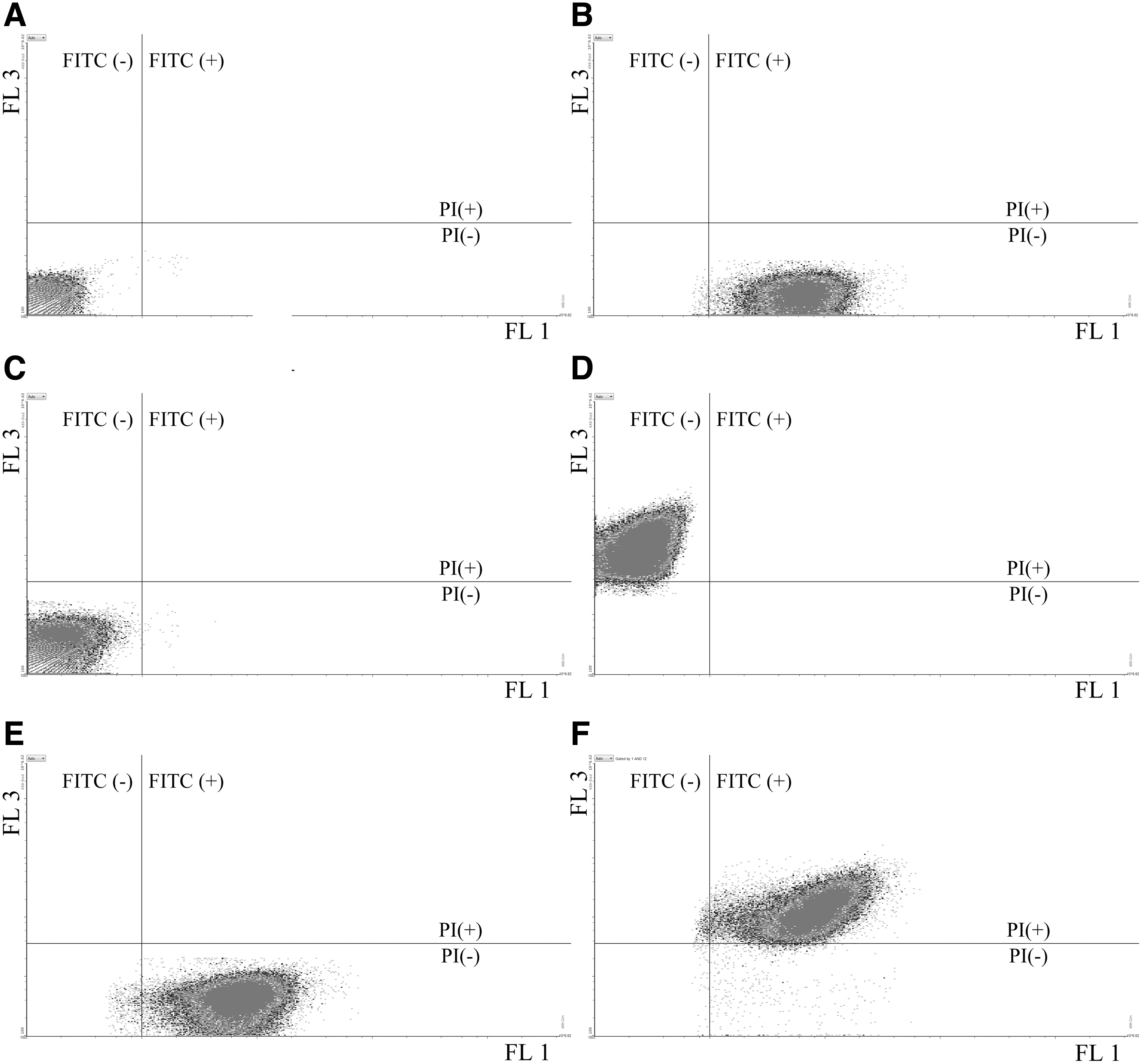
Staining of Staphylococcus aureus cells.
Inclusivity/exclusivity testing
A total of 47 bacterial strains (Table 1) were stained with the FITC-labeled anti-S. aureus antibody to evaluate the inclusivity and exclusivity of the FCM-based method. All 10 S. aureus strains were recognized by the FITC-labeled antibody, whereas none of the 37 non-S. aureus strains was. This result suggested that the FCM-based method showed a high specificity for S. aureus.
Pre-enrichment time
Six artificially contaminated samples of milk and milk powder with S. aureus at a final concentration of ∼101 CFU/mL and 101 CFU/g, respectively, were prepared and analyzed by the plate-based method after 0, 3, 4, 5, or 6 h of pre-enrichment. The number of S. aureus cells in milk and milk powder samples continuously and steadily increased between 3 and 6 h (Fig. 2). Cells could be detected by FCM after 5 h, but not after 4 h of pre-enrichment. Hence, the optimal pre-enrichment time was determined to be 5 h.
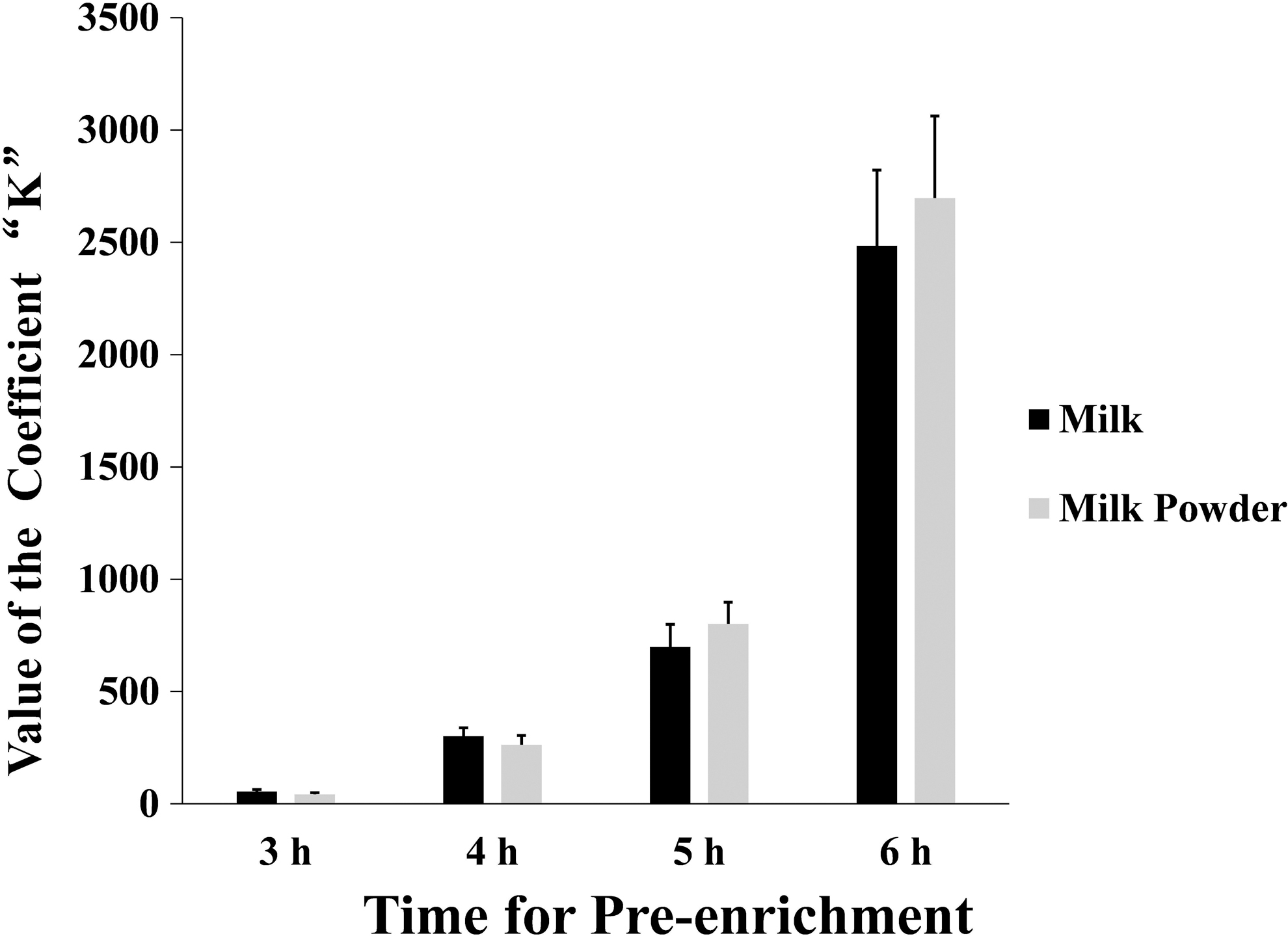
Relationship between the coefficient “K” and the time of the pre-enrichment. A total of 6 milk samples (25 mL) with ∼10 CFU/mL of S. aureus and 6 milk powder samples (25 g) with ∼10 CFU/g of S. aureus were prepared. Each of all samples was incubated in 225 mL of SCB, and then analyzed by the plate-based method after 0, 3, 4, 5, and 6 h of pre-enrichment. Each sample was analyzed in six replicates. The coefficient “K” was calculated using the equation:
Relationship between the initial cell number and the number after 5 h of pre-enrichment
One hundred twenty artificially contaminated milk and milk powder samples were prepared, using two different S. aureus strains (n = 30 each). The initial number and the number after 5 h of pre-enrichment of each sample were determined by the plate-based method. There was an obvious linear relationship between initial and final number for each of the four groups of samples (Table 2), and there was no statistically significant difference between the four regression equations. Therefore, a fitting regression equation was established by combining the data for all four groups of samples:
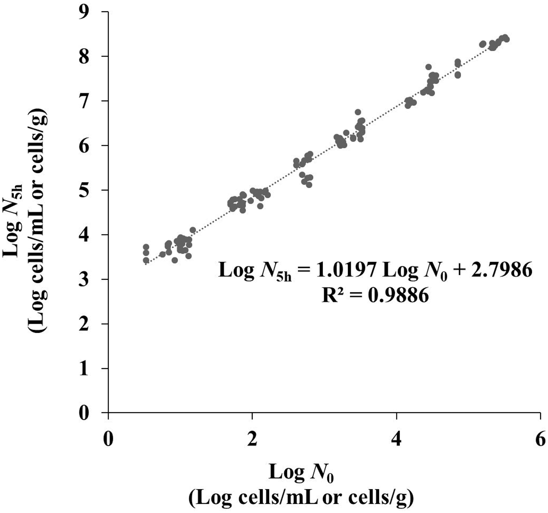
Relationship between the initial number (N 0) and the number after 5 h of pre-enrichment (N 5h). Four groups of artificially contaminated samples (Table 2) were prepared with different populations of S. aureus (S. aureus ATCC 6538P or S. aureus ATCC 25923) cells. The initial number and the number after 5 h of pre-enrichment of each sample were tested by the plate-based method, respectively. The fitting regression equation was carried out by combination of all the data from the four groups of samples. N 0, the initial number of S. aureus cells in milk or milk powder; N 5h, the number of S. aureus cells in milk or milk powder after 5 h of pre-enrichment.
where NFCM is the output of the FCM, and
Detection of S. aureus in artificially contaminated samples
Raw and powdered milk samples with S. aureus at final concentrations of 100–105 CFU/mL or CFU/g, respectively, were prepared, and then analyzed by the FCM-based method. The results are presented in Table 3. Both FCM-based method and plate-based method yielded similar results, with a regression equation of
Detection of S. aureus in real samples
To confirm the performance of FCM-based method in real samples, 75 UHT milk, 25 pasteurized milk, 66 raw milk, and 123 milk powder samples were analyzed by the FCM-based and plate-based methods. Both methods yielded negative test results for all UHT milk, pasteurized milk, and milk powder samples. As for the raw milk samples, 21 tested positive and 45 negative according to the two methods (Table 4). In each of the 21 S. aureus-positive raw milk samples, similar numbers were reported by both methods (Supplementary Fig. S3).
Detection of Staphylococcus aureus in Real Samples
A positive result means that there was an exact concentration of S. aureus in the sample higher than the LOD of the method.
A negative result means that the concentration of S. aureus is lower than the LOD of the method.
For each of 21 S. aureus-positive raw milk samples, both the FCM-based method and plate-based method yielded similar results of S. aureus counts (Fig. S3).
LOD, limit of detection; UHT, ultra-high-temperature-treated.
Discussion
PI is a red fluorescent nucleic acid stain that only penetrates cells with damaged membranes. FCM worked well in quantification of viable S. aureus cells in combination with FITC-labelled antibody and PI. However, FCM cannot detect bacterial cells when the concentrations are <102 cells/mL or cells/g, and many studies have added a pre-enrichment procedure to carry out an acceptable LOD (McClelland and Pinder, 1994; Williams et al., 2017). The current study corroborated that a pre-enrichment procedure before FCM analysis was necessary for the detection of S. aureus. Ding et al. (2017) used a nonselective medium, BHI broth, to reduce the enrichment time. On the contrary, we used a selective medium, SCB, for pre-enrichment because SCB could suppress the proliferation of nonrelated bacteria. Since the proliferation rate of S. aureus is influenced by various external conditions, such as temperature and nutrient medium (Baliavichene, 1981), as well as by its physiological status (Fakruddin et al., 2013; Dan Córdoba et al., 2020), the pre-enrichment procedure in this study was optimized to reduce the influence of external conditions on reproduction. S. aureus cells each have a different physiological status, which can lead to different proliferation rates. When high numbers of bacterial cells are incubated, postenrichment cell counts are more predictable (Williams et al., 2017), which was also confirmed in this study.
We tested different pre-enrichment times for optimization, using the coefficient “K,” which is based on the Eq. (1) It was found that ∼101 cells/mL of S. aureus in milk and 101 cells/g of S. aureus in milk powder could be analyzed by FCM after 5 h of pre-enrichment (Fig. 2). Considering the detection limit of 10–100 CFU/mL or CFU/g in milk and dairy products prescribed by the European Commission regulations (EC, 2007), the pre-enrichment time in this study was determined to be 5 h. Moreover, this method can even detect a single S. aureus cell when a suitable pre-enrichment time is used.
To analyze the correlation between initial and postenrichment bacterial cell numbers, four regression equations were established using four groups of samples (Table 2). There was no significant difference among the four regression equations, which indicated that S. aureus cells proliferate at similar rates in milk and milk powder, and different strains of S. aureus have similar reproduction rates in SCB. Consequently, a fitting regression Eq. (2) was established by combining the data from the four sample groups, which could be applied for calculating the initial number of S. aureus cells in milk and milk powder (Fig. 3). Although utilization of two strains might not be conservative enough to account for potential discrepancies between growth dynamics of other S. aureus strains, this study could be a preliminary methodology study that provide a proof of concept for the application of other strains in this method.
The results (Table 3) demonstrated that the LOD of the established method was 7.50 cells/mL in milk and 8.30 cells/mL in milk powder, and the TTR was nearly 6 h in total, including 5 h of pre-enrichment, 40 min of isolation, and 20 min of staining and analysis. The TTR of the newly established method is much shorter compared with the plate-based method (FDA, 2016). Table 5 shows the reported technologies, including immunoassay methods, PCR-based assays, biosensors, and FCM, for the detection of S. aureus. Compared with the reported technologies, the developed FCM-based method possesses a better LOD and a lower TTR, for quantitative detection of viable S. aureus cells in low numbers.
Comparison of the Characteristics of Different Technologies Mentioned in the Study
The symbol “/” means “not mentioned.”
TTR, time-to-results.
Thus, this established method could be used for rapidly monitoring the product quality, as well as reducing the time between production date and on-shelf date of dairy products, especially the pasteurized milk with a short shelf life. At present, although the FCM method may be only available with big industries or developed countries, and may not be feasible in remote areas with the underdeveloped testing facility as it requires sophisticated equipment with relatively high cost, it offers great potential for rapid and sensitive detection of S. aureus. We believe that with further development of FCM, and more utilization of the instruments and reagents, the cost would be reduced.
Conclusion
We developed a faster FCM-based method for the detection of viable S. aureus cells in milk and milk powder. Using a 5-h pre-enrichment period, this method could detect low numbers of S. aureus cells within 6 h. The LOD of the established method was 7.50 cells/mL in milk and 8.30 cells/mL in milk powder. This study provided proof of concept for the application of this assay to other pathogenic microorganisms after rational specific antibody screening and pre-enrichment optimization. With further improvement, it is believed that this method can provide an effective platform for the rapid monitoring of the microbiological quality of milk and its products.
Footnotes
Acknowledgment
The authors thank Elsevier Language Editing Services for providing language help.
Disclosure Statement
No conflict of interest, financial or other, exists.
Funding Information
This study was supported by grant 2017YFF0204602 from the National Key Research and Development Program of China, grant 2019MK114 from the Science and Technology Program of the State Administration for Market Regulation, grant AKY1958 and grant AKY1818 from the National Institute of Metrology, P. R. China, grant 2018KJ34 from Nanjing Customs Project, and grant JC2018094 from Nantong Customs Project.
Supplementary Material
Supplementary Figure S1
Supplementary Figure S2
Supplementary Figure S3
Supplementary Table S1
References
Supplementary Material
Please find the following supplemental material available below.
For Open Access articles published under a Creative Commons License, all supplemental material carries the same license as the article it is associated with.
For non-Open Access articles published, all supplemental material carries a non-exclusive license, and permission requests for re-use of supplemental material or any part of supplemental material shall be sent directly to the copyright owner as specified in the copyright notice associated with the article.