




Abstract
Developments of portable biosensors for field-deployable detections have been increasingly important to control foodborne pathogens in regulatory environment and in early stage of outbreaks. Conventional cultivation and gene amplification methods require sophisticated instruments and highly skilled professionals; while portable biosensing devices provide more freedom for rapid detections not only in research laboratories but also in the field; however, their sensitivity and specificity are limited. Microfluidic methods have the advantage of miniaturizing instrumental size while integrating multiple functions and high-throughput capability into one streamlined system at low cost. Minimal sample consumption is another advantage to detect samples in different sizes and concentrations, which is important for the close monitoring of pathogens at consumer end. They improve measurement or manipulation of bacteria by increasing the ratio of functional interface of the device to the targeted biospecies and in turn reducing background interference. This article introduces the major active and passive microfluidic devices that have been used for bacteria sampling and biosensing. The emphasis is on particle-based sorting/enrichment methods with or without external physical fields applied to the microfluidic devices and on various biosensing applications reported for bacteria sampling. Three major fabrication methods for microfluidics are briefly discussed with their advantages and limitations. The applications of these active and passive microfluidic sampling methods in the past 5 years have been summarized, with the focus on Escherichia coli and Salmonella. The current challenges to microfluidic bacteria sampling are caused by the small size and nonspherical shape of various bacterial cells, which can induce unpredictable deviations in sampling and biosensing processes. Future studies are needed to develop rapid prototyping methods for device manufacturing, which can facilitate rapid response to various foodborne pathogen outbreaks.
Introduction
Foodborne pathogens, such as certain serotypes of Escherichia coli and Salmonella, have always been one of the major threats to public health (Dewey-Mattia et al., 2017). The outbreak caused by these bacteria in food supply chain may bring about hardship to local clinics, for which they may be unprepared for, so the early detection and close monitoring of bacteria in food matrices have become critical tasks, especially in places with a lack of resources. From the detection perspective, many practical issues, such as a complex microflora from food and environments, can affect biosensor sensitivity by decreasing target signals or increasing background signals, and in turn delaying the proper response to a pathogen outbreak (Vanegas et al., 2017; Puiu and Bala, 2020; Campbell et al., 2021).
One approach to improve sensitivity is to increase the concentration of pathogenic bacteria in a presampling step before detection. For example, the amount of foodborne Salmonella as low as 1 CFU (colony-forming unit) is regulated by laws to be infectious, yet the biosensing methods reported by literature have shown that it is challenging to either reach sensitivity better than 10 CFU/mL or validate those methods that achieved it (Shen et al., 2021). Cultivation-based and polymerase chain reaction (PCR) methods are current benchmarks for cell-related diagnostics and detections (Sande et al., 2020; Yim et al., 2021). However, the required processes can be time consuming and dependent on skilled professionals and specific laboratory equipment (Yim et al., 2021).
Immuno-based methods such as enzyme-linked immunosorbent assay (ELISA) are another major category for cell detection/screening, with the advantage of rapid detection time and fewer requirements for instrumentation. Other methods based on mass spectrometry (MS), loop-mediated isothermal amplification (LAMP), and next-generation sequencing have been investigated and have shown their potential for improving sensitivity. However, they are still dependent on various predetection sampling processes to improve bacterial concentration or purity (Vanegas et al., 2017; Sande et al., 2020; Kim and Kim, 2021).
Therefore, sorting/separating various biospecies in solution and simultaneously enriching target bacterial concentration is increasingly demanded for high-sensitivity and high-selectivity biosensing tasks by regulatory agencies and industry.
Microfluidic devices or structures have been increasingly implanted as part of biotechnological instruments or are commercially available as independent systems (Akyazi et al., 2018; Gale et al., 2018). An illustration of a microfluidic system containing a bacteria sample in a complex matrix is shown in Figure 1, with the simplified introduction of cell body-based sampling techniques. In a straight channel, the inertial forces generated by fluid drag all components to the outlet, forming a parabolic velocity gradient at the channel cross-section (Bazaz et al., 2020a).

Bacteria in complex sample matrix flowing through a straight microchannel, with typical active or passive enrichment techniques used for bacteria sorting.
Most microfluidic systems function under laminar flow, usually with a Reynolds number smaller than 2000 (Nakagawa et al., 2015; Zhao et al., 2020); therefore, multiple fluidic layers of laminar flow can be generated along the channel cross-section by different designs of the microchannel geometry, as shown in Figure 1F and G (Bazaz et al., 2020a).
Additionally, external physical fields, such as electrical or magnetic fields, acoustic waves, or optical beams, can be applied to the microchannel and generate various gradients along or across the channel, which can be used for particle sorting or enrichment, based on the physical properties of the target particles, such as certain bacterial bodies as shown in Figure 1A (Narayanamurthy et al., 2020; Sivaramakrishnan et al., 2020). This review will briefly introduce the basic knowledge of microfluidics by citing other high-quality review articles, then focus on the research articles in the past 10 years, so that the readers can have a quick grasp of microfluidics in general while following up with the most recent progress in the specific area of bacterial enrichment.
Bacteria Sampling Methods Transforming from Conventional Instruments to Microfluidic Devices
Microfluidic devices in industrial instrumentation were first reported in 1960s and 1970s, when the first continuous inkjet printer was developed with controlled release of charged ink droplets, and photolithography started to be used to fabricate inkjet nozzles in silicon wafer (Convery and Gadegaard, 2019). Additionally, in the 1950s, Richard Feynman advocated scientific research at nanometer scale, which has now become the popular fields of nanoscience and nanotechnology, the emerging technical supports for microfluidics (Conlisk, 2013). These pioneer research works at micro- and nanoscale are the foundation of the modern semiconductor industry. When it comes to fluid behavior at microscale, microfluidics has been established to study flowing systems with a channel width/height scale between 100 nm and 100 μm (Convery and Gadegaard, 2019).
Microfluidic systems have been frequently compared with microelectronic systems because of their similar mathematic interpretations, but fluidic systems (including different liquid phases and species they contain) are more variable regarding the size and shape of each component. Moreover, in the bioanalytical area, the need for miniaturizing chromatography instruments has stirred the development of capillary electrophoresis and field-flow-fractionation (FFF), which integrate separation and detection within a streamline channel system (Giddings, 1966; Jorgenson and Lukacs, 1981). Although the channel diameter in these instruments may not be strictly below 100 μm, they have shown the future potential of microfluidic devices from a biosensing perspective.
Microfluidic devices have been referred to by different terms, such as microelectromechanical systems, miniaturized total analysis systems, or lab-on-a-chip (LOC) devices (Castillo-Leon, 2015; Faustino et al., 2016). In the last decade, needs for portable/wearable sensors and point-of-care (POC) diagnostics stimulated research and commercialization of microfluidic devices, which makes it difficult to summarize every aspect in this rapid developing field (Akyazi et al., 2018; Niculescu et al., 2021; Zhang et al., 2021).
Even in the very specific area of microfluidic sampling, pioneering studies have reported various microsystems based on gene amplification techniques, such as PCR and LAMP, or particle (cell body)-based enrichment methods, such as passive and active microfluidics (Fig. 1). This article will focus on particle-based sampling methods, but most methods introduced in later sections (Classification of Microfluidic Enrichment Devices, and Fabrication of Microfluidic Sampling Devices section) can be directly coupled to PCR or other molecular biology methods as a presampling process to improve the quality of gene extraction (Ohlsson et al., 2016; Jin et al., 2018; Azizi et al., 2019; Fu et al., 2021).
Moreover, LOC systems based on the modification of the fluid phase, including viscoelastic microfluidics, droplet microfluidics, and ferrofluidics, are also an important direction for cell sorting. In viscoelastic microfluidics, the fluid viscosity is changed by synthetic polymer powders, such as polyvinylpyrrolidone or polyethylene oxide to produce non-Newtonian viscoelastic fluids, which apply both elastic and inertial lift forces to the sample particles, and in turn achieve a higher efficiency of separation (Zhou et al., 2019; Kumar et al., 2021; Liu et al., 2021). Similarly, ferrofluidics cell sorting relies on doping ferromagnetic nanoparticles into the sample solution to bind and move the target sample through magnetophoresis (Soares et al., 2019), so it provides a unique method to manipulate fluid with a relatively simple setup (Yu et al., 2020).
Using different droplet-generating techniques (Zhu and Wang, 2017), droplet microfluidics can encapsulate microorganisms in droplets for cell sampling (Kaminski et al., 2016; Pryszlak et al., 2021). These methods are usually combined with active or passive microfluidic methods to further improve the specificity and sensitivity of sample detection, specifically bacteria cells. Examples of these fluid-modifying methods for bacteria sampling can be found in the literature (Dong et al., 2016; Kaminski et al., 2016).
When choosing enrichment methods to differentiate cells and increase the target cell concentration for detection, different techniques have their own advantages and issues. The techniques mostly used for bacteria/cell enrichment are listed in Table 1.
Comparing Enrichment Methods for Bacterial Sampling (or Particle-Based Sampling in General)
LAMP, loop-mediated isothermal amplification; LOC, lab-on-a-chip; LOD, limit of detection; PCR, polymerase chain reaction.
Among these techniques, cell culture and PCR are considered the standard methods in biological laboratories (Sande et al., 2020). Filtration and chromatography methods are widely used in analytical laboratories. Magnetic particles used for immunomagnetic separation (IMS), and micro/nanostructures used for cell trapping are recently benefited by the rapid developing of nanotechnologies to make them more available in research laboratories (Chen et al., 2017; Whang et al., 2018).
Drying methods have also evolved to use more miniaturized devices for more localized control of the process, such as microevaporation, based on porous membrane devices to introduce pressure gradient across the liquid/gas interfaces and capillary effect for the evaporation of liquid (Table 1). Although these techniques utilize different biological and physical mechanisms, all of them have been adapted into microfluidic devices over the last 10 years.
In general, microfluidics is the best choice when developing portable devices for field deployable or POC detections. It has shown significant advantages over conventional instruments used for biosensing. First, microfluidic devices are miniaturized with less sample consumption, better integration for multifunction or high-throughput machinery, and lower cost. Second, fluids and samples inside microfluidic channels show unique biophysical properties that can be used to mimic physiological conditions for target biomolecules at the micrometer or single-cell level. Third, microchannel structures can improve quantitative control and measurement for living cell samples, with reduced background and increased functional interface.
Classification of Microfluidic Enrichment Devices
Based on different biophysical mechanisms used, these enrichment or cell sorting devices can be classified into two large groups, active microfluidic devices and passive microfluidic devices. Active microfluidic devices usually contain electrodes, magnets, optical fiber, or actuator to generate physical force fields for electrophoresis, dielectrophoresis, magnetic mobility, optical effects, or acoustophoresis (Sivaramakrishnan et al., 2020).
Passive microfluidic devices rely on hydrodynamic forces generated by inertial fluidic flow or secondary flow along different microchannel geometries, such as sidewall openings, arrays of micropillars, drastic change of channel cross-section, and so forth (Narayanamurthy et al., 2020; Zhao et al., 2020). Therefore, instrumentations and setups of passive devices are simpler than those of active devices, and presumably more flexible when they are integrated into portable biosensors.
Active microfluidic sampling
The active devices require the application of extra physical force or field to activate the enrichment mechanisms, such as electrophoresis, dielectrophoresis, magnetic mobility, optical effects, and acoustophoresis, with some examples shown in Figure 2. These active enrichment devices provide precise system control, rapid enrichment time, and high resolution of separation/purification. Especially, electrophoresis and dielectrophoresis have been coupled to a wide range of biosensing methods, since the electronic devices used to provide these mechanisms are already commercialized and supported by a well-developed semiconductor industry (Fig. 2A) (Salari and Thompson, 2018).
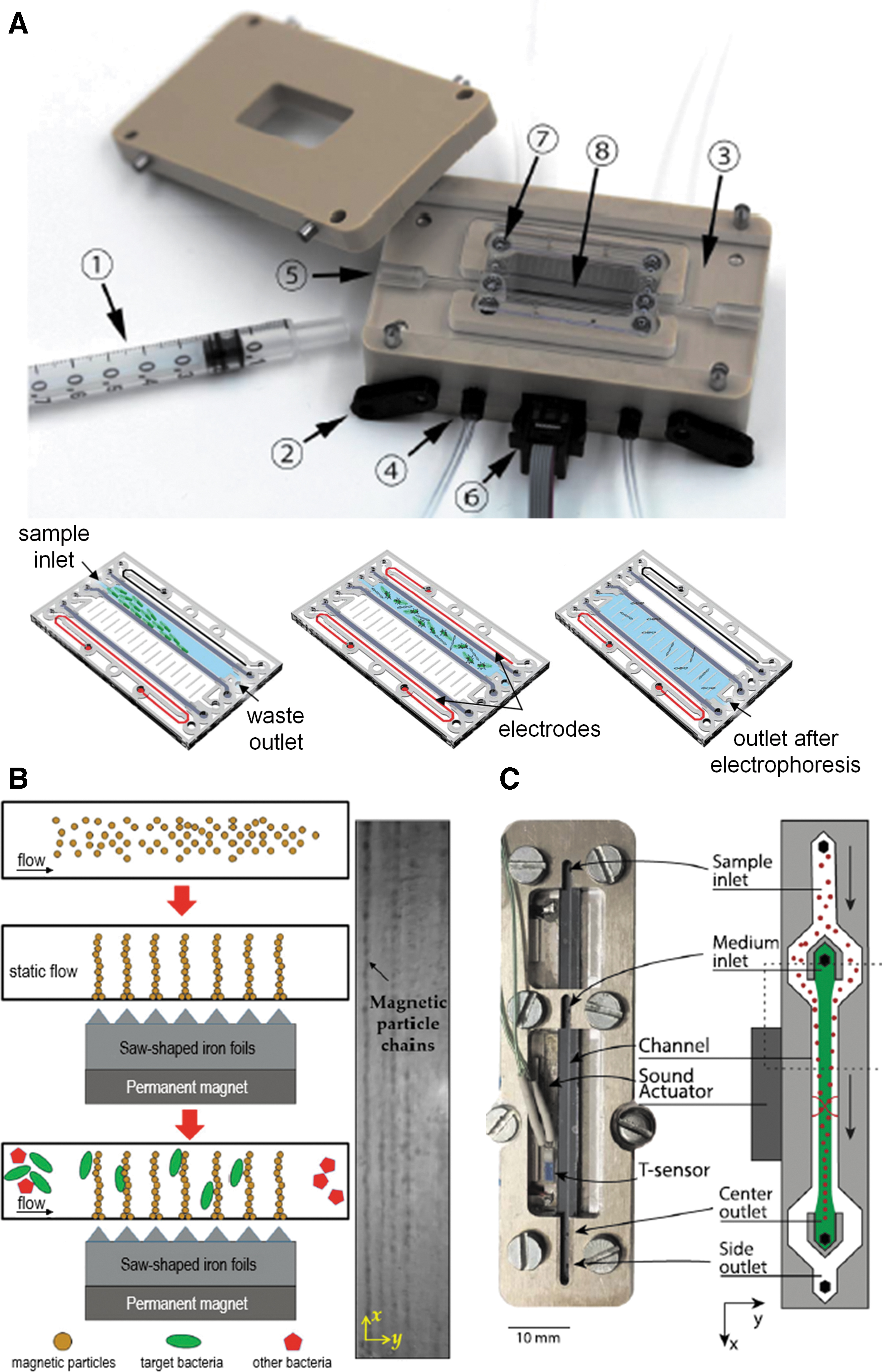
Examples of active microfluidic devices that have been used for bacteria sorting.
Electrophoresis, such as gel electrophoresis and capillary electrophoresis, is among the most widely used bioanalytical techniques for separation and enrichment of biospecies (Islam et al., 2012; Kartsova et al., 2021). Microfluidic electrophoresis uses instruments reduced in size down to the micrometer scale and can be easily coupled to other microelectronic devices (Ou et al., 2020). Therefore, biosensing systems operating with microfluidic electrophoresis have been increasingly reported. These systems include those that utilize optical sensors such as ultraviolet-visible (UV-Vis) absorbance (to detect protein fingerprinting of bacteria) (Markuszewski et al., 2003; Kartsova et al., 2021), fluorescence (Islam et al., 2017), and chemiluminescence (Liu et al., 2003).
Additionally, electrochemical sensors such as amperometry (Islam et al., 2012), cyclic voltammetry (CV) (Ino et al., 2009), and capacitively coupled contactless conductivity detection (Chau et al., 2020; Takekawa et al., 2021), surface sensors such as surface-enhanced Raman spectroscopy (SERS) (Chen et al., 2019), and mass spectra (MS) (Jender et al., 2020) have also been employed.
Another method applying an electric field to separate sample is dielectrophoresis, based on a nonuniform electric field applied to polarizable particles (including biospecies) (Fernandez et al., 2017; Kudr et al., 2017). It has been coupled to UV-Vis spectroscopy (Rahmani et al., 2018), fluorescence (Shangguan et al., 2015), Raman (Schröder et al., 2013), photoconductive electrodes and microscopy (Lin et al., 2010), amperometry (Goto et al., 2020), impedance (Wang et al., 2017, 2018), capacitance-based surface stress biosensors (Sang et al., 2016), trapping of biomolecules in nanopipette (key component of scanning ion conductance microscopy) (Ying et al., 2004; Chen et al., 2012), SERS (Cheng et al., 2014), surface plasmon resonance (SPR) (Galvan et al., 2018), quartz crystal microbalance (QCM) (Fatoyinbo et al., 2007), and antibiotic susceptibility tests (Chung et al., 2012; Wang et al., 2020).
The major challenge of these two methods is that the overall sampling effect essentially depends on the charges or dipole distribution on the sample, so the sampling process could be interfered with by other charged species in a complex matrix.
Immunomagnetic methods have been increasingly used in research laboratories, because they need the least amount of instrumentation compared with other active microfluidic methods, and the emerging nanotechnologies have made magnetic particles more available (Fig. 2B). IMS is based on the magnetophoresis of magnetic particles that capture the bacteria and carry them to desired locations. In a simple experimental setup, immunomagnetic particles (IMPs) are added into sample solution, and then the IMP–bacteria conjugates are extracted with a magnet, so it is easy to operate as long as the IMP is available.
Therefore, it has been increasingly used with various biosensing techniques, such as colorimetry (Jo et al., 2020), fluorescence (Kim et al., 2015), CV (Altintas et al., 2018; de Oliveira et al., 2018), amperometry (Laczka et al., 2011), impedance (Varshney et al., 2007), SERS (Walter et al., 2011), QCM (Han et al., 2011), and MS (Zhao et al., 2019) as a presampling process. The activity of the binding reagent (such as antibody or aptamer) coated on the surface of magnetic particles may be interfered with by the microenvironment on the particle surface and induce variations to the recovery rate of bacteria in quantitative analysis, but it still has the great potential for field-deployable sampling and biosensing tasks.
Acoustophoresis refers to the focusing or trapping of sample particles by acoustic waves (Fig. 2C). The acoustic method is less used in biosensing applications, because the design and operation require very specific knowledge of the acoustophoretic properties of samples and microchannels (Evander and Nilsson, 2012; Şahin et al., 2020). It has been used for biosensing studies with light scattering, fluorescence, Raman, and MS (Björn et al., 2009; Evander and Nilsson, 2012; Ngamsom et al., 2016), but its applications in cell/bacteria sampling are not as many as those of other active sampling methods.
Compared with other active techniques, the optofluidic method has even greater potential for miniaturized instrumentation, since its detection sensitivity can reach single-molecule level, with highly integrated optical, mechanical, and fluidic structures (Atajanov et al., 2018). The mechanical body of the optic fiber can function as both cell manipulation tool and signal sensing probe (Seeger et al., 2021). The controls on fluidics, target cells, and signaling can be achieved within a seamless and miniaturized flowing system, making optofluidic systems a great platform for label-free detection (Hunter et al., 2019; Lee et al., 2021).
Optofluidic devices are supported by the state-of-the-art technologies from fiber optics, laser, and nanoscale fabrication. Currently, most optofluidic methods are still under development, but they are among the most promising techniques for portable biosensors (Song et al., 2017).
Passive microfluidic sampling
The other type of microfluidic sampling method is passive microfluidics, which only utilizes fluidic movement inside microchannels for bacteria enrichment, therefore, no additional instrumentation or external energy input is required. These devices have simpler configuration, smaller size, and lower cost than active devices, since no additional machinery or power source is needed.
Typical methods under this category are deterministic lateral displacement (DLD), inertial focusing, laminar flow vortex, and hydrodynamic filtration. DLD and hydrodynamic filtration have been better investigated than others, with more biosensor applications (Yamada and Seki, 2005; Jusková et al., 2020; Yin et al., 2021). Inertial focusing and vortex-based particle separation are relatively new, yet they have drawn increasing attention from biomedical and environmental scientists, since these presampling methods are good choice for reusable sampling devices (compared with disposable chips), and make these biomedical/clinical devices “more economical, accessible, and sustainable,” which are especially important for field-deployable tasks or in places that lack resources (Natu et al., 2020).
Passive methods are based on careful design of microfluidic channels and precise control of flow rates, so the device development requires more background knowledge on hydrodynamics. Currently, most of these studies are conducted in research laboratories, and the biosensing technique mostly used for passive methods is optical imaging (Cruz et al., 2019; Narayanamurthy et al., 2020). The major challenges for passive enrichment devices are: (1) enrichment or separation efficiency can be very low for small particles, such as bacteria and submicron particles; (2) particle focusing and separation are very sensitive to flow rate and microchannel geometries, so careful designs and tests are needed; (3) many details of the hydrodynamic mechanisms behind the particle enrichment are still unknown, so it is difficult to predict the performance of a device for an unknown sample.
Examples of passive microfluidic sampling are shown in Figure 3. DLD and hydrodynamic filtration can also be classified into a more general category as micropillar array methods, because they utilize the pathways determined by micropillars or branch sidewall openings for size exclusion separation. The micropillars can be designed to have various shapes to influence the laminar flow and particle movement in the microchannel, to function as either filter barrier, lateral displacing machinery, or single-cell trap (Fig. 3A).

Examples of passive microfluidic devices that have been used for bacteria or cancer cell sorting/enrichment.
The major issues for this type of microfluidic devices are: (1) it is easily clogged, and difficult to be cleaned; (2) fabrication of these barrier structures with high aspect ratio is very challenging. For example, the height and shape of micropillars generated by photolithography may accumulate deviations or defects at micrometer scale, which in turn induce undesired flow behaviors or mechanical responses to the device, and eventually shorten the lifespan and increase the overall cost.
The inertial focusing is a relatively new method, which is based on the particle sorting along laminar flow generated by different channel geometries, and more details of its mechanism and applications can be found in the literature (Cruz et al., 2019; Zhao et al., 2020; Kalyan et al., 2021). Sometimes, an active method is combined with a passive method to enhance the overall sorting effect, especially in inertial focusing, as shown in Figure 3B. In this study, the sample needs the additional step of magnetic particle binding before entering the helix channel, but 3D printing fabrication was used to build additional multilayer structures above the substrate, so here the microfluidic part is still in a compact configuration.
The laminar flow can also generate vortices in microfluidic chamber when carefully designed. Currently, vortex sorters have been investigated for cancer cell detection (Fig. 3C) (Che et al., 2016), but it is more challenging to be used for separation of smaller cells, such as pathogenic bacteria. Although inertial focusing and vortex-based separation still need more theoretical investigations, they have great potential to be implanted into label-free biosensing platforms such as SERS and SPR for portable field detections.
Fabrication of Microfluidic Sampling Devices
The fabrication of devices at micro- or nanoscale essentially follow two directions, top–down and bottom–up, and they are combined in different ways for microfluidics (Gao et al., 2014; Scott and Ali, 2021). For example, micromilling and other micromachining methods use a more conventional top–down approach to make many layers of the devices from bulk materials, and bond these layers together to form the enclosed channel structures (Jung et al., 2007). Photolithography also builds device layer by layer, but repeats the circle of bottom–up deposition and top–down etching for each layer, and achieves very high spatial resolution (Martinez-Duarte and Madou, 2012). Soft lithography is of very high efficiency to make repeatable patterns, involving multiple steps of stamping or injection molding (Raj and Chakraborty, 2020). Paper and similar fiber materials are usually produced by a bottom–up approach. These are ideal choices for making disposable devices with extremely low cost, but they need to be assembled together for fluidic control (Lim et al., 2019).
One method worth more extensive study is the additive manufacturing, or 3D printing method (Bhattacharjee et al., 2016). It is a bottom–up approach in general, yet using precise control to form the right structure at the right location, and eventually build the device voxels by voxels through a continuous process (Wang and Pumera, 2020).
These three major fabrication methods are illustrated in Figure 4. Photolithography is a well-developed industry standard for semiconductor devices as well as microfluidic devices, with high spatial resolution and reliable quality control, yet it requires a cleanroom for its critical processes. On the contrary, soft lithography can be achieved with common laboratory equipment, so it has been used to make microfluidic devices with relatively low spatial resolution, which are mostly disposable. The biggest limitation is that the master template is still made by photolithography or other high-resolution methods to meet the basic requirements on spatial resolution.
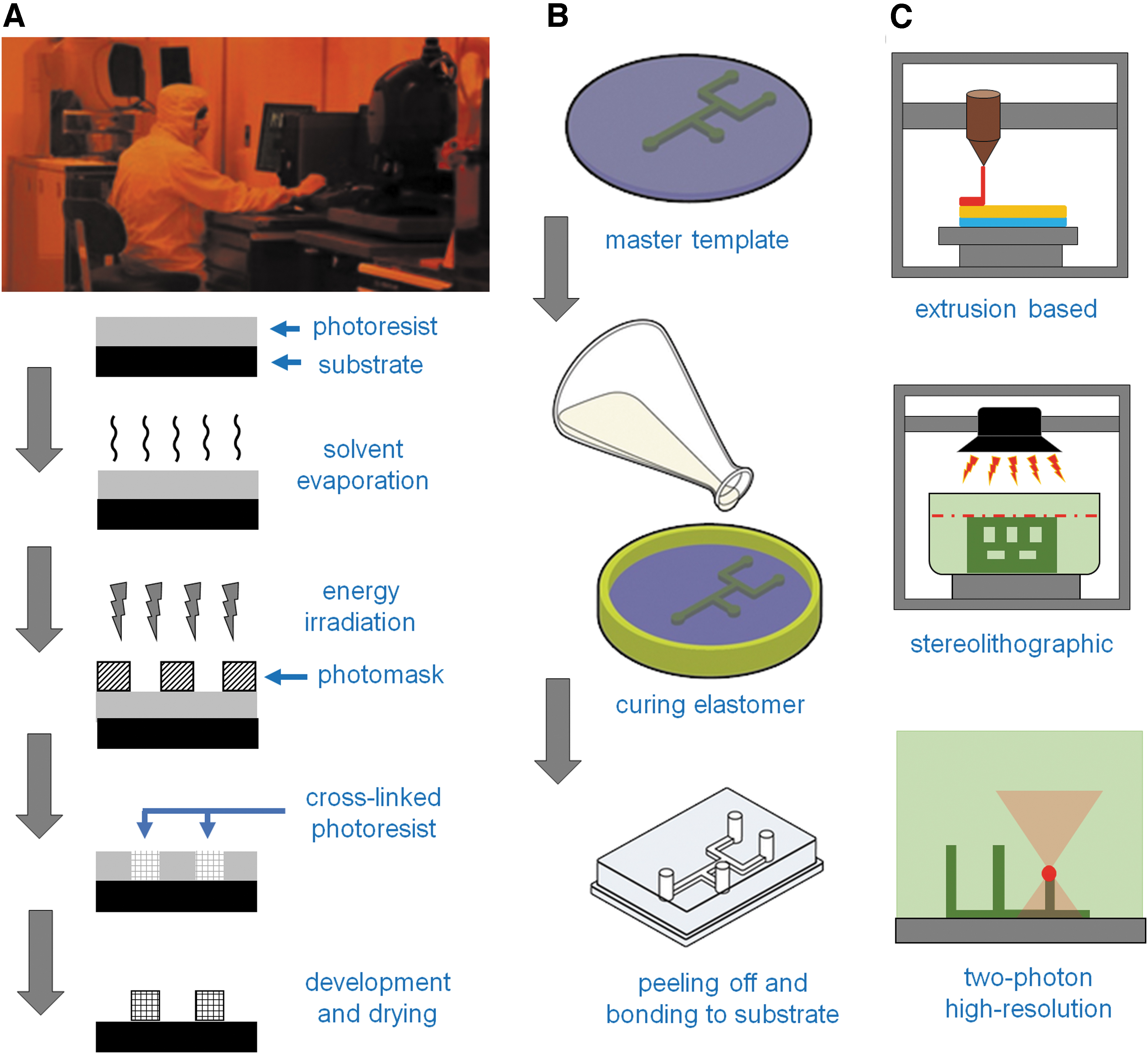
Typical methods used for microfluidic fabrication.
The 3D printing methods are under rapid development, with increasing applications in microfluidic fabrications (Lee et al., 2015; Jusková et al., 2020; Šakalys et al., 2021). Currently, the spatial resolution, printing speed, and instrumental cost have to be compromised with each other when printing functional devices (Macdonald et al., 2017), but novel techniques have been consistently making breakthroughs in high-resolution 3D printing (Bazaz et al., 2020b).
Out of research laboratories, micromachining techniques have been widely used in various manufacturing areas, so it is easier to access instrumental and technical supports from industry. Photolithography is a highly specific and highly professional area, so most of the fabrications are done by collaboration with large cleanroom facilities. Soft lithography and paper microfluidics have lower barriers to individuals, and it is a good choice for places that lack resources (Nguyen et al., 2018).
The 3D printing combined with computer design and simulation is certainly the best choice for rapid prototyping, which is a powerful tool to shorten the time for product design and manufacturing (Lim et al., 2014; Thomas et al., 2018). These techniques provide more freedom and flexibility to fabricate microfluidic devices, not in a remote and centralized factory, but in the individual laboratory that confronts a specific biosensing challenge (Naderi et al., 2019).
Applications in Bacteria Sorting and Enrichment
For pathogenic bacteria sampling and detection, microfluidic methods have provided miniaturized instruments and potential to improve sensitivity. Recent publications on microfluidic sampling for E. coli and Salmonella are shown in this study to discuss the progress and future directions. Literature has shown that E. coli is widely used as the model bacterium for microfluidic studies. For bacteria detection from food matrices or clinical samples, nonpathogenic E. coli has been used since most research work is limited to proof-of-concept studies; nonetheless, passive and active microfluidic methods have been coupled with a wide range of biosensing techniques and tested, such as electrophoresis, dielectrophoresis, IMPs, and acoustophoresis.
Figure 5 shows three studies using different microfluidic methods for E. coli sampling. The device in Figure 5A combined dielectrophoretic-FFF and fiber optics together in one streamline device. The active sampling in Figure 5B utilizes a multilayer design to provide a compact device, which matches the configuration of SERS instrument very well, but limits its application in field task because of the additional electric equipment used for electrophoresis.

Recent published studies using microfluidic devices for Escherichia coli enrichment and detection.
For other bacteria, such as Salmonella, most publications are still proof-of-the-concept studies, using fluorescent imaging or other optical detection instruments in a research laboratory (Fig. 6). It represents one major limitation for microfluidic application, especially for bacteria sampling, which is the lack of real application in field detection with real-world samples.
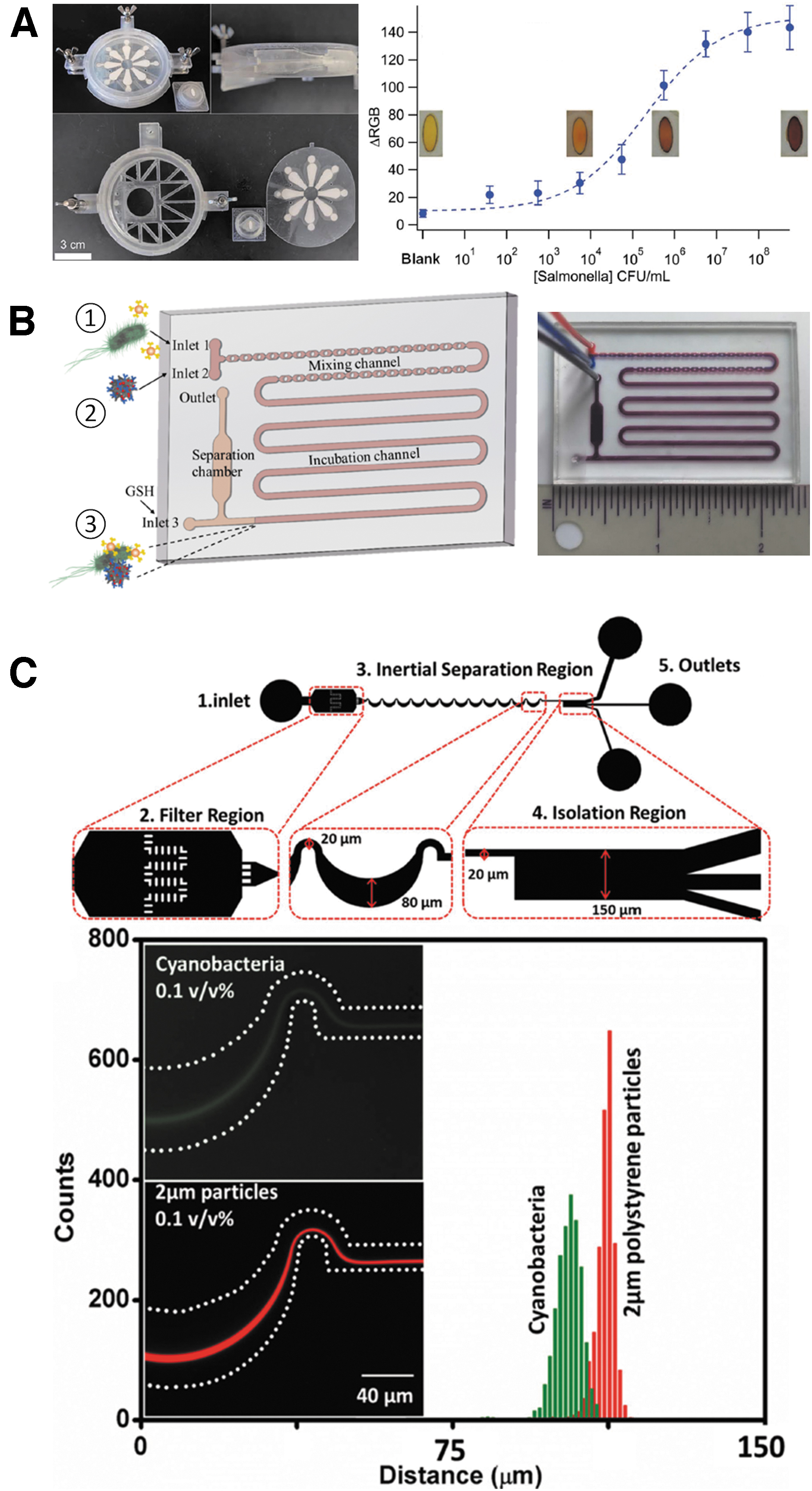
Recent published studies using microfluidic devices for enrichment and detection of Salmonella, cyanobacteria, and other bacteria.
The integration of microfluidic sampling device and label-free biosensing techniques, such as electrochemical sensor, SERS, and SPR, can provide the opportunity to make real portable biosensor with both functions of sample enrichment and rapid detection. The device in Figure 6A represents another direction, low cost, and easy-to-operate biosensing by smartphone. It could become a convenient method for rapid screening if the key parameters of target bacteria are well known for the sampling and biosensing system. Both the studies shown in Figure 6B and 6C used hybrid microfluidic systems that combine different mixing, sampling, and/or signal amplifying processes together in one continuous flow system, which is the right direction to develop portable devices for field detection.
A summary of different bacteria detections with their corresponding sensitivity or limit of detection (LOD) is shown in Table 2. This table only incudes the passive and active microfluidic enriching and the biosensing methods used for bacterial cell body biosensing, so the detections based on PCR or other molecular biology techniques are not included.
List of Publications in Recent 5 Years on Escherichia Coli, Salmonella, and Other Bacteria Detections, Using Passive and/or Active Microfluidic Sampling Methods Coupled to Cell Body Based Biosensing Techniques, with Corresponding Limit of Detection Value for Each Bacteria Sample
CFU, colony-forming unit; LOD, limit of detection; UV-Vis, ultraviolet-visible.
As can be seen in this table, some methods can obtain sensitivity values below 103 CFU/mL for bacteria in food sample, which is already difficult to reach in literature and even more challenging in practice (as shown in Table 2). A recent trend is combining multiple sampling methods together, such as magnetic separation and inertial focusing, to achieve high efficiency of enrichment (Yao et al., 2020). For the detection of bacteria in real food matrix, most LOD values are ranging from 102 to 104 CFU/mL, still showing large deviations, which is mainly caused by various interfering species in the food matrices, and it is exactly the challenge for detection in real-world samples.
Challenges and Prospective
In microfluidic systems, the major challenges for the sampling of pathogenic bacteria are (1) most of them have a size smaller than 5 μm, (2) their shapes are not spherical, and (3) bacteria can easily form aggregation. Therefore, it is difficult to predict their behaviors in hydrodynamic systems, and even more difficult to precisely control the movements of live bacteria. When developing high-throughput sampling devices for real-world unknown samples, it is important to distinguish various bacteria from the sample matrix and living cells from dead cells, so both theoretical and applied studies are required to focus on sorting particles of submicron size or below and with nonspherical shapes.
The hydrodynamic theories on microfluidics are not the focus of this review, but it is important to understand basic hydrodynamic behaviors of the bacteria sample when they are moving through the microfluidic device. With the advances of simulation hardware and software, computer simulation has been widely used to estimate the performance of various microfluidic designs (Bazaz et al., 2020a). Because the system is operating under laminar flow instead of turbulence flow, it is relatively easy to estimate the fluidic conditions if bacteria are simplified as spherical particle, but it has become increasingly critical to estimate the behavior of nonspherical bacteria in microfluidic theories and simulations since these studies will help reduce the overall cost of the device development in the laboratories (Kim and Klapperich, 2010; Naderi et al., 2019).
In summary, the outbreaks of various foodborne pathogens and other public health crisis have stimulated the fundamental research and real-world applications of POC and LOC devices. The combination of computer simulation and 3D printing fabrication could provide a more efficient way for rapid prototyping of microfluidic sampling and biosensing systems. In the future, these microfluidic devices may become critical in field tasks to monitor pathogenic bacteria in different stages of the food supply chain (also at a very specific point during production, transportation, or distribution), and provide rapid/local response to pathogenic bacteria in complex food matrix, potentially before the situation becomes a large-scale outbreak.
Footnotes
Disclosure Statement
No competing financial interests exist.
Funding Information
No funding was received for this article.