
Abstract
To investigate the epidemiology of Shiga toxin–producing Escherichia coli (STEC) in dairy cattle, 975 samples (185 feces, 34 silage, 36 cattle drinking water, 360 raw milk, and 360 teat skin swabs) were collected from two dairy farms in Baoji and Yangling, Shaanxi Province, China, and were screened for STEC. Whole-genome sequencing was used to analyze the genomic characteristics and potential transmission of STEC isolates. A total of 32 samples were contaminated with STEC, including 4.0% (19/479) in Farm A and 2.6% (13/496) in Farm B. Compared with adult cows (4.5%), nonadult cows had a higher rate (21.3%) of STEC colonization. A total of 14 serotypes and 11 multilocus sequence typing were identified in 32 STEC isolates, among which O55:H12 (25.0%) and ST101 (31.3%) were the most predominant, respectively. Six stx subtypes/combinations were identified, including stx1a (53.1%), stx2g (15.6%), stx2d, stx2a+stx2d, stx1a+stx2a (6.3%, for each), and stx2a (3.1%). Of 32 STEC isolates, 159 virulence genes and 27 antibiotic resistance genes were detected. Overall, STEC isolates showed low levels of resistance to the 16 antibiotics tested (0–40.6%), with most common resistance to ampicillin (40.6%). The phylogenetic analysis confirmed that STEC in the gut of cattle can be transmitted through feces. The results of this study help to improve our understanding of the epidemiological aspects of STEC in dairy cattle and provide early warning and control of the prevalence and spread of the bacterium.
Introduction
Shiga toxin–producing Escherichia coli (STEC) is an important foodborne pathogen, which can cause serious diseases, that is, watery diarrhea, hemorrhagic colitis (HC), and hemolytic uremic syndrome (HUS) (Mekata et al., 2014; Peng et al., 2019). The Shiga-like toxin (Stx) encoded by stx1 or/and stx2 genes is one of the main associated causes of pathogenicity of STEC (Ballem et al., 2020). The stx2-positive STEC is significantly associated with HC and HUS, especially subtypes stx2a, stx2c, and stx2d, and stx1-positive STEC is frequently implicated in the development of diarrhea (Amezquita-Lopez et al., 2018; Dong et al., 2017). At present, 13 different subtypes have been identified, including the 3 stx1 subtypes (stx1a-stx1d) and 10 stx2 subtypes (stx2a-2k) (Elsayed et al., 2021; Scheutz et al., 2012).
Ruminants, especially cattle, are considered to be the most important reservoirs for STEC (Borczyk et al., 1987; Gyles, 2007). As cattle lack the vascular globular globotriaosylceramide receptor required for Shiga toxin binding, it does not normally cause any disease (Persad and LeJeune, 2014). However, this pathogen can be excreted in their gastrointestinal tract through feces, causing contamination of the farm environment, raw milk, vegetables, and water (Farrokh et al., 2013; Hussein and Sakuma; 2005; Hussein, 2007). Contaminated raw milk and dairy products have been recognized as important vectors for the transmission of STEC to humans (Hussein and Sakuma, 2005; Jones et al., 2019). Worldwide, outbreaks of STEC by the intake of contaminated raw milk have been reported in Germany, the United States, and Canada (Borczyk et al., 1987; Martin et al., 1986; Mylius et al., 2018). Notably, Rosario et al. (2021) reported that STEC isolates were isolated from pasteurized dairy products in Brazil. Thus, monitoring the prevalence STEC from cattle is important for tracing the source of contamination, managing outbreaks, and reducing the risks for human infection (Mylius et al., 2018; Wang et al., 2011; Xiong et al., 2012).
Up to now, most studies have focused on the occurrence of STEC in feces from cattle, while there is a lack of elaboration on the prevalence and transmission of STEC in dairy cattle, raw milk, and the farming environment (Han et al., 2022; Peng et al., 2019; Tong et al., 2021; Zhang et al., 2022). Herein, the main aims of this study were to (1) investigate the prevalence, molecular characteristics, and antibiotic resistance of STEC from dairy cattle, raw milk, and farm environment and (2) assess the potential for fecal transmission of STEC isolates to the farm environment and raw milk.
Materials and Methods
Sample collection, isolation, and identification of STEC
From April 2019 to October 2020, 975 samples were collected from two dairy cattle farms (A and B) in Baoji and Yangling, Shaanxi Province of China, including 479 and 496 samples from Farm A (∼1000 cattle) and B (∼300 cattle), respectively (Supplementary Table S1). These samples were obtained from dairy cattle (feces, raw milk, and teat skin swabs) and farm environment (silage and drinking water for cows in shared troughs). Among them, feces samples from the recto-anal junction of calves (1–6 months), heifers (6–18 months), sick cattle (more than 24 months), and lactating cows (more than 24 months) were collected using sterile cotton swabs. Isolation and identification of STEC were performed by following the GB4789.6-2016 “Microbiological examination of food for Escherichia coli” (National Food Safety Standards of China) with some modifications. First, 25 g of sample was suspended in 225 mL of E. coli broth (EC) (Qingdao Hope Bio-Technology Co. Ltd.), and enriched 12–16 h at 42°C at 120–150 rpm. The enrichment broth was transferred into 20 mL sterile EC broth at a ratio of 1:100 and was incubated for 12–16 h at 42°C and at 180–220 rpm. STEC-positive samples were identified by screening for targeted genes stx1 and stx2 using polymerase chain reaction (PCR), as shown in Supplementary Table S2. Positive or suspected samples were subjected to 10-fold serial gradient dilutions, and 100 µL of 10−4–10−7 diluents were spread onto a MacConkey agar plate. Forty colonies were selected from plates with 30–300 colonies with pink or peach color (some samples with fewer colonies were all selected). These colonies were transferred to Luria–Bertani (LB) agar plates. PCR amplification was performed for each five mixed colonies; if the PCR identification was positive, single colonies were detected separately. Finally, a single colony was selected for storage and further analysis.
DNA extraction and whole-genome sequencing
The DNA of STEC isolates was extracted using the bacterial genomic DNA extraction kit (Accurate Bio-Technology Co. Ltd.). Genome sequencing was performed on an Illumina NovaSeq at the Beijing Novogene Bioinformatics Technology Co., Ltd. Libraries were constructed using the NEBNext® Ultra™ DNA Library Prep Kit for Illumina (New England Biolabs). The raw data were cleaned by Fastp v0.19.4. The clean reads were assembled using SPAdes (http://cab.spbu.ru/software/spades/) to obtain scaffold sequences with default parameters.
Genotyping, virulence and antibiotic resistance gene screening, and phylogenetic analysis
The O:H serotype and sequence type (ST) of each isolate were determined by comparing assemblies to the EcOH and multilocus sequence typing (MLST) databases with SRST2 v0.2.0, respectively (Inouye et al., 2014). The stx subtype was performed by using an in-house stx subtyping database with ABRicate v1.0.1 as described previously (Bai et al., 2022). The presence/absence of virulence factor and antibiotic resistance genes (ARGs) was annotated by comparison with data on the VFDB (http://www.mgc.ac.cn/VFs/) and ResFinder (https://cge.food.dtu.dk/services/ResFinder/) databases, using the ABRicate v1.0.1 with 80% identity and minimum length coverage of 50% as thresholds. To perform phylogenetic analysis, all STEC isolates together with three reference genomes of major STEC serotypes were used to identify the core single-nucleotide polymorphisms (SNPs) using Snippy v4.4.5. The obtained SNPs were filtered to avoid recombination using Gubbins v2.4.1 (Croucher et al., 2015). A final phylogenetic tree was generated by the maximum likelihood method using FastTree v1.4.3 (Price et al., 2010). The visualization of phylogenetic tree and carrying of genetic traits were displayed using iTOL v6 (Letunic and Bork, 2021). Transmission events were defined as isolates with core-genome SNP divergence of <20 (Sun et al., 2019).
Antibiotic susceptibility
The antimicrobial susceptibility of STEC isolates was performed using the agar dilution method as previously described (Zhang et al., 2021). The results were interpreted according to the Clinical and Laboratory Standards Institute (CLSI, 2020), Antibiotic and European Committee on Antibiotic Susceptibility Testing (http://www.eucast.org). The minimum inhibitory concentration of 16 antibiotics is listed in Supplementary Table S3, including ampicillin (AMP), amoxicillin-clavulanic acid (AUG), cefoxitin (FOX), cefotaxime (CTX), ceftazidime (CAZ), ceftriaxone (AXO), gentamicin (GEN), streptomycin (STR), kanamycin (KAN), trimethoprim-sulfamethoxazole (SXT), chloramphenicol (CHL), tetracycline (TET), ciprofloxacin (CIP), nalidixic acid (NAL), azithromycin (AZM), and polymyxin B (PB). Multidrug resistance (MDR) was defined as resistance to at least three classes of antibiotics. E. coli ATCC 25922 was used as positive control.
Statistical analyses
All statistical analyses were performed using R v3.6.1. The differences in the detection rate of categorical variables were conducted using χ2 test. The differences in the number of ARGs and resistance to antibiotics were conducted using the Wilcoxon or Kruskal–Wallis tests. p-Value <0.05 was considered a significant difference.
Results
Prevalence of STEC
In total, 3.3% (32/975) samples were contaminated with STEC, including 4.0% (19/479) in Farm A and 2.6% (13/496) in Farm B (Fig. 1A). There were no significant differences (p > 0.05) in the detection rates of STEC-positive samples between Farm A and Farm B. Furthermore, 32 STEC isolates were collected from 32 STEC-positive samples (1 isolate per sample). The detection rate of STEC isolates from feces samples (11.4%, 21/185) was the highest, followed by silage (2.9%, 1/34), cattle drinking water (2.8%, 1/36), raw milk (1.4%, 5/360), and teat skin swabs (1.1%, 4/360) (Fig. 1B). In addition, we found that STEC was more commonly isolated from young dairy cattle, among which 21.3% of the nonadult cows were colonized by STEC (calves [24.4%, 10/41] and heifers [17.6%, 6/34]), whereas 4.5% of the adult cows (lactating cattle [4.9%, 4/82], and the sick cattle [3.6%, 1/28]) were found to carry this pathogen (Fig. 1C).
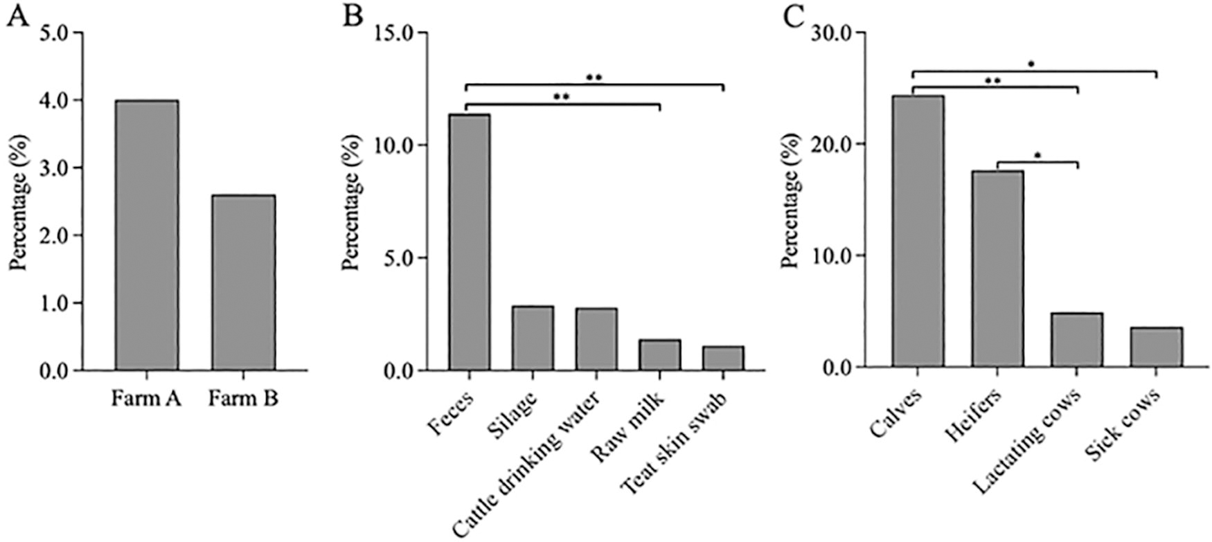
Prevalence of Shiga toxin–producing Escherichia coli (STEC) from dairy cattle, raw milk, and farm environment in Shaanxi Province, China.
O:H serotypes, MLST, stx subtyping, and virulence genes
A total 14 O-serogroups and 11 H-types were identified, among which O55:H12 was the most commonly detected (25.0%, 8/32), followed by O109:H16 and OgN12:H32 (12.5%, 4/32 for each), O136:H12 (9.4%, 3/32), O70:H31, O71:H10, and O100:H19 (6.3%, 2/32 for each), O2:H27, O8:H8, O26:H11, O54:H26, O111:H8, O156:H25, and O168:H8 (3.1%, 1/32 for each) (Table 1). Four serotypes were detected in the 13 isolates from Farm B, of which O55:H12 and O26:H11 serotypes were present only in Farm B.
Characteristics of 32 STEC Isolates from Dairy Cattle, Raw Milk, and Farm Environment in Shaanxi Province, China, in This Study
Stx subtype genes were not detected.
The presence of virulence genes eae, ehxA, efa1, paa, astA, ompA, and fimA is shown in this table, and the presence of other virulence genes is shown in Supplementary Table S2.
STEC, Shiga toxin–producing Escherichia coli.
A total of 11 STs were found in 32 STEC strains (Table 1). Among them, ST101 (31.3%, 10/32), ST329 (21.9%, 7/32), and ST10 (15.6%, 5/32) were the dominant STs. ST329 and ST101 were the predominant STs from Farm A (31.6%, 6/19) and Farm B (61.5%, 8/13), respectively.
Three stx subgroups were identified among STEC isolates, namely, stx1 (n = 20), stx2 (n = 10), and stx1+stx2 (n = 2) (Table 1). In particular, stx2-positive and stx1+stx2-positive isolates were only found from Farm A. Four stx genetic subtypes were identified in 29 (90.6%) of 32 STEC isolates, including 1 subtype of stx1 and 3 subtypes of stx2 (Table 1). All the stx1-positive strains belonged to the stx1a subtype. As for stx2-positive strains, the most common subtype was stx2g (50.0%, 5/10), followed by stx2d (20.0%, 2/10), stx2a+stx2d (20.0%, 2/10), and stx2a (10.0%, 1/10). In addition, the subtype of the stx1+stx2-positive strains was stx1a+stx2a.
In total, 159 virulence genes were identified among the 32 STEC isolates (Table 1 and Supplementary Table S2). The outer membrane protein A-encoding ompA was detected in all strains. Other main virulence genes were found as follows: fimA that encodes type 1 fimbriae (68.8%, 22/32), astA that encodes enteroaggregative heat-stable toxin (65.6%, 21/32), ehxA that encodes hemolysin (37.5%, 12/32), eae that encodes intimin and paa that encodes porcine attach and effacing associated (9.4%, 3/32 for each), and efa1 that encodes the enterohemorrhagic E. coli factor for adherence (6.3%, 2/32). In addition, STEC isolates with the same serotype exhibited similar or identical virulence gene profiles. For instance, all O55:H12 and OgN12:H32 strains carried astA, fimA, and ompA genes. Similarly, all O109:H16 strains carried astA and ompA genes. In addition, the O26:H11, O111:H8, and O156:H25 isolates carried the eae gene that encodes intimin. The detection rates of astA, fimA, and ehxA genes between Farm A and Farm B isolates were found to have significant (p < 0.05) differences. Among them, the detection rates of astA and fimA genes from Farm B were significantly (p < 0.05) higher than those from Farm A (Supplementary Table S4).
Antibiotic susceptibility, antibiotic resistance genes, and plasmids
The result of antibiotic susceptibility indicated that 53.1% (17/32) isolates were resistant to at least one antibiotic, and 31.3% (10/32) of isolates were of MDR. Overall, STEC isolates showed low levels of resistance to the 16 antibiotics tested (0–40.6%), including AMP (40.6%, 13/32), CTX and AXO (37.5%, 12/32 for each), GEN, KAN, TET, and CHL (25.0%, 8/32 for each), STR (21.9%, 7/32), NAL (9.4%, 3/32), and AUG and FOX (3.1%, 1/32 for each). All isolates were susceptible to CAZ, SXT, CIP, AZI, and PB. Compared with Farm B, Farm A had more STEC isolates that were resistant to a variety of antibiotic agents. Farm A-derived STEC isolates were more frequently resistant to STR, TET, and CHL than Farm B-derived isolates (Fig. 2A and Supplementary Table S5). However, no significant differences in the number of resistance antibiotics between the isolates from Farm A and Farm B were observed (Fig. 2B).
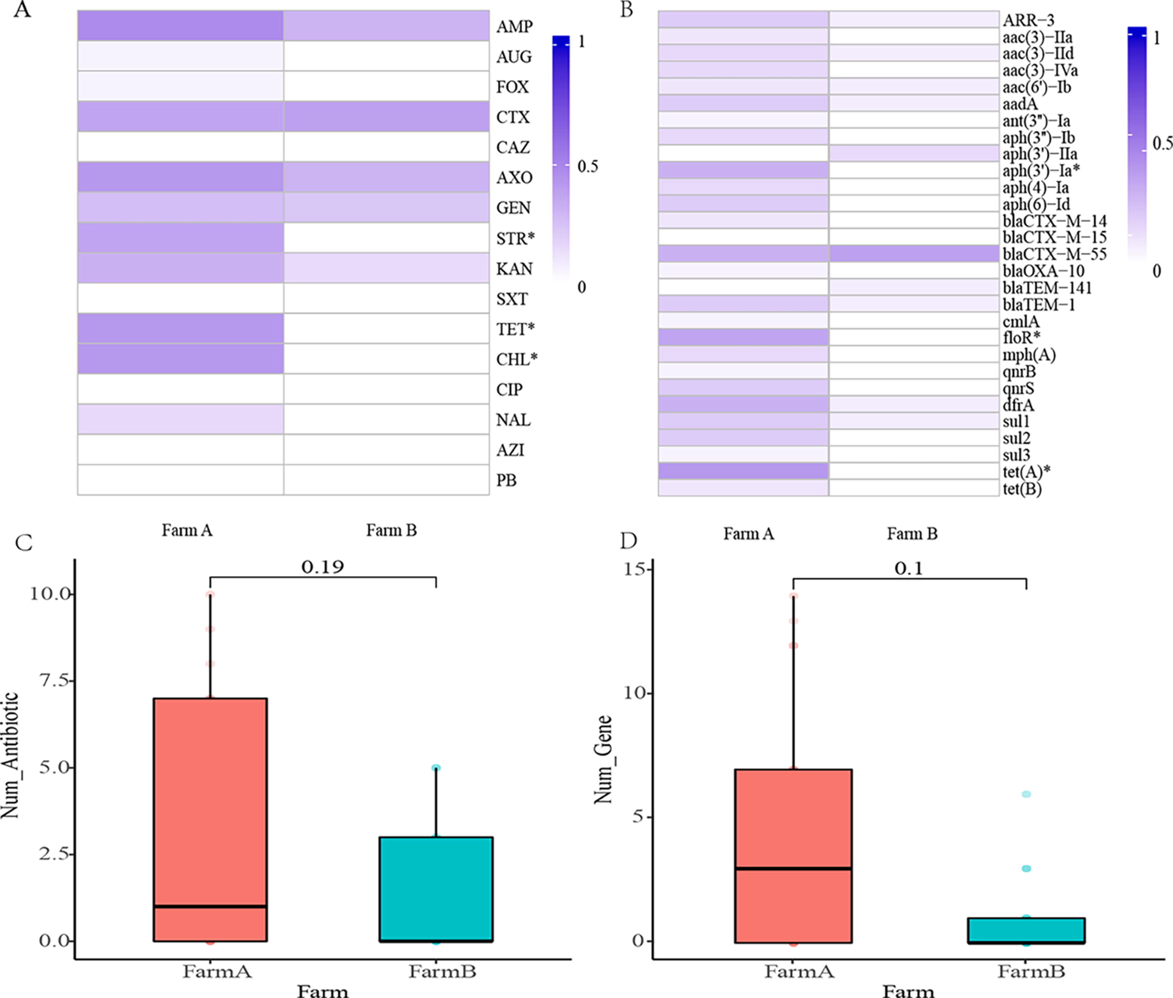
Antimicrobial resistance of Shiga toxin–producing Escherichia coli (STEC) from dairy cattle, raw milk, and farm environment in Shaanxi Province, China. Each cell in the heat map indicates the percentage of strains containing antibiotic
A total of 27 antibiotic resistance genes were detected. These antibiotic resistance genes were mainly involved in resistance to chloramphenicol (n = 2), aminoglycoside (n = 11), trimethoprim (n = 1), sulfonamides (n = 3), macrolide (n = 1), rifampin (n = 1), quinolones (n = 2), tetracyclines (n = 2), and β-lactam (n = 6). The most common resistance gene was blaCTX-M-55 (34.4%, 11/32), followed by tet(A) (31.3%, 10/32), dfrA and floR (21.9%, 7/32 for each), aph(3′)-Ia (18.8%, 6/32), ARR-3, aadA, blaTEM-1 , and sul1 (15.6%, 5/32 for each), and aac(3)-IId, aph(6)-Id, qnrS, and sul2 (12.5%, 4/32 for each). STEC isolates from Farm A carried 25 of the 27 antibiotic resistance genes, whereas the isolates from Farm B carried only 10 genes. In addition, significantly higher (p < 0.05) rates of aph(3′)-Ia, floR, and tet(A) in STEC isolates from Farm A than Farm B were found (Fig. 2C and Supplementary Table S6). No significant difference in the number of resistance genes was observed between isolates from Farm A and Farm B (Fig. 2D).
STEC isolates harbored 19 different plasmid replicons. All STEC isolates contained at least one and up to seven plasmid replicons, except three isolates from Farm B (Fig. 3). The most prevalent plasmid replicons were IncFIB (AP001918) (78.1%, 25/32), IncFIC (FII) (62.5%, 20/32), and IncI1 (34.4%, 11/32). Other plasmid replicons detected were Col (MG828) (21.9%, 7/32), IncHI2 and RepA (18.8%, 6/32 for each), Col440I, ColRNAI, and IncHI2A (15.6%, 5/32 for each), Col156 and IncFII (pHN7A8) (12.5%, 4/32), IncFIA, IncFII (pAMA1167-NDM-5), and IncQ1 (6.3%, 2/32 for each), and IncB/O/K/Z, IncFIA(HI1), IncR, IncX1, and p0111 (3.1%, 1/32 for each).

Genomic analysis of Shiga toxin–producing Escherichia coli (STEC) isolates from dairy cattle, raw milk, and farm environment in Shaanxi Province, China.
Phylogenetic analysis
To investigate the genetic relationship among dairy cattle, farm environment, and raw milk isolates, a maximum-likelihood phylogenetic tree was established using 185,251 core SNPs extracted from 35 STEC, including 32 isolates in this study and 3 STEC genomes downloaded from GenBank (Fig. 3). The results suggested that isolates with the same serotype or ST were more probable to cluster together. Genetic correlation showed that there were signs of STEC isolates spreading in the same cattle farm, which was demonstrated by isolates located on similar developmental branches. From Farm B, STEC isolate F1 recovered from heifers was closely related to F11 (calves), F5 (sick cows), and SX0704 (lactating cows) (6, 5, and 7 SNP difference, respectively). These isolates showed highly consistent characteristics in serotype, ST, stx, antibiotic resistance profiles, resistance gene profiles, and plasmid profiles. In addition, STEC isolate SX0702 from lactating cow feces at Farm B was closely related to the D3 isolate from teat skin swab of lactating cow (6 SNP differences).
Discussion
In this study, our results revealed that the overall detection rate of STEC in the dairy cattle, raw milk, and farm environment was 3.3% (32/975). The detection rate of STEC in cattle feces (12.3%) was higher than the other samples and was consistent with previous surveys in other regions of China, such as Jiangsu (12.9%), Xinjiang (12.8%), and Qinghai (11.68%) (Bai et al., 2013; Han et al., 2022; Tong et al., 2021). In contrast, the detection of STEC from feces was slightly higher than that in Hubei, Anhui, Hunan, and Henan (7.41%) and Shanghai (4.41%) (Peng et al., 2019; Zhang et al., 2022). Several studies in the rest of the world have shown the presence of STEC from the feces of healthy cattle, with estimates ranging from 1% to 54% (Ballem et al., 2020; Jaakkonen et al., 2019; Vasco et al., 2021; Venegas-Vargas et al., 2016). Multiple factors may be implicated in intrafarm prevalence, such as diet, farm size, geographical location, farm sanitation, as well as the age of animals (Ballem et al., 2020; Vasco et al., 2021). Animal age is one of the major factors affecting the prevalence of STEC (Ballem et al., 2020; Vasco et al., 2021). In this study, STEC was significantly enriched in young cows, mainly because of more diversity in the composition of the gut microbiota as the animals matured (Mir et al., 2016).
In addition, STEC isolates were detected in the farm environment and in raw milk samples but at a lower rate than in feces. This further indicates that STEC is mainly colonized in the intestines of cattle (Browne et al., 2018; Han et al., 2022; Vasco et al., 2021). Notably, our study found a small SNP difference (SNP < 10) between two STEC isolates from the lactating cow feces and teat skin swab of lactating cow at Farm B, respectively. Also, the same STEC strain (SNP < 10) was isolated from fecal samples of four different cows. During the sample collection, we observed that the cattle were resting lying on the bedding materials most of the time. Ma et al. (2017) proposed that the soil environment was the main depository of STEC, especially in bedding materials. Once STEC is present in the soil, it may survive for a long-term phase (Ma et al., 2017; Ramaite et al., 2022). Based on the phylogenetic tree results, we inferred that the STEC in the gut of cattle can be transmitted through feces and contaminate the farm environment, even raw milk. Although the detection rate was low, the presence of STEC in raw milk increased the risk of transmission through the food chain (Ranjbar et al., 2018; Rosario et al., 2021). For O55:H12 STEC strains of Farm B, we found genetic differences (SNP > 20) between STEC in the farm environment and STEC in feces. Also, some O55:H12 STEC strains from the farm environment showed to be MDR, probably because of minor genetic mutations to adapt to the different environment within Farm B (Dong et al., 2017). Therefore, monitoring the prevalence of STEC in cattle can provide early warning and control of the prevalence and spread of the bacterium.
The serotype is often used to assess the virulence of STEC. In this study, O26 and O111 out of the major 7 STEC serogroups were detected. Although not all O26 and O111 STEC strains have been found to be responsible for human disease, it is worth noting that these strains harbored simultaneously eae and ehxA genes and exhibited MDR, which may pose a threat to public health (Haugum et al., 2014; Hua et al., 2021; Ranjbar et al., 2018). Furthermore, uncommon serotypes are routinely discovered in ruminants from other regions with the advent of whole-genome sequencing (WGS) (Huang et al., 2021; Zhang et al., 2022). In this study, we found that O55:H12 and O109:H16 were the most prevalent serogroups, different to other studies, in which the prevailing were O8:H16 from the Qinghai–Tibetan Plateau in China (Bai et al., 2013), O39 from the Shandong Province in China (Dong et al., 2020), O29:H12 in Northern Portugal (Ballem et al., 2020), and O5 in India (Mahanti et al., 2015). At present, there are no reports of O55:H12 and O109:H16 STEC causing human disease. However, STEC strains of types O55 and O109 with the same O serotype have been reported to cause a series of outbreaks, such as 43 cases of O55 STEC in England between 2014 and 2018 (Hua et al., 2021; Sawyer et al., 2021). The emergence of cases caused by uncommon serotype STEC indicates that water and foodstuffs contaminated with ruminant feces may pose a threat to human safety.
Both stx1 and stx2, which are required to encode Shiga toxin, are the most essential and prevalent virulence factors of STEC (Ballem et al., 2020). In this study, stx1-positive STEC isolates were frequently detected from Farms A and B and only included stx1a subtype. Epidemiological studies have shown that the stx1-positive STEC was observed with high prevalence from cattle in China, and stx1a was the only present subtype (Peng et al., 2019; Tong et al., 2021). Previous studies have discovered that the majority of STEC isolated from diarrheic calves only produce stx1, while stx2-positive strains are the predominant type in healthy calves (Kohansal and Ghanbari Asad, 2018). Notably, stx1a subtype STEC was prevailing in diarrhea patients in China (Bai et al., 2016). In addition, stx2a, stx2d, and stx2g subtypes were detected in this study, which were associated with HC and HUS (Fuller et al., 2011; Prager et al., 2011). It is also believed that Shiga toxin by itself is not adequate to trigger serious disease but should be accompanied by other toxins to lead to more severe diseases. The intimin-encoding eae gene is an important adhesin and a marker of high virulence and severe disease (diarrhea, bloody diarrhea, and HUS), especially when accompanied with stx2a (Huang et al., 2021). Differently, we found that the eae gene only existed in stx1-positive isolates, which was consistent with the results of other studies in China, Spain, and India (Blanco et al., 2004; Han et al., 2022; Mahanti et al., 2015; Zhang et al., 2022). Furthermore, about other essential adhesion-encoding genes and hemolysin gene, a high proportion of lpfA (84.4%), ompT (62.5%), iha (25.0%), and ehxA (40.6%) genes were detected in our eae-negative STEC isolates. These genes promote adhesion and colonization and provide the chemical requirements for STEC growth and survival (Jajarmi et al., 2017; Han et al., 2022). The correlations of those subtypes and main virulence genes in STEC were detected in dairy cows and farm environments, increasing the potential risk to cause severe diseases and outbreaks.
The antibiotic resistance of STEC has become a worldwide issue. The antibiotics, including β-lactam, tetracycline, chloramphenicol, and aminoglycoside, are widely used to eliminate or deal with bacterial infections on dairy farms, which has produced high rates of resistance and even the emergence of MDR bacteria (Angappan et al., 2021; Cai et al., 2021; Elsayed et al., 2021; Mahanti et al., 2015; Peng et al., 2019). In addition, the high prevalence of resistance against trimethoprim, sulfamethoxazole, and fluoroquinolones of STEC isolates was reported in China (Peng et al., 2019). In this study, the isolates from Farm B were sensitive to streptomycin, tetracycline, and chloramphenicol antibiotics. These disparities could be attributed to the type and frequency of antibiotics used in different dairy farms (Montso et al., 2019). Moreover, studies have demonstrated that STEC can obtain antibiotic resistance genes through plasmid horizontal transfer when cultured in cow feces, storm water, or rumen fluid in vitro assay (Mir and Kudva, 2019; Poole et al., 2017). STEC could survive for several months in the rectoanal junction and feces of cattle, increasing the probability that STEC acquire or spread resistance genes (Mir & Kudva, 2019). Notably, we identified one raw milk-derived STEC isolate from Farm A that was resistant to the 10 antibiotics analyzed. Other studies also have reported the presence of MDR STEC isolates in raw milk, dairy products, and retail meat (Hu et al., 2021; Ranjbar et al., 2018). In addition to the use of antibiotics in the feeding process, infected staff in milking, dairy production, and meat processing have been implicated as important contributors to the contamination of food with MDR STEC isolates (Momtaz et al., 2013; Ranjbar et al., 2018).
Conclusion
This study demonstrated the presence of STEC contamination in dairy cattle, raw milk, and farm environment samples in Shaanxi Province, China. The intestinal tract of cattle was the reservoir for STEC. Animal age was an important factor impacting the prevalence of STEC. The STEC colonizing the gut of cattle can be transmitted through feces and contaminate farm environments and cow udder. Although direct contamination of raw milk has not been confirmed, the low detection rate of STEC in raw milk suggests that there is a risk of transmission of this bacterium along the food chain. Further molecular characterization of STEC is needed in the future to fully assess the virulence potential and ability of STEC to cause human disease.
Footnotes
Authors’ Contributions
P.Z.: Conceptualization, investigation, methodology, formal analysis, and writing—original draft. L.L.: Conceptualization, formal analysis, and writing—original draft. H.S.: Formal analysis and writing—original draft. M.Z.: Conceptualization, data curation, and methodology. T.W.: Conceptualization and data curation. G.C.: Investigation and language. Y.W.: Investigation. L.B.: Investigation, formal analysis, resources, and project administration. X.W.: Conceptualization, data curation, investigation, formal analysis, resources, project administration, and writing—reviewing and editing.
Disclosure Statement
The authors of the article declare that they have no conflicts of interest.
Funding Information
This research was supported by the Sichuan Natural Science Foundation (No. 2023NSFSC0178) and the National Natural Science Foundation of China (No. 31871894 and 31271858).
Supplementary Material
Supplementary Table S1
Supplementary Table S2
Supplementary Table S3
Supplementary Table S4
Supplementary Table S5
Supplementary Table S6
References
Supplementary Material
Please find the following supplemental material available below.
For Open Access articles published under a Creative Commons License, all supplemental material carries the same license as the article it is associated with.
For non-Open Access articles published, all supplemental material carries a non-exclusive license, and permission requests for re-use of supplemental material or any part of supplemental material shall be sent directly to the copyright owner as specified in the copyright notice associated with the article.