
Abstract
Alpha-hemolysin (Hla) is a major virulence factor secreted by Staphylococcus aureus (S. aureus), which can lyse a variety of mammalian cells and help bacteria evade the host immune system or antibiotics, posing a safety hazard to human health. Therefore, it is critical to establish a quick-responsive and sensitive method for Hla detection to ensure food safety. In this work, a dual-mode immunoassay was developed with both colorimetric and fluorescent readouts for discriminative detection of Hla. The proposed sensing system consists of p-phenylenediamine (PPD) and fluorescein, where fluorescein functions as a fluorescent reporter, and PPD serves a dual function as a colorimetric reporter and fluorescence quencher. Subsequently, the reaction system of this method was optimized, and the detection limit, sensitivity, and specificity were evaluated. Under optimal conditions, the proposed method possesses excellent analytical performance in the range from 0.5 to 500 ng/mL with a limit of detection as low as 0.5 ng/mL. Noteworthy, this method was successfully employed for the detection of Hla in milk with good selectivity and high accuracy. Overall, the dual-mode immunoassay provides a superior platform for the on-site, quantitative, and accurate detection of Hla in food samples.
Introduction
S
In addition, hla-positive S. aureus is one of the most significant contributors to bovine mastitis, increasing the risk of contamination of milk and dairy products (Bergonier et al., 2003; O'Reilly et al., 1986). Previous studies have reported that hla gene was the most often discovered virulence gene from raw milk in Xinjiang (96.9%), Korea (100%), and Egypt (54.9%) (Jung and Lee, 2022; Ren et al., 2020; Sadat et al., 2022). S. aureus strains can be eliminated through pasteurization, but its toxins can linger and cause foodborne diseases (Wang et al., 2012; Xing et al., 2016). With the increased consumption of dairy products, the rapid detection methods to assess Hla contamination in milk are needed to reduce foodborne disease outbreaks (Dai et al., 2019; Sumner, 2021).
To date, there is a relative lack of assays for Hla, mainly including Western blot (WB), enzyme-linked immunosorbent assay (ELISA), biosensors, etc. (Reddy et al., 2013; Weston et al., 2020). Although WB is a highly specific method for protein detection, it is costly, cumbersome, time consuming, and requires specific instrumentation, which dramatically limits its applications in quantitative and field testing.
Biosensors such as electrochemical sensors, erythrocyte-camouflaged biosensors, and surface plasmon resonance have been introduced for the detection of Hla in serum samples (Anderssonet al., 2020; Kim et al., 2021; Vakyly et al., 2022; Weston et al., 2020). These methods require complex material synthesis processes, professional signal transduction instruments, and skilled technicians, and have not been proven in complex food samples. Hence, it is necessary to develop a rapid and sensitive method for Hla detection in foods.
In recent years, ELISA draw more substantial research attention because of its high-selective, simple operation, cost-effectiveness, rapidness, and visual identification without special instruments, and it has been widely applied to the detection of various toxins (Chen et al., 2021). However, the traditional single-model signal method inevitably has systematic errors due to environmental factors, leading to the uncertainty of the results (Bao et al., 2021; Han et al., 2020; Hu et al., 2023).
Fluorimetry is often used to enhance the detection sensitivity in biological systems due to its simplicity, intuitiveness, high sensitivity, and extraordinary photostability (Chu et al., 2020; Yan et al., 2017; Yang et al., 2021). The coupling of fluorescence and colorimetric immunoassay could effectively guarantee the accuracy and efficiency of the detection (Wang et al., 2020).
As a benzene derivative, p-phenylenediamine (PPD) can be used as chromogenic substrate, and its oxidation product is 2,5-diamino-N, N′-bis(p-aminophenyl)-l,4-benzoquinone di-imine (PPDox) (Jiao et al., 2000; Shi et al., 2021). Compared with the commonly used chromogenic substrate 3,3′,5,5′-tetramethylbenzidine (TMB), PPD has superior water solubility, and its oxidation product PPDox is more stable than TMBox, which is beneficial to the accuracy of detection results (Chen et al., 2019).
Fluorescein sodium is an organic substance with stable fluorescence and quantum yield of ∼0.92, whose fluorescence spectrum overlaps with absorption spectrum of PPDox, which lead to fluorescence quenching (Sehrawat et al., 2022; Sun et al., 2018). At the same time, the color of the solution changes from colorless to amaranth with the oxidation of PPD. This strategy provides both highly sensitive fluorescence analysis and visualization of the assay. Therefore, a fluorescence and colorimetric dual-mode immunoassay based on the inner filter effect (IFE) strategy for highly sensitive detection of Hla was proposed, in which fluorescein functions as a fluorescence reporter, and PPD performs a dual role as a colorimetric reporter and fluorescence quencher.
Materials and Methods
Plasmids, strains, and growth conditions
The plasmids and strains used in this study are listed in Supplementary Table S1. S. aureus strain HN85 is hla positive and was cultured in trypticase soy broth (TSB) medium at 37°C. The Escherichia coli strains carrying the protein expression plasmid (pCold I) were cultured in ampicillin (100 μg/mL)-containing Luria-Bertani (LB) medium at 37°C.
Chemicals and materials
QuickCut enzyme (NdeI and BamHI), pCold I plasmid, and 2 × PrimeSTAR max premix were obtained from Takara Biomedical Technology Co., Ltd. (Dalian, China). The ClonExpress MultiS one-step cloning kit was obtained from Vazyme Biotech Co., Ltd. (Nanjing, China). Isopropyl-beta-d-thiogalactopyranoside (IPTG) and Peroxidase AffiniPure Goat Anti-Rabbit IgG were purchased from Shanghai DEEYEE Biological Co., Ltd. (Shanghai, China).
Freund's complete adjuvant and Freund's incomplete adjuvant were obtained from Sigma-Aldrich Co., Ltd. (St. Louis, MO). Ni-NTA His Bind Resin was obtained from 7 Sea Biotech Co., Ltd. (Shanghai, China). Anti-alpha-hemolysin antibody[8B7]-N-terminal was purchased from Abcam Co., Ltd. (Cambridge, the United Kingdom). Ampicillin, phosphate-buffered solution (PBS, 0.01 mM, pH 7.2–7.4), Triton X-100, and dialysis membranes were purchased from Beijing Solarbio Science and Technology Co., Ltd. (Beijing, China).
PPD and fluorescein sodium salt were procured from Aladdin Biochemical Technology Co., Ltd. (Shanghai, China). The recombinant proteins of S. aureus beta-hemolysin (Hlb), gamma-hemolysins (HlgA, HlgB, and HlgC), and delta-hemolysin (Hld) were prepared by the Microbiology Laboratory in the College of Food Science and Engineering, Northwest A&F University (Yangling, China).
Apparatus and characterization
Bacterial fragmentation was performed using Bioruptor® Pico sonication device (Diagenode). Absorption and fluorescence spectra were performed using the spark™ Multimode Microplate Reader (Tecan) and a Hitachi F-7000 fluorescence spectrophotometer instrument (Tokyo, Japan), respectively. WB images were obtained using a Champ Chemi 610 Plus chemiluminescence imaging system (Tanon, Shanghai, China). Agarose gel electrophoresis and sodium dodecyl sulfate polyacrylamide gel electrophoresis (SDS-PAGE) images were acquired using a GE1DOC XR+ (Bio-Rad).
Cloning, expression, and purification of recombinant Hla protein
The Hla nucleotide sequence was amplified from the genomic DNA of S. aureus HN85 strain, using primers listed in Supplementary Table S2. The Hla gene was cloned into the pCold I plasmid digested by NdeI and BamHI through Gibson assembly (Gibson et al., 2009), which was transformed into E. coli BL21 (DE3) cells.
Positive clones of E. coli pCold I-hla-BL21 (DE3) were confirmed through polymerase chain reaction (PCR) and single sequencing (Supplementary Material S1). Next, the expression of the transformants with an N-terminal 6-His tag was induced by 0.1 mM IPTG at 16°C, and the recombinant Hla proteins were purified by Ni-NTA His Bind Resin. Finally, the purity and specificity of Hla were analyzed by SDS-PAGE and WB, respectively.
Hemolytic assay
The hemolytic activity of Hla was detected by the method described by Chang et al. (2023). In brief, different concentrations (80, 40, 20, 10, 5, 2.5, 1.25, 0.63, 0.32, 0.16, 0.08, and 0.04 μg/mL) of Hla were mixed with equal volumes of 6% (vol/vol) rabbit, mouse, or sheep erythrocytes, respectively, in the 96-well plates and cultured statically at 37°C for 3 h. After centrifugation (800 × g, 5 min), the optical density (OD) measurements of 100 μL supernatant were performed at 405 nm. Positive and negative controls were PBS and 0.1% Triton X-100, respectively. The percentage of hemolysis was calculated as follows:
where A represents OD value of sample tested, N represents OD value of negative control, and P represents OD value of positive control.
Preparation of rabbit polyclonal antibody against Hla
The antibody production protocol was reviewed and approved by the Animal Ethics Committee of Northwest A&F University (Approval No. XN2023-0515; Approval date March 3, 2023). Rabbit polyclonal antibody (pAb) against Hla was produced as described by Wardenburg et al. (2007), with some modifications. The 14-week-old New Zealand White rabbits (Chengdu Dossy Experimental Animal Co., Ltd., Chengdu, China) were subcutaneously injected with 20 μg of Hla emulsified in Freund's complete adjuvant for the initial vaccination.
Subsequently, four booster immunizations were performed with 20, 30, 30, and 60 μg of Hla emulsified by Freund's incomplete adjuvant at biweekly intervals. One week after the last inoculation, the whole blood was collected from rabbits by carotid cannulation and centrifuged at 3000 × g for 10 min. The antiserum was purified through the ammonium sulfate-octanoic acid combined method (CA-AS) and Protein A agarose (Smart-Lifesciences, Changzhou, China) (Naveed, 2019). Finally, the purity, specificity, and sensitivity of the pAb were identified by SDS-PAGE, WB, and indirect ELISA, respectively.
Establishment of the colorimetric and fluorescence immunoassay for Hla
Immunoassay procedure
The assay procedure for the dual-mode immunoassay was carried out as follows (Fig. 1): First, 96-well microplates were coated with 100 μL/well of anti-Hla mAb (1 μg/mL in 50 mM carbonate buffer) and incubated overnight at 4°C. The plates were washed three times with 200 μL/well PBST (PBS containing 0.05% Tween-20), and then blocked with 200 μL/well of blocking buffer (PBST containing 5% milk powder) for 2 h at 37°C to reduce nonspecific binding. After three washes, samples were added at 100 μL per well and incubated at 37°C for 1 h.

Schematic illustration of dual-signal detection of Hla based on IFE. Hla, alpha-hemolysin; IFE, inner filter effect.
After a reapplied washing step, 100 μL/well of anti-Hla pAb (2.5 μg/mL) was introduced and incubated at 37°C for 1 h. The plates were washed again, after which the HRP-Ab (100 μL/well, diluted 1:5000 in 0.01 mol/L PBS) were added and incubated for 1 h at 37°C. After the final washing, the substrate buffer was introduced (100 μL/well) and incubated for another 15 min at room temperature to perform the enzymatic reaction. The fluorescence intensity (excitation, λ = 490 nm, emission, λ = 515 nm) and OD values at 530 nm were recorded by a TECAN multimode microplate reader.
Optimization of experimental conditions
The optimal pH of the substrate buffer was determined by referring to the following procedures. First, 30 μL PPD (10 mM), 30 μL H2O2 (10 mM), 90 μL HRP (100 ng/mL), and 30 μL phosphate buffer (0.2 M, pH = 2.0, 3.0, 4.0, 5.0, 6.0, 7.0, 8.0, 9.0, 10.0, and 11.0) were mixed into ultrapure water in a final volume of 300 μL. After incubation for 15 min, the OD530nm values were determined using a multimode microplate reader to assess the effect of pH on the color reaction.
According to the above procedure, the final concentrations of PPD (0.5, 1.0, 1.17, 1.33, 1.5, and 1.67 mM) and H2O2 (0.5, 1, 1.33, 1.67, 2, 3, and 4 mM) in the substrate solution were adjusted, and then the OD530nm value was utilized as a criterion to select the optimal conditions. As the key experimental parameters of immunoassay, the concentrations of anti-Hla mAb (0.25, 0.5, 1, 2.5, 5, and 10 μg/mL), pAb (0.25, 0.5, 1, 1.25, 2.5, 5, 10, and 20 μg/mL), and HRP-Ab (1:1000, 1:2500, 1:5000, 1:7500, 1:10,000, and 1:15,000) were optimized to improve the sensitivity of the method.
The effect of these parameters was evaluated by the P/N values (where P and N represent the OD530nm values of positive and negative samples, respectively) and the fluorescence intensity ratio I/I0 values (I and I0 were the fluorescence intensities of test samples and blank samples, respectively).
Evaluation of the established dual-mode immunoassay
To determine the cutoff values for the dual-mode immunoassay, a total of 32 supernatants of hla-negative S. aureus were detected with the assay. Based on the OD530nm values and I/I0 values at 515 nm obtained by measuring this set of samples, the means (
Hlb, Hld, HlgA, HlgB, and HlgC were utilized to assess the specificity of the dual-mode immunoassay. The extent of crossreactivity was evaluated based on the difference in the OD530nm values and the I/I0 values at 515 nm.
To determine the sensitivity of the dual-mode immunoassay, 12 different concentrations of Hla (0, 0.1, 0.5, 1, 2.5, 5, 10, 25, 50, 100, 500, and 1000 ng/mL) were tested. Calibration curves were obtained by plotting the linear relationship between the OD530nm values or the I/I0 values at 515 nm and the Hla concentration. And the limit of detection (LOD) was calculated as 3 SD/slope (SD represents the standard deviation of the blank samples, slope is the slope of the linear regression equation).
Recovery analysis
To investigate the practicability of the dual-mode immunoassay in food samples, different concentrations of Hla (0, 1, 5, 10, 25, 50, 100, and 250 ng/mL) were spiked into Hla-negative pasteurized milk samples to carry out the recovery experiments. Each sample was performed in triplicate. The recovery of Hla was calculated by referring to the method described by Godefroy et al. (2018): Recovery (%) = [(concentration detected in spiked samples)/(added concentration)] × 100%.
Statistical analysis
The experimental data were exhibited as mean (
Results and Discussion
Expression, purification, and hemolytic activity of Hla
The results of PCR (1100 bp, including the 882 bp Hla sequence with the signal peptide removed and 217 bp plasmid sequence) and sequencing confirmed that the E. coli pCold I-hla-BL21 (DE3) expressing strain was successfully constructed (Fig. 2A). After purification by Ni-NTA His Bind Resin, the Hla recombinant protein with a molecular weight of ∼35 kDa (including plasmid fusion sequence) was successfully purified (Fig. 2B). Then, the purified protein was analyzed by WB using both anti-His and anti-Hla antibodies. As expected, specific bands were clearly observed at ∼35 kDa (Supplementary Fig. S1), indicating good antigenicity of the protein (Gouaux, 1998).
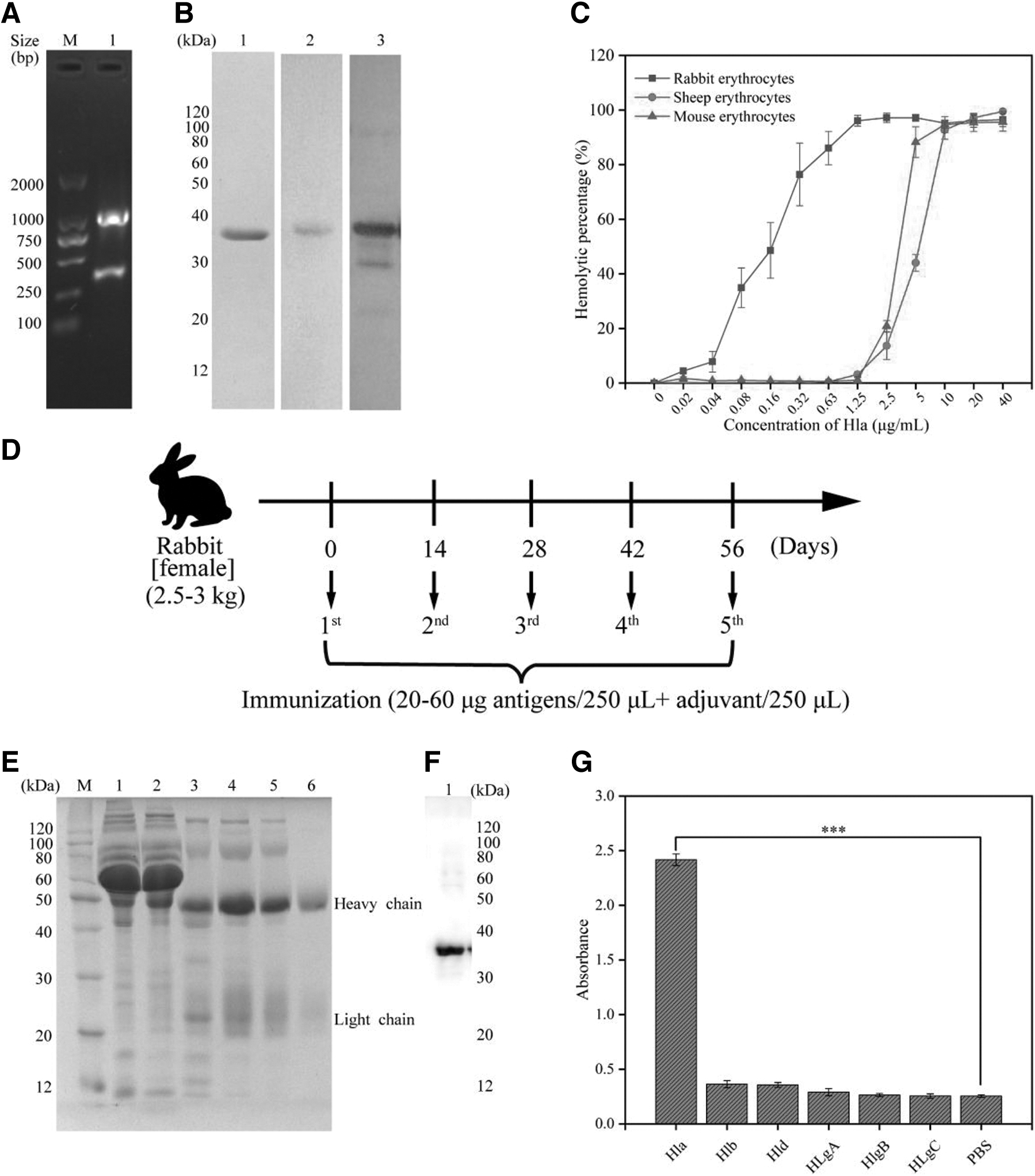
The hemolysis results of Hla on different species (sheep, rabbit, and mouse) erythrocytes are shown in Figure 2C, in which Hla was most sensitive to rabbit erythrocytes, followed by mouse and sheep erythrocytes. The species specificity for erythrocytes of Hla was attributed to the host cell A Disintegrin And Metalloprotease 10 (ADAM10) content, a high-affinity receptor (Berube and Wardenburg, 2013; Wilke and Bubeck Wardenburg, 2010). In conclusion, Hla with high purity and activity was successfully prepared in this study, which provided an antigen basis for the preparation of rabbit polyclonal antibodies.
Specificity of antibodies
SDS-PAGE analysis revealed the presence of two bands on the gel: a clear band of ∼50 kDa (heavy chain) and a faint band of ∼25 kDa (light chain) (Fig. 2E). WB result demonstrated that the rabbit anti-Hla pAb was able to recognize Hla with a specific signal of 35 kDa at a dilution of 1:5000, which corresponds to the expected size of Hla (Fig. 2F). Indirect ELISA results further revealed that anti-Hla pAb did not crossreact with Hlb, HlgA, HlgB, HlgC, and Hld, indicating the high specificity of pAb in the detection of Hla (Fig. 2G).
Feasibility of the PPD/fluorescein system for immunoassay
According to the principle of dual-signal method, whether PPDox can quench the fluorescence of fluorescein was the key factor (Hu et al., 2022; Sun et al., 2018; Zhu et al., 2022). PPD is a colorless compound, which could be oxidized to purple-red PPDox by H2O2 in the presence of HRP (Jiao et al., 2000). As described in Figure 3A, PPDox showed a broad absorption band with an absorption peak around 530 nm.
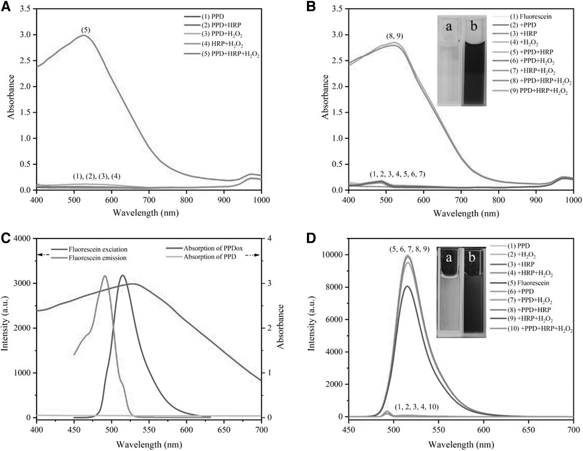
The characteristic excitation/emission peak of fluorescein is 490/515 nm, which almost coincided with the absorption spectrum of PPDox, and IFE-induced fluorescence quenching can occur efficiently (Sjoback et al., 1995; Sun et al., 2018; Wang et al., 2019). It is noted that neither obvious color change nor characteristic peak was observed by the addition of H2O2 or HRP alone to PPD solution. In addition, the coexisting solutions of PPD/HRP, PPD/H2O2, or HRP/H2O2 did not affect the fluorescence intensity of fluorescein (Fig. 3C, D).
In contrast, the fluorescence intensity dropped considerably in the simultaneous presence of PPD, H2O2, and HRP, indicating the fluorescence quenching by PPDox (Fig. 3D). Furthermore, PPDox had no significant absorption spectral change upon addition of fluorescein, which revealed no interaction or complex formation. Given the above discussion, the PPD/fluorescein/H2O2 system as a substrate and HRP as the signaling output enzyme can be used for the fluorescent and colorimetric identification of Hla.
Optimization of experimental conditions
Previous studies have indicated that pH values can significantly interfere with the catalytic decomposition of H2O2, and PPD could be oxidized by H2O2 under HRP catalysis in mildly acidic and neutral solutions (Jiao et al., 2000; Shi et al., 2021; Sun et al., 2018). Similarly, our research showed that PPD could be rapidly oxidized by HRP at pH 5, 6, and 7, while the oxidation process was almost inhibited under strong acidic and basic conditions (Shi et al., 2021; Sun et al., 2018) (Supplementary Fig. S1A).
Among them, the colorimetric signal of the assay performed best at pH 6, and the chromogenic reaction could be completed within 15 min. Due to both the concentrations of PPD and H2O2 affect the color signal in this experiment, they were also optimized (Supplementary Fig. S1B, C), and were, respectively, chosen as 1.5 and 1 mM for the following experiments.
Consequently, phosphate buffer (20 mM, pH 6.0) containing PPD (1.5 mM), H2O2 (1 mM), and fluorescein (5 μM) was selected as substrate buffer. Aiming to further improve the analytical performance of this method, the concentrations of mAb, pAb, and HRP-Ab were examined. Considering the reagent cost and the potential disturbance from negative samples, the concentrations of mAb (1 μg/mL), pAb (2.5 μg/mL), and HRP-Ab (1:5000) were chosen for the assay procedure (Supplementary Fig. S1).
Analytical performance
Under the optimal conditions, 32 Hla-negative samples were tested using the developed immunoassay to determine the cut-off values for the assay. As shown in Figure 4, the mean values (
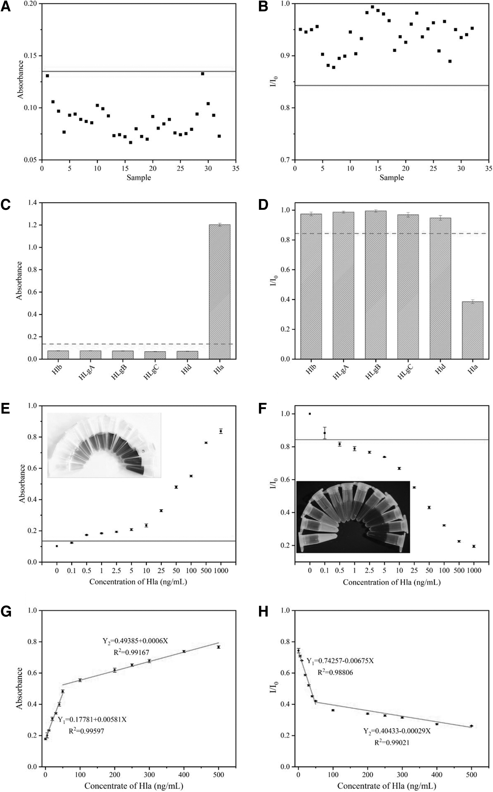
The selectivity of the established method was assessed by measuring the signal changes with Hla and other S. aureus hemolysins (including Hlb, Hld, HlgA, HlgB, and HlgC). As shown in Figure 4C, except for Hla, the OD530nm values of the other five hemolysins were <0.1349, and the I/I0 values were >0.8439. The results strongly illustrate that this method could only specifically recognize Hla without cross-reactivity, which may be attributed to the high selectivity of the antibodies.
To evaluate the sensitivity of the developed assay, both fluorescence and colorimetric modes were used to quantitatively determine the concentration of Hla. With the concentration of Hla increasing from 0 to 1 μg/mL, the OD530nm value increased accordingly, while the I/I0 value at 515 nm gradually decreased (Fig. 4). When the Hla concentration reached 0.5 ng/mL, the OD530nm was >0.1349 and the I/I0 value was <0.8429. On this basis, the calibration curves of fluorescence method and colorimetric method were constructed for the quantitative determination of Hla, respectively.
In colorimetric detection method, a linear relationship between the OD530nm value and the Hla concentration was well calibrated in the range of 0.5–50 and 50–500 ng/mL with the linear regression equation of Y = 0.17781 + 0.00581X (R 2 = 0.99597) and Y = 0.49385 + 0.0006X (R 2 = 0.99167), respectively. For fluorescence detection, the relative fluorescence intensities (I/I0 at 515 nm) of the system negatively correlated with Hla concentration in the range of 0.5–50 and 50–500 ng/mL. And the calibration curve could be calibrated as Y = 0.74257 − 0.00675X (R 2 = 0.98806) and Y = 0.40433 − 0.00029X (R 2 = 0.99021), respectively. The different slopes of the two linear ranges may be due to the different reaction rates within the two concentration ranges.
In addition, we listed the detection methods for Hla and their related parameters in the last decade (Table 1). Among them, all methods except one were mainly used for serum and culture supernatant samples, rather than food samples. However, the method has a high limit of determination (6.62 μg/mL) (Weston et al., 2020). It has been reported that the acute toxin concentration of α-hemolysin in human blood ranges from 100 to 250 ng/mL (3–7.5 nM), which also limits its wider application in food detection (Vakyly et al., 2022). Compared with the complex signal output strategies described in other documents, the dual-mode immunoassay established in this study can combine the characteristics of high sensitivity, wide linear range, and simplicity, which provides more options for the detection of Hla in food.
Comparison of Various Analytical Methods to Detect Hla
A single-stranded DNA molecular recognition element.
ELISA, enzyme-linked immunosorbent assay; Hla, alpha-hemolysin; LOD, limit of detection; MRE, molecular recognition element; PDA, polydiacetylene.
Determination of Hla in milk
The practicality and accuracy of the dual-mode immunoassay were evaluated by detecting pasteurized milk samples spiked with different concentrations of Hla (1, 5, 10, 25, 50, 100, and 250 ng/mL). Based upon the standard calibration curve (Table 2), the calculated recovery rates of colorimetric detection for Hla-spiked milk samples ranged from 98.5% to 106.5% with relative standard deviations (RSDs) <16.15%.
Detection of α-Hemolysin in Spiked Samples
Found = mean ± SD.
Recovery (%) = (found concentration)/(spiked concentration) × 100%.
RSD (%) = (SD/mean) × 100%.
RSD, relative standard deviation; SD, standard deviation.
Meanwhile, the recoveries range of fluorescence detection mode was 96.3–103.8%, with RSDs of <11.72%. Furthermore, there were no significant differences between the averages of the fluorescence and colorimetric detection results. These results revealed that the dual-mode immunoassay can be applied to the quantitative determination of Hla in milk samples with good accuracy and reliability.
Conclusions
In this study, a novel dual-mode immunoassay for determination of Hla was constructed based on the IFE between PPDox and fluorescein. The proposed method served as the efficient approach for quantitative detection of Hla by colorimetric and fluorescent dual modes, with the advantages of rapidity, convenience, good specificity, and high sensitivity. Meanwhile, the proposed method was characterized with good recovery, high accuracy, and strong practicality in milk samples, demonstrating its ability to resist matrix interference to some extent. In summary, this study provides a simple and efficient strategy for quantification of Hla.
Footnotes
Authors' Contributions
X.X. and P.Z. contributed to conceptualization, methodology, formal analysis, investigation, data curation, writing—original draft, and writing—review and editing. F.R. and G.C. were involved in writing—review and editing. X.W. provided supervision and writing—review and editing.
Disclosure Statement
No competing financial interests exist.
Funding Information
This research was supported by the Sichuan Natural Science Foundation (No. 2023NSFSC0178) and National Natural Science Foundation of China (Nos. 31871894 and 31271858).
Supplementary Material
Supplementary Material S1
Supplementary Figure S1
Supplementary Table S1
Supplementary Table S2
References
Supplementary Material
Please find the following supplemental material available below.
For Open Access articles published under a Creative Commons License, all supplemental material carries the same license as the article it is associated with.
For non-Open Access articles published, all supplemental material carries a non-exclusive license, and permission requests for re-use of supplemental material or any part of supplemental material shall be sent directly to the copyright owner as specified in the copyright notice associated with the article.