




Abstract
Both Klebsiella pneumoniae and Chryseobacterium cause an increasing number of diseases in fish, resulting in great economic losses in aquaculture. In addition, the disease infected with Klebsiella pneumoniae or Chryseobacterium exhibited the similar clinical symptoms in aquatic animals. However, there is no effective means for the simultaneous detection of co-infection and discrimination them for these two pathogens. Here, we developed a duplex polymerase chain reaction (PCR) method based on the outer membrane protein A (ompA) gene of Klebsiella pneumoniae and Chryseobacterium. The specificity and validity of the designed primers were confirmed experimentally using simplex PCR. The expected amplicons for Klebsiella pneumoniae and Chryseobacterium had a size of 663 and 1404 bp, respectively. The optimal condition for duplex PCR were determined to encompass a primer concentration of 0.5 μM and annealing temperature of 57°C. This method was analytical specific with no amplification being observed from the genomic DNA of Escherichia coli, Vibrio harveyi, Pseudomonas plecoglossicida, Aeromonas hydrophila and Acinetobacter johnsonii. The limit of detection was estimated to be 20 fg of genomic DNA for Chryseobacterium and 200 fg for Klebsiella pneumoniae, or 100 colony-forming units (CFU) of bacterial cells in both cases. The duplex PCR was capable of simultaneously amplifying target fragments from genomic DNA extracted from the bacteria and fish liver. For practical validation of the method, 20 diseased fish were collected from farms, among which 4 samples were PCR-positive for Klebsiella pneumoniae and Chryseobacterium. The duplex PCR method developed here is time-saving, specific, convenient, and may prove to be an invaluable tool for molecular detection and epidemiological investigation of Klebsiella pneumoniae and Chryseobacterium in the field of aquaculture.
Introduction
Klebsiella pneumoniae is a Gram-negative emerging opportunistic zoonotic pathogen that causes a variety of diseases (Wang et al., 2020). Globally, Klebsiella pneumoniae has emerged as a major clinical and public health threat (Wang et al., 2021) and it has a wide range of hosts including pigs, dogs, calves and aquatic animals (Bidewell et al., 2018; Das et al., 2019; Lalruatdiki et al., 2018; Lin et al., 2023; Sartori et al., 2019). After infection with Klebsiella pneumoniae, the Indian major carp (Cirrhinus mrigala) showed hemorrhage and reddish lesions, while the Chinese mitten crab Eriocheir sinensis exhibited significant structural changes in the hepatopancreas (Das et al., 2019; Wang et al., 2022). Recently, pathogenic Klebsiella pneumoniae K2 was isolated from the ark clam Tegillarca granosa and farmed American bullfrogs (Lin et al., 2023; Xu et al., 2022). Therefore, Klebsiella pneumoniae can be considered an emerging pathogen of aquatic animals, potentially leading to threatening the aquaculture industry (Wang et al., 2022).
Chryseobacterium is a bacterial genus from the family Flavobacteriaceae that consist of 120 validly published species (Loch and Faisal, 2015), some of which are pathogenic to humans, land animals and fishes (Mwanza et al., 2022). Over the last decade, several species of the genus Chryseobacterium have been reported to cause bacterial gill disease and systemic hemorrhagic septicemia in puffer fish (Campbell et al., 2008), rainbow trout (Ilardi et al., 2009; Zamora et al., 2012), Atlantic salmon (Kampfer et al., 2011) and golden mahseer (Shahi et al., 2018). Notably, with the increase of reported cases of fish infected with Chryseobacterium, it is now considered an emerging pathogen of freshwater and marine fishes around the world (Abraham et al., 2017; Loch and Faisal, 2015; Shahi et al., 2018). Interestingly, a number of Chryseobacterium strains that are more frequently associated with human infections or human consumables were recently recovered from fish (Loch and Faisal, 2015). Thus, the genus Chryseobacterium also represents a great threat to public health (Mwanza et al., 2022).
Rapid and exact identification of pathogens is crucial for the successful treatment of infections. In the past, the common routine procedure for pathogen identification was based on time-consuming and unreliable morphological observations as well as physiological and biochemical characterization of bacteria (Bhatia and Basu, 2007; Deurenberg et al., 2017). Although, the indirect ELISA has been adapted to detect Klebsiella pneumoniae infection in goats (Chen et al., 2021), its application in the diagnosis of fish diseases was restricted due to a lack of available enzyme-labeled secondary antibodies, and the difficulty of collecting serum samples from fish (Adams and Thompson, 2008; Fernandez-Alvarez et al., 2018). Therefore, it is necessary to establish rapid and specific detection methods for fish.
PCR has been used for early and quick diagnosis of bacterial diseases in fish (Hellebo et al., 2017; Seidlova et al., 2021). Notably, bacterial diseases of fish are mostly co-infections (Cardoso et al., 2021; Kotob et al., 2016). However, the common PCR assays that usually only detect a single species of pathogen in a single reaction system are not suitable for the detection of co-infections in fishes (Byers et al., 2002). By contrast, multiplex PCR-based (mPCR) assays have the advantage of detecting multiple pathogens in a single PCR reaction for the detection of pathogenic bacteria in fishes (Tsai et al., 2012; Zhang et al., 2014). Additionally, the fish infected with the Klebsiella pneumoniae or Chryseobacterium show similar clinical symptoms (Das et al., 2019; Bernardet et al., 2005). Recently, co-infection with Klebsiella pneumoniae and Elizabethkingia miricola (Elizabethkingia was split from the genus Chryseobacterium) (Alyami et al., 2020; Bernardet et al., 2005; Loch and Faisal, 2015) has been reported in Pelophylax nigromaculatus (Li et al., 2023). However, there is still no duplex PCR method for the simultaneous detection and discrimination of Klebsiella pneumoniae and Chryseobacterium in fish.
In this study, a reliable duplex PCR using four specifically designed primer pairs and optimized PCR conditions was developed for the detection and differentiation of Klebsiella pneumoniae and Chryseobacterium. The genus-specific duplex PCR was further confirmed to be suitable for the analysis of clinical samples.
Materials and Methods
Bacterial strains and genomic DNA extraction
Klebsiella pneumoniae strain KPLYC2 (identified by the 16S rRNA, GenBank: MT953921) and Chryseobacterium sp. CHLYC3 (identified by the 16S rRNA, GenBank: OP954499) were isolated from the liver of large yellow croaker infected in a farm in Zhejiang province, China. Strain CVCC4080 was purchased from China Institute of Veterinary Drug Control (Beijing, China). Strain ATCC700603 was stored in our laboratory. The genomic DNA was extracted from the isolated strains using a Rapid Bacterial Genomic DNA Isolation Kit (Sangon Biotech, China) according to the manufacturer’s instructions. The concentration and quality of genomic DNA were determined using a NanoDrop 2000 spectrophotometer (Thermo Scientific, MA, USA), after which it was stored at −20°C until further use.
Primer design
The ompA gene sequences of Klebsiella pneumoniae (Kp52.145 strain, Accession: FO834906.1) and Chryseobacterium (Chryseobacterium sp. strain C-17, Accession: CP087131.1) published in GenBank were used for primer design in Premier 5.0 software. For the detection of Klebsiella pneumoniae, a primer pair (Forward: 5′-CGCGGATCCATGAAAAAACGCGTACTTATG-3′; Reverse: P2 5′-CCGCTCGAGTTACTGCAGCGGGCTGAG-3′) was designed to specifically amplify a 663-bp region of ompA. The full length of the ompA of KP was designated as KP-ompA, the corresponding primers were designated as KP-ompA-F and KP-ompA-R. For Chryseobacterium, the primer pair (Forward: 5′-CGCGGATCC ATGCAAGATTCAATAGCGGTG–3′; Reverse: 5′−CCGCTCGAGTTATTTAGCTTCGAAATAAAC–3′) was targeted to a 1404-bp region of the ompA. The full length of the ompA gene of Chryseobacterium was designated as ompA, the corresponding primers were designated as ompA-F and ompA-R. The specificity of the designed primers was examined by Primer-BLAST against the NCBI database. All primers were synthesized by Shanghai Sangon Biological Engineering Technology & Services Co., Ltd (Shanghai, China).
Optimization of the duplex PCR protocol
The duplex PCR protocol was optimized according to a previous study (Yang et al., 2018). The primer concentrations and the annealing temperatures were optimized using the Beyo Multiplex PCR kit (Beyotime Biotechnology, China). The concentrations of the reaction mixture were applied as follows: 10×PCR buffer, 0.2 mM dNTPs Mixture, Taq DNA Polymerase, various concentrations of each of the forward and reverse primers and 1 μL template DNA with a concentration of 0.2 ng/μL, after which nuclease-free water was added to adjust the total reaction volume to 20 μL. Primers with different concentrations of 0.125, 0.25, 0.5 and 1 μM were prepared to optimize the assay. The following thermal cycling conditions were used: 1 cycle of 94°C for 5 min and 30 cycles of 94°C for 30 s, 30 s for annealing at a different temperature, and 1 min at 72°C, followed by a final extension step at 72°C for 10 min. Annealing temperatures of 55°C, 56°C, 57°C, 58°C and 59°C were tested. The PCR products were analyzed by 1.0% agarose gel electrophoresis and imaged using a gel imaging analysis system (FR-980B, Shanghai Furi Technology Co., Ltd, China). The optimized PCR reactions were identified based on the clearest and cleanest amplification products.
Cloning of the PCR products
The PCR products with expected sizes were cloned using the pUCm-T Vector Rapid Cloning Kit (Sangon Biotech, China) according to the manufacturer’s instructions and electroporated into competent cells of DH5-α. Positive clones were determined by PCR and the plasmids were extracted using a DiaSpin Plasmid Mini-Prep Kit (Sangon Biotech, China). The correct plasmids were further confirmed by DNA sequencing and named pUCm-T-KP-ompA and pUCm-T-ompA, respectively. Finally, the sequences of ompA were submitted to the NCBI database.
Specificity analysis of duplex PCR
The specificity of the duplex PCR was evaluated using genome DNA extracted from Klebsiella pneumoniae, Chryseobacterium, Escherichia coli, Vibrio harveyi, Pseudomonas plecoglossicida, Aeromonas hydrophila, and Acinetobacter johnsonii. The negative-control was DNA extracted from the liver of healthy large yellow croaker fish (Larimichthys crocea). The extracted genomic DNA was quantified and adjusted to 60 ng/μL. The PCR products were electrophoresed in a 1.0% agarose gel and imaged.
Sensitivity detection of the duplex PCR
The detection limit of the duplex PCR was determined using two conditions, as reported previously (Zhang et al., 2014). First, a mixture of genomic DNA from Klebsiella pneumoniae and Chryseobacterium at concentrations of 20 ng, 2 ng, 200 pg, 20 pg, 2 pg, 200 fg, 20 fg, and 2 fg was prepared. The maximum dilution was considered the minimum sensitivity for duplex PCR. Second, cultures of Klebsiella pneumoniae and Chryseobacterium were subjected to tenfold serial dilution to achieve culture concentrations ranging from 101-105 CFU/ mL. Then, the DNA templates for PCR were prepared from those cultures to determine the detection limit of the duplex PCR assay. The PCR products were electrophoresed in a 1.0% agarose gel and imaged.
Repeatability evaluation of duplex PCR
Repeatability characterizes the level of agreement among replicates of a sample tested under the same conditions. The repeatability evaluation was performed according to a previous study (Cellier et al., 2015), with minor modifications. Briefly, this test was performed using strains calibrated at 107 CFU/mL in molecular biology-grade water that were diluted to 3 different concentrations of 106,104 and 103 CFU/mL. Genomic DNA was isolated from the corresponding cell suspensions and used as the template. Finally, the repeatability was assessed based on three replicates using the corresponding genomic DNA.
Analysis of simulated samples
The applicability of the established duplex PCR was tested on spiked fish tissue samples according to a previous method (Zhang et al., 2014). Samples comprising 30 mg of zebrafish liver were homogenized and separately spiked with 103 CFU of each target strain or a mixture of the two strains. The DNA was then extracted using the Ezup Column Animal Genomic DNA Purification Kit and used as template for the duplex PCR.
Application of the duplex PCR on field-collected samples
A total of 20 dying diseased large yellow croaker fish with the symptoms of tardiness, social withdrawal, eating disorders, and abdominal bloating were collected from the culture ponds in Taizhou, Zhejiang Province, China. Bacterial DNA was isolated from the diseased fish according to a previous study (Das et al., 2019). Briefly, the fish carcasses were cleaned with 75% ethanol, the livers were aseptically removed and cut into pieces, which were placed into sterile 1.5 mL tubes containing 1 mL of normal saline. Each tissue sample was weighed to extract the DNA using the Ezup Column Animal Genomic DNA Purification Kit according to the manufacturer's instructions. The optimized duplex PCR was applied in the detection. For confirmation, the positive PCR amplicons were cut from the gel, purified, T-A cloned and sequenced at Sangon (Shanghai, China). Finally, these two sequences of ompA from Klebsiella pneumoniae and Chryseobacterium were searched against the NCBI database.
Results
Determination of the optimal duplex PCR conditions
According to the agarose gel electrophoresis results, the clearest and cleanest amplificons were obtained at a primer concentration of 0.5 μM and an annealing temperature of 57°C (Fig. 1). Therefore, all subsequent PCR reactions were conducted under these conditions.
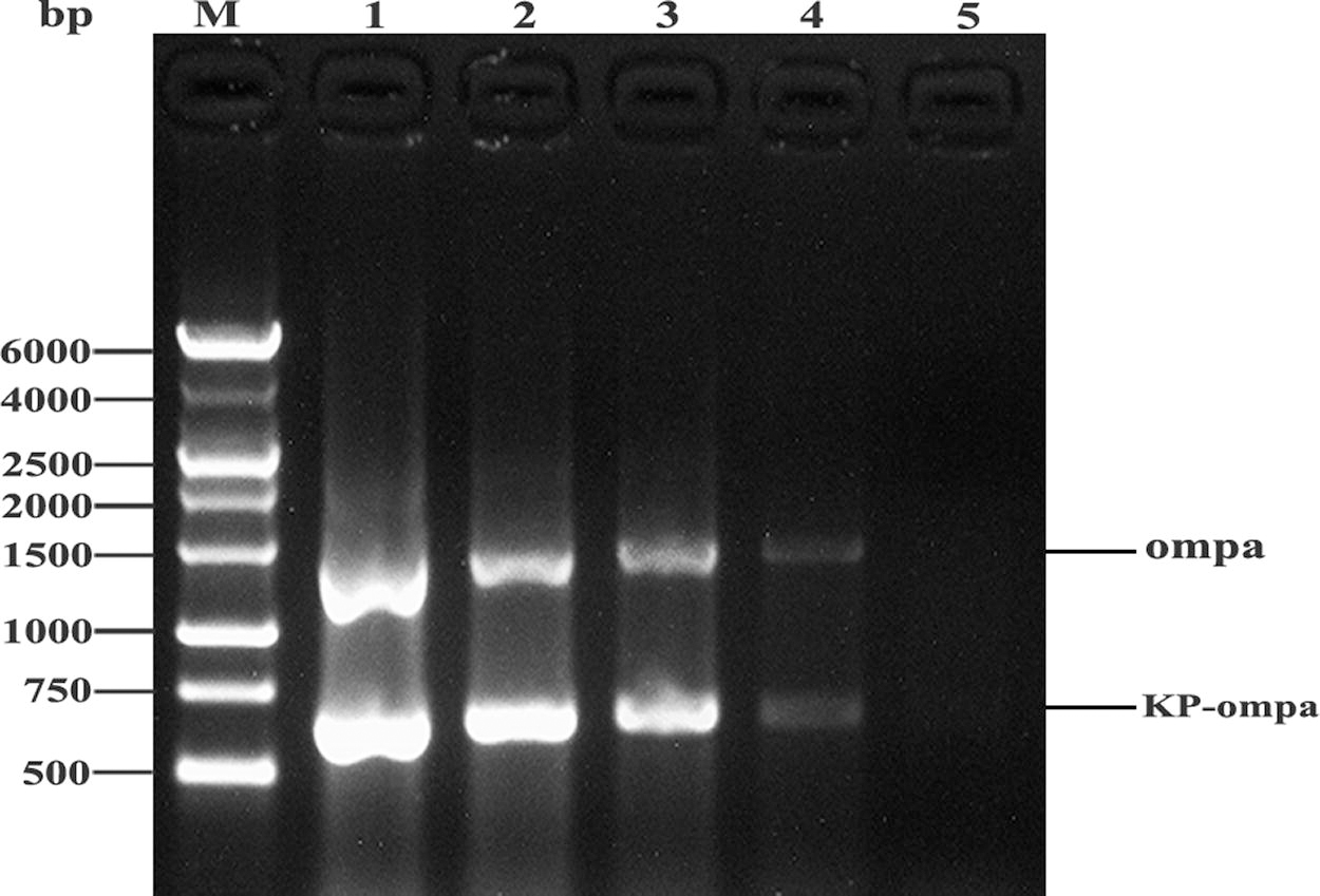
The results of duplex PCR optimization for Klebsiella pneumoniae and Chryseobacterium. M:1kb DNA Marker I; lanes1-4: Duplex PCR using 1, 0.5, 0.25 and 0.125 μM primers (KP-ompA-F/KP-ompA-R for Klebsiella pneumoniae, ompA-F/ompA-R for Chryseobacterium); lane 5: negative control.
Analytical specificity of the duplex PCR
The specific primer pairs amplified a 663 bp fragment for Klebsiella pneumoniae, a 1404 bp band for Chryseobacterium (Fig. 2). No cross amplification was observed with the genomic DNA of Escherichia coli, Vibrio harveyi, Pseudomonas plecoglossicida, Aeromonas hydrophila, or Acinetobacter johnsonii under the same conditions. No PCR product was observed in the negative control (without genomic DNA) using the two primer pairs (Fig. 2). The specificity of amplification was further confirmed by cloning and sequencing. The amplified ompA sequences were 100% identical with those of Klebsiella pneumoniae (GenBank Accession: OQ079160) and Chryseobacterium (GenBank Accession: OQ079161), respectively.
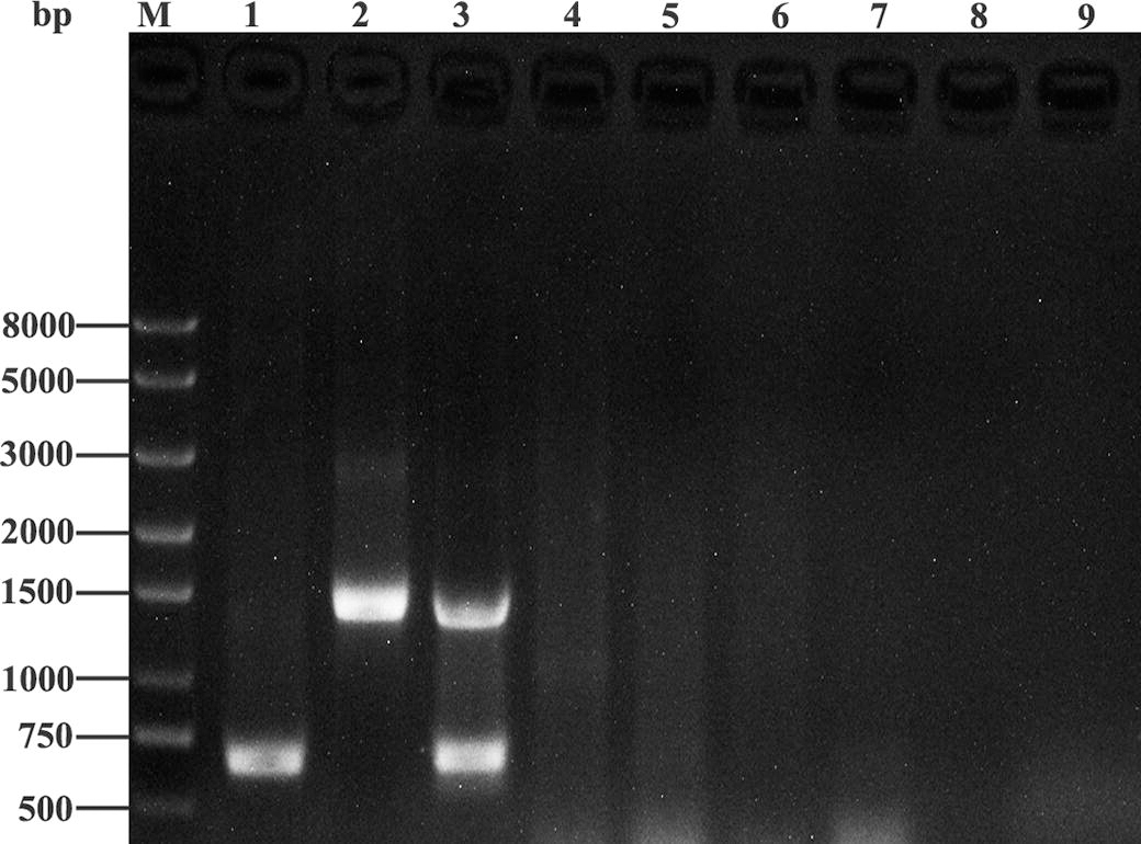
Specificity of duplex PCR using primers specific for Klebsiella pneumoniae and Chryseobacterium. M: Q8000 DNA Marker. Lanes 1–8: template genomic DNA extracted from Klebsiella pneumoniae, Chryseobacterium, or both Klebsiella pneumoniae and Chryseobacterium, Escherichia coli, Vibrio harveyi, Pseudomonas plecoglossicida, Aeromonas hydrophila, Acinetobacter johnsonii; lane 9: negative control.
Detection limit
The diagnostic limitations of the duplex PCR were evaluated by serially diluting genomic DNA or bacterial cell suspensions and using them as PCR templates. The minimal detectable amount of the genomic DNA template was 20 fg for Chryseobacterium and 200 fg for Klebsiella pneumoniae, while the minimal detectable number of bacterial CFUs was determined to be 100 for both strains (Fig. 3).
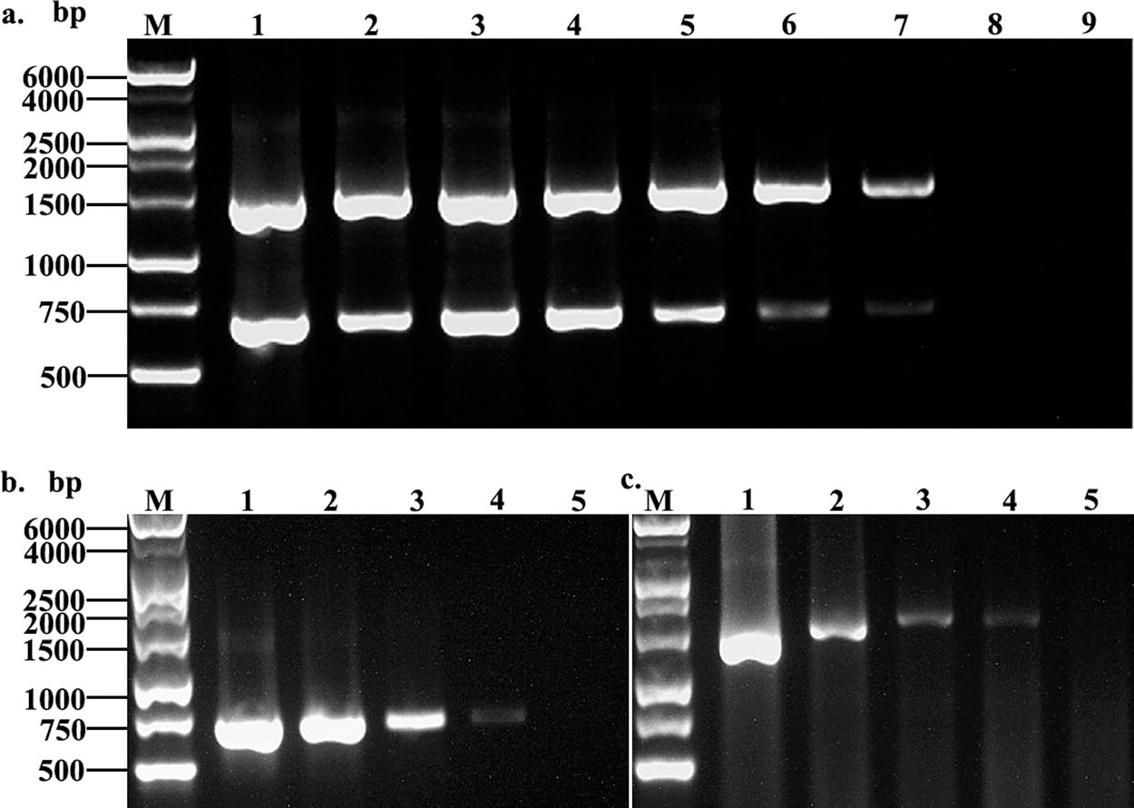
Detection limit of the duplex PCR for Klebsiella pneumoniae and Chryseobacterium using genomic DNA
Repeatability assay
The target bands with the same size could be amplified from the genomic DNA isolated from cell suspensions of different concentrations and their respective duplicate samples (Fig. 4). This result indicated that the established duplex PCR had good repeatability.

Duplex-PCR reproducibility assay using genomic DNA isolated from the target strains at different cell concentrations
Fish tissue and simulated bacterial samples
To test the method under more realistic conditions, the mixed genomic DNA extracted from fish liver and bacteria was used as the template for the duplex PCR analysis. The diagnostic efficiency was maintained as indicated by the successful generation of the expected amplicons, which suggests that there was no interference from fish DNA (Fig. 5).
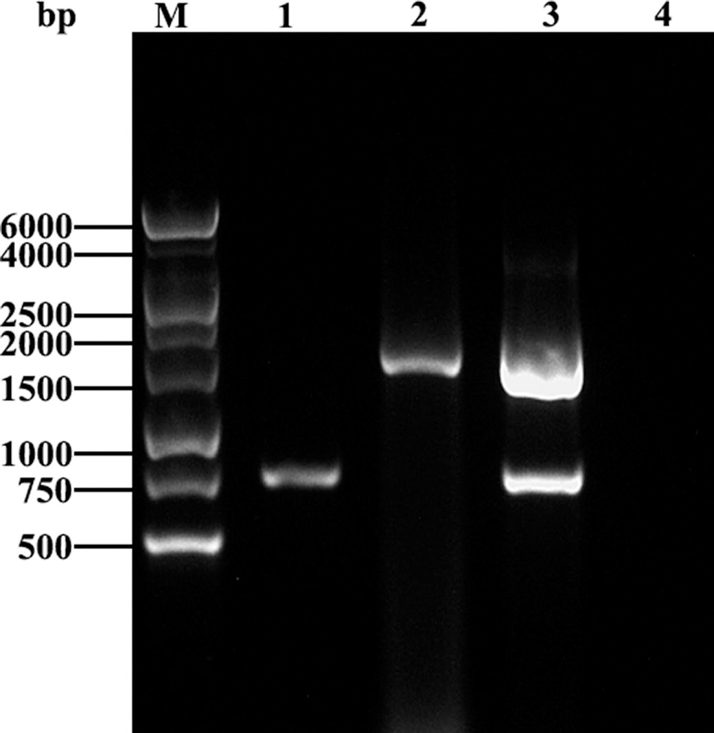
Agarose gel electrophoresis of the duplex PCR products obtained using the genomic DNA of each bacterium or both together, mixed with liver tissue from zebrafish. Lane 1, mixture of genomic DNA from the Klebsiella pneumoniae and liver tissue; lane 2, mixture of genomic DNA from the Chryseobacterium and liver tissue; lane 3, mixture of genomic DNA of both Chryseobacterium and Klebsiella pneumoniae, combined with liver tissue; line 4: negative control (without template); M: 1 kb DNA Marker I.
Application of duplex PCR in clinical samples
To assess the duplex PCR using actual clinical samples, 20 diseased large yellow croaker fish collected from farms in Zhejiang, China were analyzed for infection with Klebsiella pneumoniae and Chryseobacterium. A total of four samples were found to be infection with both Klebsiella pneumoniae and Chryseobacterium, while three samples were only positive for Klebsiella pneumoniae and one for Chryseobacterium. All other samples were negative (Fig. 6). When the samples were assayed using simplex PCR, the results were the same, and the PCR products were further confirmed by sequencing. The amplified ompA were 100% identical to the pUCm-T-KP-ompA and pUCm-T-ompA, respectively. All samples were retested twice, each analysis yielded the same, consistent result, confirming that the duplex PCR was capable of sensitively and specifically detecting Klebsiella pneumoniae and Chryseobacterium infections in field samples.
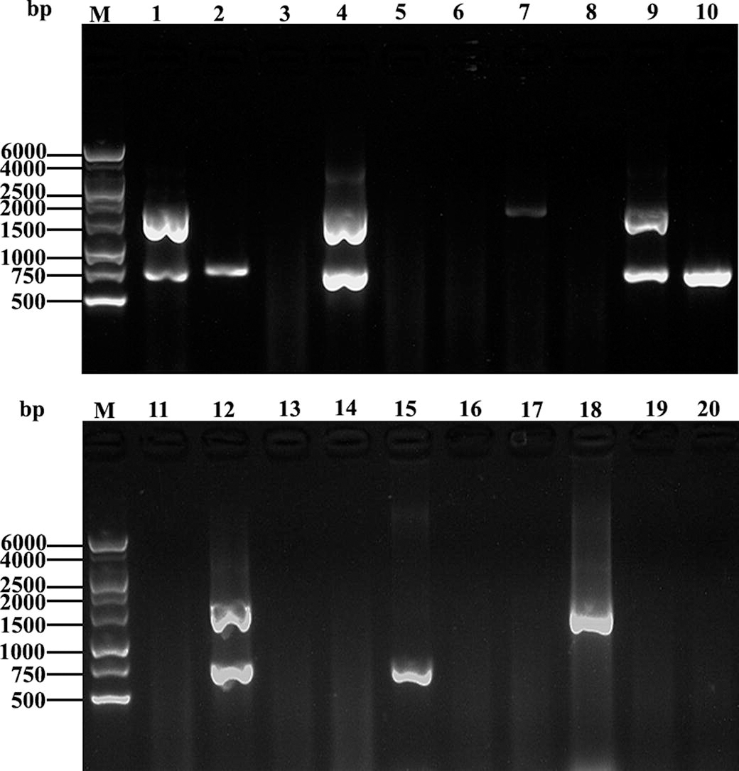
Testing of clinical samples using duplex PCR. M: 1 kb DNA Marker I. Lines 1–20: the genomic DNA was extracted from clinical samples comprising liver tissue of diseased fish.
Discussion
Bacterial diseases of fish are mostly mixed infections with similar clinical symptoms (Cardoso et al., 2021; Kotob et al., 2016). The fish infected with the Klebsiella pneumoniae or Chryseobacterium show similar clinical signs such as lethargy, anorexia, abnormal swimming patterns or spinning, ulcerative skin lesions, abdominal distension, fluid accumulation in the coelomic cavity, gill necrosis, septicemia, and mortality (Bernardet et al., 2005; Das et al., 2019; Wu et al., 2021).
In recent years, the morbidity and mortality caused by Klebsiella pneumoniae and Chryseobacterium infection in fishes was increased, resulting in severe economic losses and arousing widespread concern (Bruce et al., 2020; Wang et al., 2022). Rapid and accurate diagnosis is the key prerequisite for effective treatment and control of disease. Traditional detection methods are time-consuming and labor intensive, whereby serological diagnostic methods are also limited by the difficulties of collecting serum from fish and a lack of enzyme-labeled secondary antibodies, they are not suitable for large-scale diagnosis of infectious disease in fish.
Multiplex PCR was considered as one of the ideal methods for the diagnosis of fish diseases for co-infection disease (Adams and Thompson, 2008; Zhang et al., 2014). The first step to develop an effective PCR is to select a suitable target sequence. A previous study established a multiplex PCR based on the amplification of 16S and 50S rDNA fragments, fimbrial genes, the toxR gene, and heat shock protein 60 gene for simultaneous detection of Chryseobacterium meningosepticum, Edwardsiella tarda, Vibrio parahaemolyticus, V. vulnificus and Aeromonas hydrophila (Tsai et al., 2012). However, there was no mention of related methods for diagnosing Klebsiella pneumoniae and Chryseobacterium infections in fish. Additionally, previous methods mostly rely on the amplification of target sequences with similar size, which makes it difficult to discriminate them.
In this study, the ompA gene was selected as the target sequence for the detection of Klebsiella pneumoniae and Chryseobacterium. The outer membrane protein A (ompA) is a major structural component of the outer membrane of Gram-negative bacteria, where it functions as a multifaceted molecule with many diverse roles (Freudl and Cole, 1983; Liao et al., 2022). In addition, the ompA is relatively small and conserved among Gram-negative bacteria, but the interspecies difference is large (Confer and Ayalew, 2013; Freudl and Cole, 1983; Zhang et al., 2012). Notably, the ompA of Chryseobacterium was 1404 bp longer than that of Klebsiella pneumoniae with 663 bp. Thus, differences in gene length could be used for easy discrimination of PCR results. Errors may occur if targeting primers to an unsuitable region of the target, especially for larger genes (Guo et al., 2021). The functionality and specificity of the two primer pairs was firstly determined using simplex PCR (Supplementary Fig. S1). The results of single PCR showed that the primers amplified the target bands with the appropriate size, which indicated that the designed primers were specific to the ompA sequences of Klebsiella pneumoniae and Chryseobacterium, respectively.
A suitable annealing temperature is another important factor affecting the results of duplex PCR (Lorenz, 2012). Here, we tested annealing temperatures from 55°C to 59°C (Supplementary Fig. S2), and 57° C was determined to be the optimal annealing temperature (Fig. 1). Whether the primers could cross-react with other pathogenic bacteria directly determines the accuracy of PCR detection (Huang et al., 2018). The correct number of specific bands was detected in both Klebsiella pneumoniae and Chryseobacterium, while there was no cross-reaction with 5 other potentially interfering pathogens (Fig. 2). The duplex approach developed here therefore has high specificity. The detection limit of duplex PCR was 20 fg of genomic DNA for Chryseobacterium and 200 fg for Klebsiella pneumoniae (Fig. 3), which is comparable to a previously reported isothermal recombinase-aided amplification assay for Klebsiella pneumoniae alone (Hou et al., 2022). However, the duplex PCR established in this study has the capacity to detect both Klebsiella pneumoniae and Chryseobacterium while clearly distinguishing them. Notably, duplex PCR has a significant advantage over single pathogen detection systems when analyzing large numbers of samples, saving substantial time. In addition to, the cost of duplex PCR is relatively low and the method is easy to operate, and the addition of a sequencing step can improve the accuracy of this method. The results of duplex PCR were visualized by agarose gel electrophoresis, which is widely used in the field of diagnosis and identification of foodborne animal pathogens (Kundave et al., 2018; Reddy and Rao, 2022; Wan et al., 2019). However, considering the commercial application and increased selectivity needed for a diagnostic method, real-time PCR with a relatively short PCR product is also widely used in the field of disease diagnosis. Therefore, our follow-up work will be focused on the development of a quantitative fluorescence PCR method.
Out of the 20 samples, 4 fish were found to have co-infections with both Klebsiella pneumoniae and Chryseobacterium. Three samples showed infection with Klebsiella pneumoniae alone, while one was only infected with Chryseobacterium (Fig. 6). For further validation, the amplified PCR products were purified and sequenced. It was found that the amplified sequences were 100% identical to the corresponding ompA sequences in GenBank. Overall, the results indicated that duplex PCR may find practical applications win endemic areas where Klebsiella pneumoniae and Chryseobacterium co-occur in fishes. However, obviously diseased fish were collected for detection in this study, which still leaves the question if the developed duplex PCR can detect Klebsiella pneumoniae and Chryseobacterium DNA prior to the appearance of clinical symptoms. At the same time, the ompA gene could also be amplified from the genomic DNA of Klebsiella pneumoniae species complex CVCC4080 (Supplementary Fig. S3). Therefore, the isolate could not be identified as a specific strain of Klebsiella pneumoniae based only on the ompA gene sequence. Whole genome sequencing of the strain should be performed to determine which specific strain caused the infection. The same is true for Chryseobacterium. However, for bacterial pathogen diagnosis, this duplex PCR method could provide help for drug treatment, but requires further analysis for the use of vaccines, as these may be strain-specific.
Conclusions
In conclusion, we constructed a duplex PCR targeting the ompA gene for the co-detection and discrimination of Klebsiella pneumoniae and Chryseobacterium. This method is specific with no cross reaction with other pathogens. We further demonstrated that the duplex PCR could also identify the corresponding pathogens in field-collected samples. This duplex PCR was therefore confirmed as a simple and efficient diagnostic method for the simultaneous detection of Klebsiella pneumoniae and Chryseobacterium, which may be an ideal tool for epidemiological studies in endemic regions.
Footnotes
Authors' Contributions
G.W.H: Investigation (lead), project administration (lead), writing-original draft (lead) and writing-review and editing (lead). L.F.Y: Formal analysis (lead), methodology (lead), writing-review and editing (equal), supervision (equal), project administration (equal). X.L: Investigation (supporting), data curation (supporting), writing-review and editing (equal). Y.J.M: visualization (lead), formal analysis (supporting), data curation (supporting), and writing-review and editing (equal). J.Y.Y visualization (supporting) and editing (equal), writing-review and editing (supporting l).
Disclosure Statement
The authors do not have any competing or financial interest associated with the publication of this article.
Funding Information
This study was supported by Zhejiang Provincial Natural Science Foundation of China (Grant No. LTGN23C180001), Science and Technology Plan Project of Taizhou (Grant No. 22nya08) and National College Students Innovation and Entrepreneurship Program (Grant No. 202210350030).
Supplementary Material
Supplementary Figure S1
Supplementary Figure S2
Supplementary Figure S3
References
Supplementary Material
Please find the following supplemental material available below.
For Open Access articles published under a Creative Commons License, all supplemental material carries the same license as the article it is associated with.
For non-Open Access articles published, all supplemental material carries a non-exclusive license, and permission requests for re-use of supplemental material or any part of supplemental material shall be sent directly to the copyright owner as specified in the copyright notice associated with the article.
