
Abstract
The escalating prevalence of colistin-resistant Escherichia coli in poultry has emerged as a significant concern. This study aimed to assess the occurrence of the mcr-1 gene in colistin-resistant E. coli isolates from poultry samples. A cross-sectional study was conducted at National Avian Disease Investigation Laboratory, Nepal, on 210 chicken meat samples, including liver, heart, and spleen. E. coli was isolated and identified by conventional cultural methods. Antibiotic resistance pattern was assessed by the Kirby–Bauer disc diffusion method. The mcr-1 gene was detected by conventional polymerase chain reaction. The average viable count in chicken meat samples was log 6.01 CFU (colony-forming unit)/g, whereas the average coliform count was log 3.85 CFU/g. Coliforms were detected in at least one sample from 48.01% of total samples. The prevalence of E. coli in all meat samples was 39.52%. Liver accounted for the largest fraction of E. coli isolates (45.45%). Cefepime was the most effective antibiotic. Among all isolates, 45 (54.21%) were multidrug-resistant E. coli, 17 (20.48%) were colistin-resistant E. coli, and 11 (64.70%) harbored the mcr-1 gene. High prevalence of multidrug-resistant E. coli isolates, colistin-resistant isolates, and mcr-1 gene-carrying isolates indicates a serious concern, as it could potentially lead to colistin resistance in human pathogens through horizontal transfer of resistant genes from poultry to humans.
Introduction
Escherichia coli poses a significant threat to the poultry industry, resulting in considerable mortality rates, weight loss, and decreased egg production (Barlaam et al., 2019). One of the most common disease of poultry, colibacillosis, is caused mainly by avian pathogenic E. coli (Kołsut et al., 2017). Antibiotic resistance (ABR) has reached critical levels in human medicine, with numerous countries, including Nepal, recognizing it as a significant emerging threat to global public health and food safety (Adhikari et al., 2021; Adhikari et al., 2019; Regmi et al., 2020a; Sapkota et al., 2020). The agricultural utilization of antibiotics intended for human use, either as growth promoters or prophylactic agents in farm animals, has drawn criticism for contributing to the widespread challenge of antimicrobial resistance (AMR) (Acharya et al., 2017). Colistin, tylosin, neomycin, and doxycycline have been reported to be most commonly used antibiotics in broilers in the Kathmandu valley (Koirala et al., 2021). It is widely believed that the extensive use of antibiotics for growth promotion and therapeutic purposes has played a central role in the emergence and dissemination of drug-resistant bacteria, affecting both pathogenic and nonpathogenic strains (Dhungana et al., 2019).
Colistin, classified as a polymyxin antimicrobial agent, belongs to a group of bioactive compounds that were originally isolated from the soil-dwelling organism Paenibacillus polymyxa. Polymyxins A, B, C, D, and E are included in this group, but only polymyxin E (colistin) and polymyxin B are used in human medicine. Colistin is a mixture of two bacterial pentacationic lipopeptides, polymyxin E1 and E2. Although the exact mechanism of action of colistin is not fully understood, it involves binding to lipopolysaccharides and phospholipids in the outer membrane of Gram-negative organisms, resulting in membrane disruption and cell death. Colistin exhibits activity against a broad range of Gram-negative bacteria (Devkota et al., 2018).
Until now, resistance to polymyxin has been primarily attributed to chromosomal changes, such as mutation, and there has been no association with horizontal gene transfer (Moraes Mello et al., 2018). However, the emergence of mcr-1 genes carrying colistin-resistant E. coli isolates has become a major concern in the health sector, as they are found in humans, animals, and environmental settings (Muktan et al., 2020). To date, 10 slightly different variants of the mcr gene (mcr-1 to mcr-10) have been identified, with mcr-1 being the most prevalent circulating strain (Hussein et al., 2021).
In Nepal’s poultry farming industry, the widespread use of antibiotics as growth promoters, coupled with excessive dosing to accelerate growth, presents a current problem (Pokharel et al., 2019; Pokharel and Adhikari, 2020). Poultry harboring colistin-resistant E. coli can serve as reservoirs for drug-resistant genes, which have the potential to be transferred to other pathogenic organisms affecting humans (Savin et al., 2020). These resistant bacteria are present in various organs of poultry and can be easily transmitted to humans through the food chain (Dawadi et al., 2021). This study aimed to determine the prevalence of colistin-resistant E. coli carrying the mcr-1 gene in poultry farms located in Bharatpur city.
Materials and Methods
Study design and settings
A cross-sectional study was conducted at the National Avian Disease Investigation Laboratory (NADIL) in Chitwan, Nepal, for a period of 6 months. Chitwan district, situated in the central part of the country, is widely acknowledged as the capital of the poultry industry because of its significant role in meeting the national demand for poultry products. The region benefits from favorable climatic conditions and a fertile agricultural landscape, making it an ideal location for poultry farming. NADIL, a prominent facility specializing in avian research and diagnostics, operates under the Department of Livestock Services within the Ministry of Agriculture and Livestock Development of the Government of Nepal.
Sample collection, processing, and identification of E. coli
Hundred grams of meat sample was collected from each dead and/or sick chicken received in the postmortem room of the avian laboratory by the veterinary officer after complete anamnesis. The samples were submitted to the laboratory by the owners of the commercial broiler farms with flock size ranging from 500 to 2000. All the samples were from Cobb breed. The samples were collected in a sterile zip-lock plastic bag and immediately transported to the microbiology laboratory. The meat sample was ground in a sterile mortar and pestle, 1 g was inoculated into 9 mL of distilled water, and a loopful was streaked in MacConkey agar plates in triplicate. The plates were incubated at 37°C for 24 h to obtain pure colonies. Pink-colored isolated colonies on MacConkey agar plates were subjected to Gram staining and an array of biochemical tests to phenotypically confirm the E. coli isolates (Hall, 2013). One isolate was considered per sample for further laboratory investigations.
Antibiotic susceptibility test of the isolates
The antibiotic susceptibility pattern of all the isolates was determined by the Kirby–Bauer disk diffusion method following Clinical and Laboratory Standards Institute (CLSI) guidelines (CLSI, 2016) against the following antibiotics: imipenem (10 μg), ampicillin (10 μg), colistin (10 μg), ciprofloxacin (5 μg), gentamicin (10 μg), cefazolin (30 μg), cefotaxime (30 μg), and cefepime (30 μg). Isolates showing nonsusceptibility (either resistance or intermediate) to at least one antimicrobial agent in three or more of the categories were considered as multidrug resistant (MDR) (Regmi et al., 2020b).
Screening of colistin-resistant isolates
Agar dilution method was used to determine the minimum inhibitory concentration (MIC) of colistin. Concentrations ranging from 2 to 32 μg/mL were prepared in agar medium, and bacterial inoculum was applied readily onto the agar surface and the plates were incubated at 37°C for 18 h. The MIC end point was determined as the lowest concentration of antibiotics that completely inhibited the visible growth. Isolates having a MIC of ≤ 4 μg/mL were considered colistin susceptible, whereas those having MIC of >4 μg/mL were considered colistin resistant (Bista et al., 2020).
DNA extraction and PCR amplification of colistin resistance mcr-1 gene
Plasmid DNA was extracted from E. coli isolates by the alkaline lysis method, as described elsewhere (Feliciello and Chinali, 1993). The amplification process adhered to conventional techniques, using a designated primer combination, CLR5-F (5′-CGGTCAGTCCGTTTGTTC-3′) and CLR5-R (5′-CTTGGTCGGTCTGTAGGG-3′), to specifically amplify the mcr-1 gene (Liu et al., 2016) using a Prime GeNeiTM thermocycler (Bangalore, India). The polymerase chain reaction (PCR) mixture for the mcr-1 amplification was prepared with a final volume of 25 μL, consisting of 21 μL of 1× Qiagen Master Mix, 3 μL of template DNA, and 0.5 μL of each of the forward and reverse primers (0.4 μM). The PCR amplification cycle included an initial denaturation step at 95°C for 15 min, followed by annealing at 57°C for 1.5 min and extension at 72°C for 1 minute. Subsequently, the amplified products were separated via electrophoresis on a 1.2% agarose gel and visualized through ethidium bromide staining. The presence of the mcr-1 gene was ascertained by comparing the results with a positive control as well as a 100-bp DNA ladder following electrophoresis (Bista et al., 2020).
Results
Altogether, 210 nonduplicated chicken meat samples were analyzed in the study.
Coliform prevalence in chicken meat samples
Total viable count in the collected meat samples ranged from log 4.23 to log 8.76 CFU (colony-forming unit)/g. Total coliform count was found to be ranging from log 2.041 to log 6.531 CFU/g. Out of 210 chicken meat samples, 101 (48.09%) samples were found to be harboring at least one coliform. Further investigation revealed that 83 (39.52%) samples were contaminated with E. coli, followed by Klebsiella spp. (11 [5.23%]) and Citrobacter spp. (7 [3.33%]).
Comparison of bacterial load of meat sample with standards
The permissible limit of total viable count in raw chicken meat as set by the Food Safety and Standards Authority of India is ≤106 CFU/g. Liver samples obtained from the areas of Tandi and Patihani exhibited E. coli levels that exceeded the acceptable threshold, whereas heart samples collected from Gitanagar, Prembasti, Tandi, Jagatpur, Patihani, and Parbatipur showcased E. coli concentrations surpassing the permissible limit. In addition, spleen samples from Prembasti and Tandi exceeded the standard limit. The total coliform count with the International Commission on Microbiological Specifications for Foods standards for raw chicken meat with permissible limit is ≥2000 CFU/g. Liver samples from Tandi, Jagatpur, and Parbatipur exhibited total coliform counts below the standard limit. Likewise, heart samples from Gitanagar, Tandi, Jagatpur, Patihani, and Parbatipur, as well as spleen samples from Krishnapur and Jagatpur, exhibited counts below the standard limit (Fig. 1).
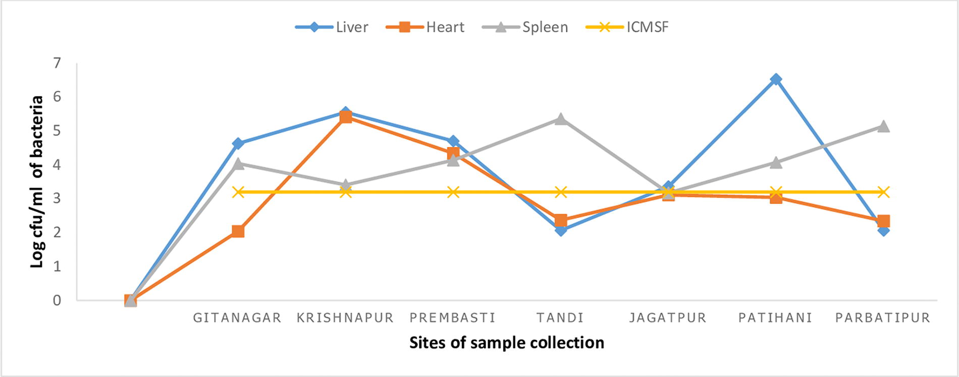
Comparison of total coliform count of meat samples collected from different locations with ICMSF standards. ICMSF, International Commission on Microbiological Specifications for Foods.
Area- and organ-specific distribution of E. coli in chicken meat
A total of 210 meat samples were collected from seven different areas in Chitwan, with each area contributing 30 samples. Among these areas, Krishnapur exhibited the highest contamination rate (66.67%) by E. coli, whereas Parbatipur had the lowest contamination rate (20%). The observed difference was statistically significant (p < 0.05). Liver samples had the highest prevalence of E. coli (45.45%), followed by heart samples (40%) and spleen samples (26%). However, this difference was not statistically significant (p > 0.05) (Table 1).
Area- and Organ-Wise Distribution of E. coli
Significant at 5% level of significance.
Antibiotic susceptibility patterns of E. coli
Cefepime was the most effective drug, inhibiting the growth of 85.54% E. coli, followed by cefotaxime (83.13%). Colistin was found to be effective against only 66 (79.51%) isolates. The least effective antibiotic was found to be gentamicin, which was resisted by 84.48% of E. coli isolates. Of the 83 isolates, 45 (54.22%) were found to be MDR (Table 2). In MIC assays, 17 E. coli isolates were found to be colistin resistant (MIC ≥ 4 μg). Overall prevalence of colistin-resistant E. coli in meat samples was 20.48% (17/83). MICs of colistin-resistant isolates ranged from 4 μg/mL to 32 μg/mL.
Antibiotic Susceptibility Patterns of E. coli
Prevalence of mcr-1 gene in colistin-resistant E. coli isolates
Among 17 colistin-resistant E. coli isolates, 11 (64.7%) were identified as carrying the mcr-1 gene. Out of these, seven isolates originated from liver samples, whereas heart and spleen samples contained two isolates each. In terms of specific locations, meat samples from Krishnapur area accounted for five (45.45%) E. coli isolates harboring the mcr-1 gene.
Discussion
Meat is considered as an essential part of the human diet. Meat contains a generous supply of nutrients conducive to the growth of bacteria. The contamination of poultry meat is very much dependent on the status of the birds prior to slaughter and on operational hygiene during poultry meat processing. During the conversion of live birds into poultry carcasses and meat, there is a significant risk of contamination from external sources, including the bird’s surroundings and the surfaces it encounters during processing. Furthermore, there is a potential for exposure to the intestinal contents of the bird, further increasing the risk of contamination. In addition, handling, storage time, and temperature conditions play a crucial role in determining the number of organisms present in the final product (Fuzihara et al., 2000).
In the current study, viable and coliform counts for the meat samples were found to range from log 4.23 to log 8.76 CFU/g and log 2.04 to log 6.53 CFU/g, respectively.
Studies have suggested the dissemination of bacteria from gut to meat during sample collection and processing of meat in the farm (Adeyanju and Ishola, 2014). As the status of the farm conditions in which birds were kept in this study was unknown, further investigations into specific conditions and practices in poultry farms are recommended. The prevalence of E. coli in the meat samples in this study was found to be 39.52%. A study done by Manish et al. (2021) revealed that 48% chicken meat samples from Lalitpur were contaminated with E. coli, whereas a study done by Saud et al. (2019) in Bhaktapur claimed that the contamination rate was 33%. During the poultry slaughter process, there is a possibility of fecal contamination on the carcasses originating from the bird’s intestinal tract. This implies that bacteria present in the spilled gut contents can be transferred as contaminants, resulting in an increased likelihood of bacterial contamination in meat samples (Adeyanju and Ishola, 2014). The bacterial load of meat samples from Krishnapur area was higher (66.67%) as compared with other places. Adhikari et al. (2020), while doing a similar study in Bharatpur, also found that the bacterial load was different from place to place. Most of the places from where samples were collected were densely populated, and the farm owners were not aware of the good hygienic practices while handling the poultry, which might be the reason behind the higher contamination of meat from Krishnapur as compared with other places (Adhikari et al., 2020). The rate of E. coli contamination was higher in liver (60.91%) as compared with heart and spleen. Numerous reports from around the world have indicated that the liver is consistently found to be heavily contaminated in chickens (Adhikari et al., 2020; Bista et al., 2020; Manish et al., 2021; Saud et al., 2019). The liver is considered to be an optimal environment for bacterial growth owing to its moist nature, abundance of nitrogenous compounds, presence of growth factors, and efficient food metabolization. These factors contribute to the liver’s suitability for the accumulation of microorganisms (Hines et al., 2010). Various factors could account for the variations in the prevalence rates of coliforms and E. coli. These factors include differences in the source of samples, the time period of sample collection, the age of the samples, the sampling procedure used, the contamination levels within poultry farm structures (such as inadequate sanitation practices), the extent of processing involved, cross-contamination of products, and variances in the methodologies used to detect these pathogens (Saleh et al., 2020). It is a well-established fact that the resistance to colistin occurs through point mutation, especially in genes that affect the synthesis of cell wall and lipid bilayer (Odwar et al., 2014). The results of our study, as determined by MIC assays, demonstrated a colistin resistance rate of 20.48% among the isolates, which is significantly higher than the 2.2% reported in the study conducted by other researchers (Karki et al., 2021). Our study revealed that 11 isolates of E. coli (64.7%) were found to contain the mcr-1 gene. This proportion is higher than the rate reported by Bista et al. (2020)—mcr-1 gene was detected in 43.9% of colistin-resistant E. coli isolates. Furthermore, one study reported an even lower rate of 22.8% for E. coli harboring the mcr-1 gene in chicken meat samples (Acharya et al., 2017). Higher prevalence of E. coli isolates harboring the mcr-1 gene has been reported from other countries as well, such as China (Zhang et al., 2017) and Pakistan (Azam et al., 2017). The presence of colistin resistance determinants, specifically the mcr-1 gene, initially identified in E. coli and subsequently in K. pneumoniae derived from raw meat, animals, and human cases in the People’s Republic of China, highlights the significant role of horizontal gene transfer in the dissemination of such resistance mechanisms. The development of colistin resistance is attributed to the presence of the mcr-1 gene, which encodes a phosphoethanolamine transferase, resulting in a diminished affinity to colistin (Liu et al., 2016).
The ABR patterns observed in this study for E. coli were in line with findings from other studies conducted elsewhere (van den Bogaard et al., 2001; Khanal et al., 2017; Samun Sarker et al., 2019). In this study, cefepime (85.54%) was the most effective drug against E. coli isolates. Similar result was reported from an earlier study done by a team of researchers led by Bista et al. (2020), where cefoxitin, a second-generation cephalosporin, was found to be inhibiting the growth of 86.8% E. coli isolates. Furthermore, in the study conducted by Saud et al. (2019), resistance to ceftazidime, a third-generation cephalosporin, was detected in just 4.3% (1 out of 23) of the tested E. coli isolates. Likewise, this study revealed that gentamicin exhibited the lowest efficacy, with 84.38% of the isolates showing resistance to this drug. A similar study performed in Central Veterinary Laboratory, Kathmandu, also reported that 76.4% of E. coli isolates were resistant to gentamicin (Bista et al., 2020). Prevalence of MDR E. coli was 54.21% in the current study, which is slightly less than previous studies done in Nepal (76.4%) (Bista et al., 2020) and the Netherlands (van den Bogaard et al., 2001). The transmission of antimicrobial genes between organisms has been facilitated by factors such as meat products and other environmental conditions, resulting in the emergence of MDR and pan drug-resistant bacteria. This has led to the challenging situation we face today. In addition, reports suggest that the environment, pets, and wildlife also play a significant role in influencing the spread of AMR genes across different reservoirs (Chen et al., 2018; Zhang et al., 2018). Our findings reveal a notable level of resistance to imipenem among the isolates under study. Imipenem, being a carbapenem, plays a crucial role in the treatment of MDR bacterial infections.
Although this study can be considered a reference for epidemiological investigations on the prevalence of the mcr-1 gene in poultry in Nepal, it is important to acknowledge its limitations. These include the inability to confirm colibacillosis and the absence of histological, molecular, and nucleotide sequencing investigations. In addition, the study focused only on E. coli load in meat samples, disregarding other pathogenic microorganisms. Although 10 variants of mcr genes (mcr-1 to mcr-10) have been identified in bacteria isolated from livestock and human worldwide, only the mcr-1 gene has been analyzed in this study, raising the possibility of missing other mcr gene types.
Conclusions
The study reveals a significant prevalence of colistin-resistant E. coli isolates, likely stemming from the extensive use of colistin in poultry feed. This high occurrence of the mcr-1 gene poses a potential risk of spreading colistin resistance to human pathogens, necessitating the search for new antibiotics. It is crucial for authorities to establish effective policies, raise awareness among poultry farmers, and enforce regulations to minimize the adverse consequences associated with inappropriate drug usage.
Footnotes
Acknowledgments
The authors are thankful to veterinary officers and staffs of National Avian Disease Investigation Laboratory for their cooperation during this study.
Authors’ Contributions
Conceptualization, P.P., S.K., S.A., S.S., O.P., and T.K.; formal analysis, S.A. and R.S.R.; investigation, A.T., S.L., and R.S.R.; methodology, A.T., S.L., and R.S.R.; supervision, P.P., S.K., S.S., O.P., and T.K.; V., P.P.; writing—original draft, A.T.; writing—review and editing, P.P., S.K., S.L., S.A., R.S.R., S.S., O.P., and T.K.
Disclosure Statement
No competing financial interests exist.
Funding Information
This research received no external funding.