




Abstract
Staphylococcus aureus contamination and prevention has always been a major concern for food industry. This work investigated the antibacterial activity and mechanisms of lauric acid (LA) against S. aureus. Results revealed 156 μg/mL was the minimum inhibitory concentration (MIC) for LA and it retarded growth rate of S. aureus. The inhibitory effect was enhanced with LA concentration. After being treated with 2 MIC LA for 24 h, the number of S. aureus decreased by 3.56 log colony-forming unit (CFU)/mL. Scanning electron microscopy profiling revealed that LA resulted in altered morphology of S. aureus cells. In addition, propidium iodide staining of flow cytometry suggested that LA treatment disrupted the cell membrane integrity. Changes in 8-anilino-1-naphthalenesulfonic acid fluorescence indicated a depolarization change in cell membrane fluidity. For practical applications, LA also displayed an antimicrobial potential in cooked chicken food model system, with 1.25–5 g/L of LA prolonging shelf life by 2 days at 4°C. Moreover, it had no adverse effect on pH values, color in cooked chicken meat, and even reduced lipid oxidation. To sum up, LA has great antimicrobial properties and is a candidate preservative for cooked meat food.
Introduction
Currently, foodborne pathogens led to an average of 1 in 10 people worldwide suffering from foodborne diseases every year (Gonzales-Barron et al., 2021). Among them, outbreaks of Staphylococcus aureus are prevalent in different countries and regions. High-protein foods are susceptible to the contamination (Huang et al., 2023; Suaifan et al., 2017), and as a major source of edible meat protein, chicken has a high prevalence of S. aureus infections (Wang et al., 2023). Symptoms of food poisoning caused by S. aureus often include nausea and vomiting, but may also be accompanied by watery diarrhea and fever (Gallina et al., 2013). How to prevent bacterial pollution while inhibiting bacterial growth has remained a major concern in the food field. Increasingly, biological preservatives of natural origin are favored because of their advantages of being green, safe, and not easily causing microbial resistance.
Lauric acid (LA, C12:0) is an amphiphilic saturated fatty acid of medium chain. It is the most abundant component in natural coconut oil. LA is listed as “Generally Recognized as Safe, GRAS” according to the Food and Drug Administration (Yoon et al., 2018). Kumar Ghosh et al. (2017) added refined LA coconut powder obtained from coconut meal to butter biscuits and discovered that it improved the sensory properties of the biscuits and decreased the oxidation degree of biscuits. Further, LA can slow down the oxidation of lipid as well as maintain the color in meat products (Hoa et al., 2022). When used as an animal feed additive, LA improved the quality of slaughtered meat and reduced the initial bacterial load of raw meat (Araújo et al., 2022; Zeiger et al., 2017).
Overall, LA is widely used, but its application to cooked meat products is rarely reported. Therefore, the objective of the present study was to explore the antimicrobial effect and damage mechanism of LA on the foodborne pathogen S. aureus and to use cooked chicken as a food model to test its feasibility in cooked meat products.
Materials and Methods
Materials
LA (≥98%, CAS: 143–07-7) was supplied by Aladdin Chemical Co. (Shanghai, China). LA was achieved through dissolution of monocaprin in ethanol and filtration with a 0.22 μm aqueous millipore membrane. Before each experiment, LA solutions were diluted and stirred for 5 min on a magnetic stirrer to different concentrations.
Bacterial strain and culture condition
S. aureus NCTC 8325-4 was kept frozen at −20°C. It was inoculated onto the Tryptic soy agar medium for activating strain and subjected to Tryptic soy broth for secondary activation. At the end of the incubation, the bacterial suspension was centrifuged at 5000×g for 5 min in a refrigerated centrifuge (JW-3021HR, Anhui Jiaven Equipment Industry Co., Anqing, China). The precipitates were washed and redissolved to acquire a bacterial concentration of 6–6.5 × 10⁹ colony-forming unit (CFU)/mL.
Antimicrobial activity of LA
Minimum inhibitory concentration and minimum bactericidal concentration
Determination of minimum inhibitory concentration (MIC) and minimum bactericidal concentration (MBC) were measured according to Moosavi-Nasab et al. (2016). Double LA series dilutions were prepared from MH broth in 96-well microtiter plates. Then, 100 μL cell culture (1 × 10⁶ CFU/mL) was inoculated in each well. MIC is the minimum concentration of antibacterial agents that inhibits the growth of S. aureus under visual observation. Subsequently, counting samples will have no bacterial growth. The lowest antimicrobial concentration corresponding to plates with the absence of colony growth was MBC (Zhang et al., 2016). MH broth bacterial suspension without antibacterial agents and MH broth liquid culture medium without bacteria were served as positive and negative controls, respectively.
Growth curve of S. aureus
The bacterial suspension (prepared in phosphate buffered saline as described in the Bacterial Strain and Culture Condition section) was centrifuged at 5000×g for 5 min and collected. After resuspending the cells in fresh MH broth to 1 × 10⁶ CFU/mL, LA of different concentrations (1/2 MIC, 1 MIC, and 2 MIC) was added. These suspensions were cultivated at 37°C and the absorbance value at 600 nm (Synergy HT, Biotek, Vermont, USA) was determined (Kang et al., 2020).
Time-kill assay of LA treatment
Briefly, the pretreatment of bacteria was the same as above. The treatment groups were supplemented with LA, and the group without LA was established as a blank control group. Then, 100 μL bacterial samples were aspirated at different time points and enumerated for viable cells.
Detection of membrane integrity by flow cytometry test
S. aureus at 1 × 10⁸ CFU/mL was mixed with LA, whereas no LA was added to the blank control. All sample groups were cultivated at 37°C for 12 h. Propidium iodide (PI) fluorescent dye (final concentration 15 µmol/L) was added. After incubation for 15 min at 37°C under dark conditions and washing, the PI-stained cells were run on flow cytometer (BD FACSCalibur, America).
Detection cell morphological change of S. aureus by scanning electron microscopy
S. aureus were treated with 1 MIC concentration of LA. The cells were fixed with 2.5% glutaraldehyde and dehydrated with sequential graded ethanol (30%, 50%, 70%, 80%, 90%, 95%, and 100%, v/v). Finally, all resulting samples were dried and coated with a layer of gold. The ultrastructure of S. aureus was then visualized with a Hitachi SU-8100 emission scanning electron microscope (Tokyo, Japan).
Changes of cell membrane fluidity of S. aureus after LA treatment
S. aureus cells were incubated at 37°C with shaking. Subsequently, 4 µM 8-anilino-1-naphthalenesulfonic acid was co-incubated with the suspension obtained at different times under dark conditions. The fluorescence intensity was measured by fluorescence spectrophotometer at excitation wavelength 385 nm and emission wavelength 473 nm.
Changes of cell membrane potential in S. aureus after LA treatment
The harvested S. aureus cells were resuspended with 5 mM HEPES buffer (containing 5 mM glucose). Then all samples were cultured at 37°C with shaking. The 3,3′-dipropylthiadicarbocyanine iodide [DiSC3(5)] and KCl solution were added at concentrations of 5 µM and 100 mM, respectively, at different time points, and kept for 15 min at 37°C in the dark. The fluorescence intensity was measured with a fluorescence spectrophotometer at the excitation wavelength of 650 nm and the emission wavelength of 672 nm.
Application of LA against S. aureus in a cooked chicken food model
Fresh chicken breasts were washed with water and cooked in an autoclave. After cooking, the samples were cut into cubes of about 5 g and soaked in different concentrations of LA solution (1.25, 2.5 and 5 g/L) for 10 s. The cooked chicken was inoculated with S. aureus to a final concentration of ∼ 4 log CFU/g. The group not treated with LA was taken as the blank control. The meat samples were dried for 20 min and placed in sealed bags at 4°C and 25°C. During experiments, chicken breasts would be taken out at fixed time points, homogenized and diluted with sterile water in a gradient. The mixture of 100 μL was coated on BP plates and enumerated.
Quality changes of cooked chicken meat with LA treatment during storage
Microbial analysis in cooked chicken meat
The cooked chicken meat was prepared as above. After treatment with different concentrations (1.25, 2.5 and 5 g/L) of LA, the samples were sealed and stored at 4°C and 25°C, respectively. For the total viable counts (TVC) analysis, the cooked chicken was taken out at a fixed time and spread on Plate count agar medium for plate counting.
Determination of pH and color in cooked chicken meat
The pH values were tested with a pH meter (PHS-3C, Rex Electric Chemical, China) based on Daszkiewicz et al. (2018) with some modifications. Color values (L* is the lightness, a* is the redness, and b* is the yellowness) were obtained by measuring three times on different parts of the sample with a chromatic meter SC-8C (Kangguang Instrument Co., Ltd., Beijing, China).
Evaluation of thiobarbituric acid reactive substances in cooked chicken meat
Cooked chicken samples were added to 25-mL trichloroacetic acid (7.5%) and filtered after shaking. The filtered sample was mixed with 5 mL of 0.02 M thiobarbituric acid (TBA) and incubated in a 90°C water bath for 40 min. After cooling rapidly, the mixture was centrifuged at 2000×g for 5 min at 25°C. Then, 5 mL of trichloromethane was added to the supernatant, shaking and layering. The absorbance from 532 to 600 nm was recorded using a multifunctional enzyme marker and the TBA reactive substances (TBARS) was calculated as follows:
Statistic analysis
One-way analysis of variance and multiple comparison analysis were performed by Excel and IBM SPSS Statistics 19 software for the data statistically. Differences were considered significant at p < 0.05. Graphs were drawn with Origin 2022 b software (Origin Lab Corporation, USA).
Results and Discussion
In vitro antibacterial activity of LA
Since log phase cells are more sensitive to stressful conditions, the use of these cells instead of more resistant stationary phase cells is justified (Lu et al., 2005). S. aureus was cultured, and the measured MIC and MBC were 156.25 μg/mL and 312.50 μg/mL, respectively. This is different from the MIC Ouattara et al. (1997) determined for LA, where their results were 250–500 μg/mL. This may be because of the fact that fatty acids are species-specific depending on the strain (Yuyama et al., 2020).
Time curves were plotted for the effect of LA on S. aureus. The control cells entered the stationary phase after 20 h. As can be seen from Supplementary Figure S1a, S. aureus was inhibited in the treatment containing 1/2 MIC LA, the maximum value was much lower than that of the control, whereas the treatments with 1 and 2 MIC LA were completely inhibited. The results of Supplementary Figure S1b and Supplementary Figure S1a are similar in that 1/2 MIC did not even significantly differ from the control group. 1 MIC and 2 MIC concentrations of LA led to a decrease in the number of S. aureus by 2.48 and 3.56 log CFU/mL, respectively. These experimental results suggested that LA inhibited S. aureus growth and in vitro with no bacterial regeneration in the time span of the experiment.
Effect of LA on membrane integrity of S. aureus
If the cell membrane is broken, PI can penetrate the cell and combine with nucleic acid emitting red fluorescence (Li et al., 2020). The PI contamination rate of control was only 8.77%, indicating that the cell membrane was intact. After 1/2 MIC, 1 MIC, and 2 MIC treatments, the PI contamination rates reached 73.33%, 80.23%, and 90.26%, respectively (Fig. 1). Based on the results, LA treatment disrupted the integrity of S. aureus cell membrane and the degree of destruction varied with concentration. It is similar to the previous studies where cell membranes are the site of action for antimicrobial free fatty acids, and some fatty acid antimicrobials exert their effects by non-specifically disrupting bacterial cell membranes (Cartron et al., 2014; Le and Desbois, 2017; Mondal and Maity, 2016).
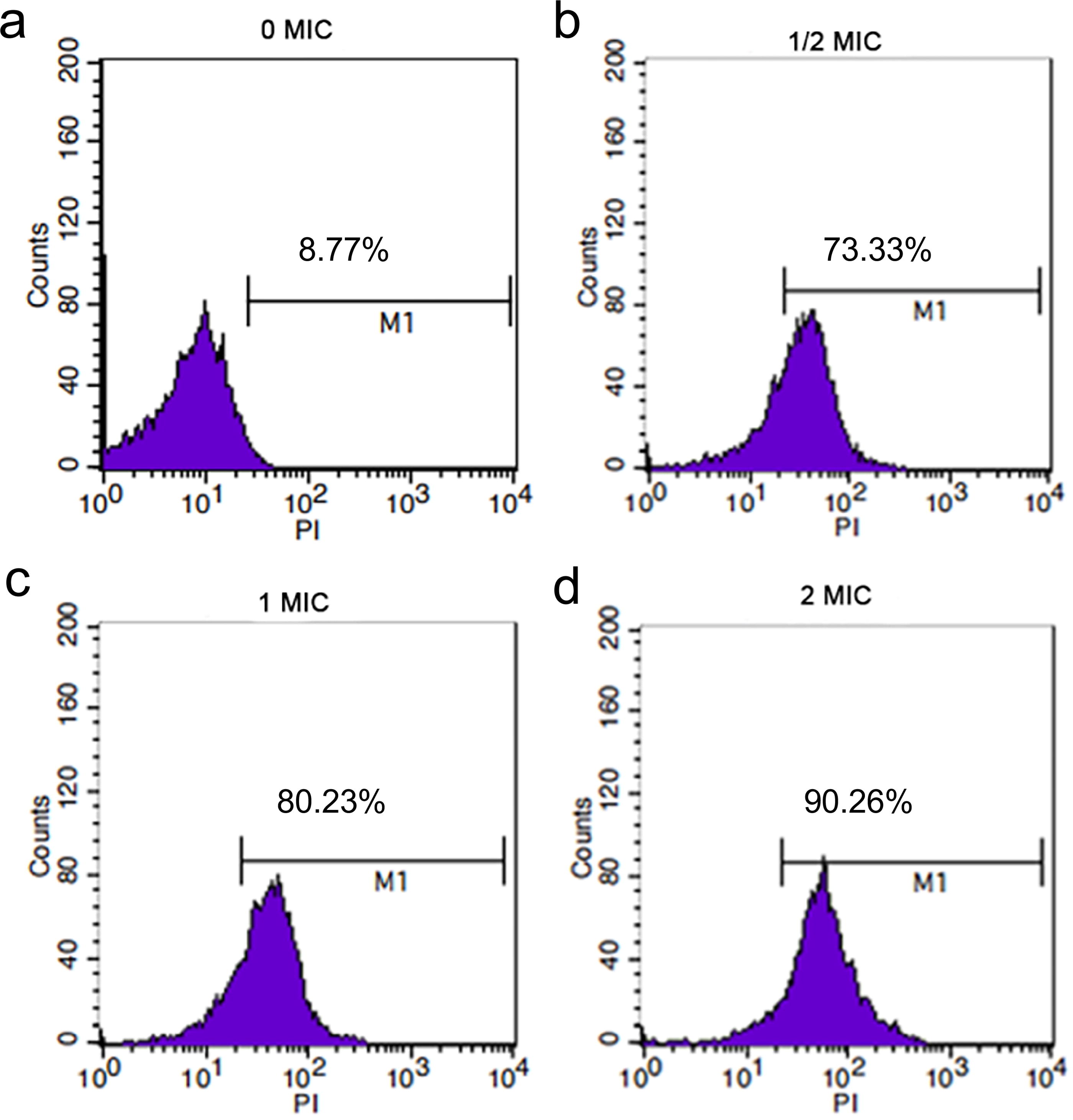
Effect of LA on cell membrane integrity of Staphylococcus aureus observed by flow cytometry.
Effect of LA on the morphology of S. aureus
Scanning electron microscopy can intuitively understand the changes in cell shape. In the control group, S. aureus cells were round, with clear and smooth cell boundaries and intact cell membranes and cell walls (Supplementary Fig. S2a). After 6 h of treatment with LA, the morphology of some bacteria was changed. The spherical shape, which was originally smooth, becomes irregular, and the cell boundaries become blurred. This can lead to impaired cell membrane function.
Effect of LA on S. aureus cell membrane fluidity
In the group treated with LA, the fluorescence intensity continued to increase within 0–8 h. The fluorescence intensity increased significantly (p < 0.05) from 32.80 ± 0.32 to 34.14 ± 0.76 after 2 h treatment with 1 MIC LA, and it is even more significant (p < 0.01) with 2 MIC LA (Fig. 2a). Regulation of the fatty acid composition of membranes is thought to be necessary to maintain their fluidity, but LA may insert itself to have an effect on the fluidity of cell membranes (Tsuda et al., 2019). Cheng et al. (2024) suggest that by inserting into the hydrophobic portion of the membrane, fatty acids can cause cell lysis or interfere with cellular metabolites disrupting the cell membrane.

Effects of LA treatment on cell membrane fluidity of S. aureus
Effect of LA on the membrane potential in S. aureus cells
3,3′-Dipropylthiadicarbocyanine iodide [DiSC3(5)] is a cationic fluorescent dye that measures the sensitivity of cell membrane potentials. As shown in Fig. 2b, the fluorescence intensity of S. aureus treated with LA changed dramatically and showed a dose-dependent manner. In addition, the control group remained stable. It indicated that LA had an influence on the membrane potential, resulting in the diffusion of extracellular sodium ions into the membrane, which reduced the potential gap of the membrane, and depolarized the cell. Parsons et al. (2012) treated S. aureus with exogenous fatty acids and observed membrane polarization and release of solutes and peptides into the medium. This is consistent with our results. In addition, the electron transport chain and oxidative phosphorylation, processes involving bacterial cell membranes, are essential for energy production. However, fatty acids have the potential to interfere with oxidative phosphorylation by lowering the membrane potential and proton gradient (Yoon et al., 2018).
Antibacterial activity of LA against S. aureus in cooked chicken
The antibacterial effect of LA on S. aureus in cooked chicken at 4°C is shown in Fig. 3a. After 1 day, the growth rate of the bacterial slowed down or started to decline after treatment. Fig. 3b shows the inhibitory effect of LA at 25°C. S. aureus showed a rapid growth state in all groups. The growth lag period of the S. aureus treated with LA was prolonged, and the number in the LA-treated group was always (p < 0.05) lower than that of control within 0–30 h.
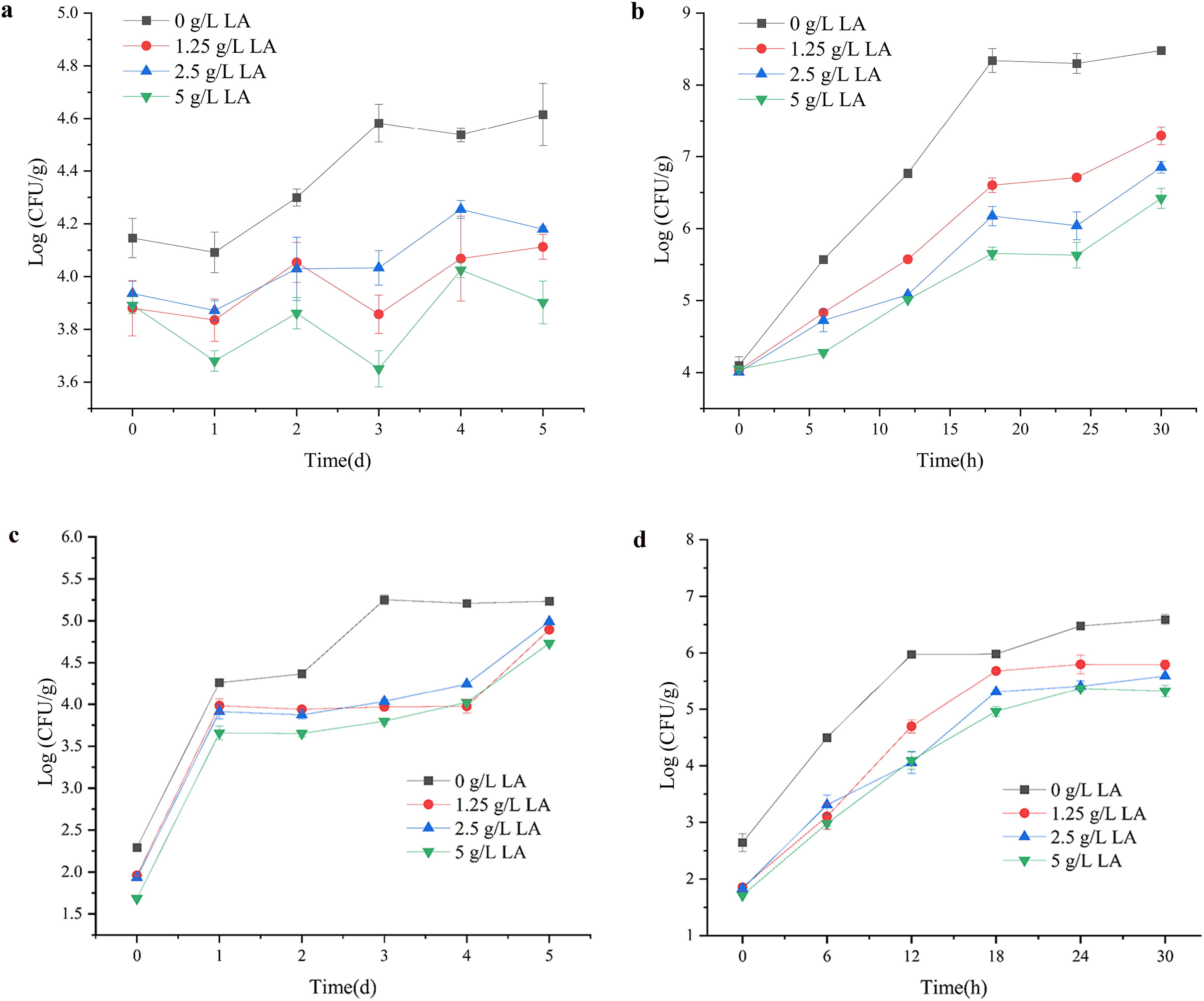
Antibacterial effect of LA treatment on S. aureus in cooked chicken at different temperatures:
LA has an inhibitory effect whether it is 4°C or 25°C (Fig. 3a and b). However, the combinatorial use of adding LA and placing it at a lower temperature may better inhibit the growth of S. aureus in cooked chicken. The inhibitory effect of S. aureus in cooked chicken was not as good as that on the culture medium system. This may be because bacteria on the surface of meat adhere firmly, reducing exposure to LA (Ouattara et al., 1997). On the contrary, LA itself is fat-soluble, and the protein and lipid components of meat can interact with its active antimicrobial components to dissipate some of them (Kim et al., 1995). Li et al. (2022) believed that the abundance of proteins in meat products helps bacteria repair damaged cells.
Effect of LA on microogranisms in cooked chicken meat during storage
TVC is suitable for characterizing microbial quality changes during storage (Kulawik et al., 2022). The effect of LA on TVC at 4°C is shown in Fig. 3c. The TVC (5.25 ± 0.06 log CFU/g) of control group on the third day exceeded 5 lg CFU/g, greater than stipulated by the Chinese National Standard for food safety (GB 2726-2016). In contrast, TVCs in the LA-treated group were less than the limit on day 5. It indicated that LA at 4°C could extend the shelf life by at least 2 days. The variation of TVC at 25°C is shown in Fig. 3d TVC in LA-treated group was lower than control group.
Results in Fig. 3c and d indicate that the application of LA at low temperatures may be better to perform its efficacy. Jiang et al. (2011) came to similar conclusions in their study, where they found that refrigerated storage improved the activity of edible coatings with added antimicrobial agents against Listeria monocytogenes on roasted turkey. In conclusion, LA under both temperature conditions had a certain inhibitory effect on the TVC in cooked chicken meat.
Effect of LA on pH and chromaticity in cooked chicken meat during storage
LA had no significant effect on the pH value of cooked chicken as shown in Table 1. L* values of all groups did not change significantly compared with the initial values over the 5d range (Table 2). However, the b* value of the control went up to 15.37 from 13.63 indicating an increase in the yellowness value. LA prevent the oxidation of certain lipids in cooked chicken, thereby reducing the degree of discoloration (Table 2). The L* value of the control stored at 25°C producing a change in chromaticity (Table 2). It is hypothesized that the change in color may be influenced by microbial growth, lipid oxidation, and pigment degradation and caused reduced acceptability of chicken meat (Hong et al., 2012; Nuñez De Gonzalez et al., 2008). Overall, the addition of LA not only did not affect the color of cooked chicken meat but also had a protective effect.
Effect of LA on pH Values in Cooked Chicken Meat During Storage at Different Temperatures (4°C and 25°C)
The values are average ± SD. Different lower case letters in the same column indicate significant (p < 0.05) differences in chicken meat PH values between treatments.
LA, lauric acid; SD, standard deviation.
Effect of LA on Chromaticity (L*, a*, and b* Values) in Cooked Chicken Meat During Storage at Different Temperatures (4°C and 25°C)
The values are average ± SD. Different lower case letters in the same column indicate significant (p < 0.05) differences in chicken meat chromaticity values between treatments.
LA, lauric acid; SD, standard deviation.
Effect of LA on TBARS values in cooked chicken meat during storage
The TBARS values of cooked chicken for the storage period at 4°C are shown in Fig. 4a. At day 5, the TBARS values of each LA-treated group were about 22.21%, 29.23%, and 28.52% lower than the control group. Moreover, the TBARS values of the LA-treated group were significantly (p<0.01) lower than those of control group, indicating that lipid oxidation could be reduced by LA. The TBARS values of all groups increased continuously at 25°C (Fig. 4b) and were lower in the LA-treated group than in control. A number of factors are associated with lipid oxidation, which is a complicated chemical process (Qi et al., 2022). LA may inhibit lipid oxidation in chicken meat by exerting an antioxidant effect on one or more of these factors, and the exact mechanism requires more research.
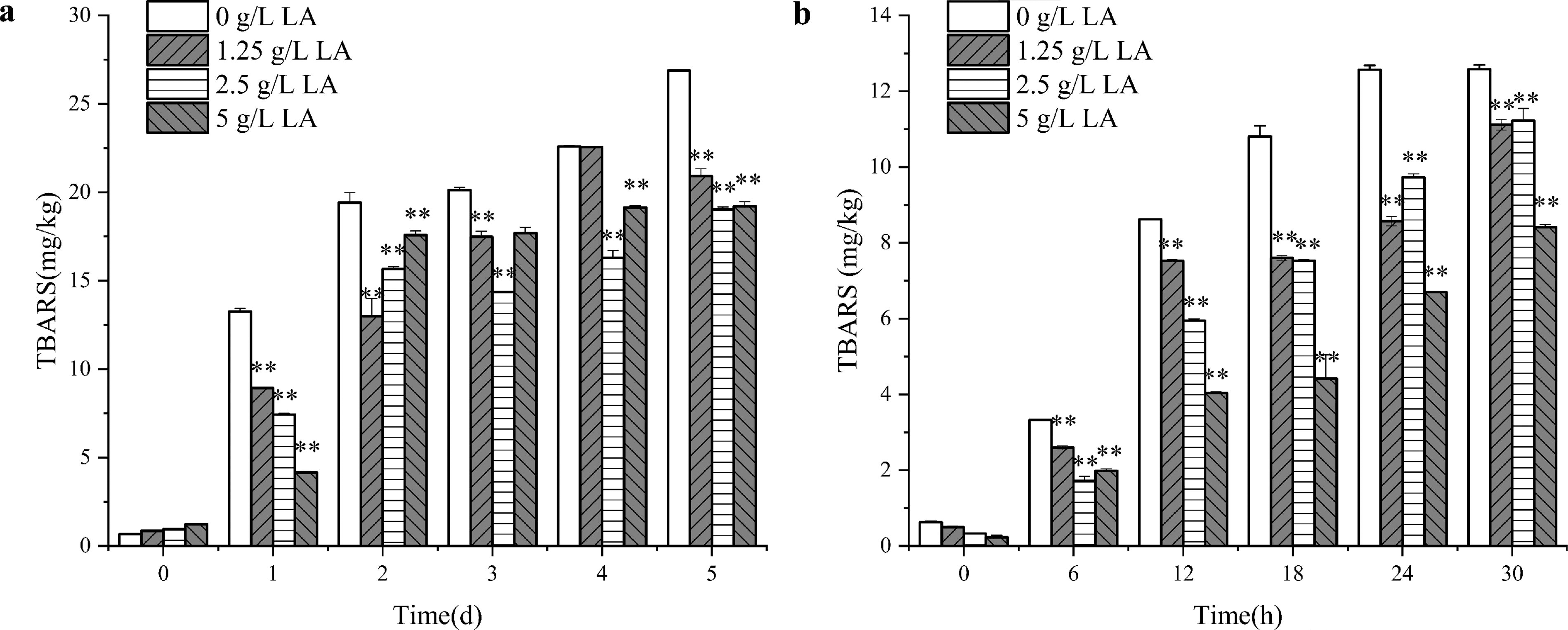
Effect of LA on TBARS in cooked chicken meat during storage at different temperatures:
Conclusions
The study investigated the antibacterial activity of LA against S. aureus and its mechanism. The results showed that LA-induced S. aureus damage through several pathways and was effective in cooked chicken meat. In conclusion, LA has the potential to be an ideal choice as a preservative in cooked meat products, improving food safety and nutritional value, and the antimicrobial effect of LA on different bacteria and its effect on food quality should be clarified in further research.
Footnotes
Acknowledgments
The authors gratefully acknowledge the fund supports.
Authors’ Contributions
L.Q.: Writing—original draft, software, data curation, and methodology. S.S.: Writing—original draft and formal analysis. L.S.: Conceptualization and data curation. X.Y.: Software and conceptualization. Y.H.: Methodology, funding acquisition, and supervision. Y.Y.: Methodology, formal analysis, funding acquisition, and supervision.
Disclosure Statement
No competing financial interests exist.
Funding Information
This work was supported by On-budget Capital Construction Fund of Jilin Province Development and Reform Commission (2023C014).
Supplementary Material
Supplementary Figures S1
Supplementary Figures S2
References
Supplementary Material
Please find the following supplemental material available below.
For Open Access articles published under a Creative Commons License, all supplemental material carries the same license as the article it is associated with.
For non-Open Access articles published, all supplemental material carries a non-exclusive license, and permission requests for re-use of supplemental material or any part of supplemental material shall be sent directly to the copyright owner as specified in the copyright notice associated with the article.
