
Abstract
The objective of this study was to determine the minimum growth temperature of Shiga toxin–producing Escherichia coli (STEC). Forty-eight strains of STEC, including E. coli O157:H7, O104:H4, O26, O45, O103, O111, O121, and O145, were inoculated into tryptic soy broth (TSB) at ca. 6.0 CFU/mL and incubated at temperatures ranging from 5°C to 11°C. The lowest temperature at which growth occurred was determined as the minimum growth temperature of the strain. The minimum growth temperature varied among strains, but the strain difference was within 2–3°C. All of the STEC strains grew at ≥10.3°C. Majority of the STEC strains (31/48) grew at 8.9°C, with some strains (10/48) being able to grow at as low as 8.0°C. None of the STEC serogroups were able to grow at ≤7.4°C. E. coli O104:H4 and O157:H7 had relatively lower minimum growth temperatures, with 8°C and average 8.4°C, respectively, whereas serogroup O26 had a higher minimum growth temperature (average 9.6°C). The results of this study provide basic but critical information on STEC growth and could be used either in fundamental research or to mitigate the risk of STEC from food products by storing them at temperatures below the minimal growth temperature.
Keywords
Introduction
Public health concern regarding non-O157 Shiga toxin–producing Escherichia coli (STEC) has risen significantly due to several major outbreaks in the United States and Europe. Notable examples in the United States within 5 years and the large outbreak caused by E. coli O104 that occurred in 2011 (European Food Safety Authority, 2011) were summarized by Walker and others (2024).
E. coli belonging to Enterobacteriaceae family are Gram-negative, facultative anaerobic, and rod-shaped bacteria. STECs are known for the Shiga toxin they produce, which can cause hemorrhagic colitis and hemolytic–uremic syndrome in humans. Many STEC outbreaks are linked to beef and dairy products, as cattle are the primary reservoir of these pathogens. E. coli O157, the STEC serotype often associated with the most severe diseases, is estimated to cause tens of thousands of illnesses and result in economic losses of hundreds of millions of dollars annually in the United States (United States Department of Agriculture, Economic Research Service, 2023).
E. coli are mesophilic bacteria that normally inhabit the gastrointestinal tract of humans and animals. Olsvik and Kapperud (1982) reported that E. coli could grow and produce heat-labile enterotoxin in culture broth (tryptic soy broth; TSB) and milk at 4°C. However, none of the subsequent reports indicated the psychrotrophic behavior of E. coli. STEC, including O157 and O111 (Paton and Paton, 1998). Once food is contaminated with STEC, it is necessary to maintain products at a temperature below the minimum growth temperature for the organism during processing and storage to prevent the growth. Therefore, knowledge of STEC’s minimum growth temperatures is critical to ensure food safety.
The USDA Food Safety and Inspection Service (FSIS) has classified six non-O157 STEC serogroups (O26, O45, O103, O111, O121, and O145), known as the “Big Six,” as adulterants, which would lead to product recalls, economic losses, and public health risks. In 2023, FSIS announced that it would begin testing all raw beef samples, including ground beef, beef trim, imported ground beef, and other raw ground beef components, for non-O157 STEC (United States Department of Agriculture, Economic Research Service, 2023). Previous studies examined how E. coli O157 respond to different environmental conditions (temperature, acidity, hydration, etc.). In the camel meat study, E. coli O157 was evaluated under refrigeration (4°C) and abusive temperature (10°C) conditions, whereas in the wheat berry study, the survival of E. coli O157 was assessed during tempering at different temperatures (15°C, 23°C, and 30°C) (Jung and Harris, 2023; Osaili et al., 2020). Palumbo and others (1995) reported the minimum growth temperatures of several STEC strains, including 14 strains of E. coli O157:H7 and 2 strains of E. coli O26:H11. Although more STEC strains were used according to the methodology of their study, the minimum growth temperatures of the rest of the STEC strains were not reported. Therefore, the objective of this study was to determine the minimum growth temperatures of different strains of the claimed six-serogroup STEC, E. coli O157:H7, and the E. coli O104:H4 isolate of 2011 German outbreak.
Materials and Methods
Bacterial cultures
The study utilized a total of 48 STEC strains as outlined by Walker et al., 2024, including E. coli O157:H7 (5 strains), E. coli O104:H4 (1 strain), E. coli O26 (7 strains), E. coli O45 (7 strains), E. coli O103 (7 strains), E. coli O111 (7 strains), E. coli O121 (7 strains), and E. coli O145 (7 strains).
Cultures were maintained individually at −70°C in glycerol-TSB mixture (80% glycerol:culture in TSB = 1:1). Fresh cultures were prepared individually by performing two serial transfers to TSB (Difco, Sparks, MD) with incubation for 18 h at 35°C. For each strain, 5 mL of the culture was centrifuged at 7000 × g for 10 min at 4°C (Beckman GS-15R, Fullerton, CA). The supernatant was discarded, and the bacterial cell pellet was reconstituted in 0.1% sterile peptone water (PW; Difco, Sparks, MD) and 10 times diluted in PW for further use as an inoculum.
Growth media preparation and inoculation
An aliquot (100 μL) of appropriate dilution of the STEC culture was inoculated into 10 mL of TSB broth to obtain ca. 6 log CFU/mL of STEC population individually and mixed well. An aliquot (200 μL) of each broth was pipetted into 2 wells of sterile microtubes (0.2 mL; Eppendorf AG, Hamburg, Germany), representing 2 replicates per strain tested, and the microtubes were placed in a 96-well aluminum block (TPS 620–5015; Cole-Parmer, Vernon Hills, IL). The blocks were stored in chilling/heating incubators (EW-44175–06; Cole-Parmer, Vernon Hills, IL) for 60 days set to target temperature of 5, 6, 7, 8, 9, 10, or 11°C. The temperatures of the incubators were stabilized for at least 24 h before use. A thermometer (CL3512A; Omega, Manchester, UK) and a data logger (TC-08; Omega, Manchester, UK) were used to monitor the temperatures of the broth in the microtubes. Actual temperatures of 5.6 ± 0.2°C, 6.6 ± 0.2°C, 7.4 ± 0.2°C, 8.0 ± 0.3°C, 8.9 ± 0.3°C, 10.3 ± 0.2°C, and 11.3 ± 0.2°C were achieved throughout the incubation period. Noninoculated TSB was incubated at 37°C for 60 days to serve as control.
Assessment of minimum growth temperature
Appropriate dilutions of the inoculated broth were prepared in PW and spread plated on tryptic soy agar (TSA; Difco, Sparks, MD), and the plates were incubated for 24 h at 37°C before colonies were counted. An initial STEC population of 6.0 ± 0.5 log/mL was achieved for each strain. Growth is considered present when there is an increase of more than 1-log CFU/mL (a 10-fold increase) during the incubation period, and no-growth is defined as an increase of less than 1-log CFU/mL or a decline in the bacterial population that was slightly modified according to Skandami and others (2007). Due to the bacteria’s optical density, E. coli will become visible to human naked eyes once it reaches 7 log CFU/mL. Development of turbidity and/or sediment in the medium indicated a ≥1 log CFU/mL growth. The minimum growth temperature was defined as the lowest temperature at which the growth was observed.
The microtubes in the incubators were observed at weekly intervals for up to 60 days. At the end of the incubation period, the samples closest to the growth boundary were vortexed, diluted, and plated on TSA to determine the bacterial population. The purity of the culture was checked by randomly picking the colonies on TSA and streaking on MacConkey Agar (BD, Sparks, MD).
Results and Discussion
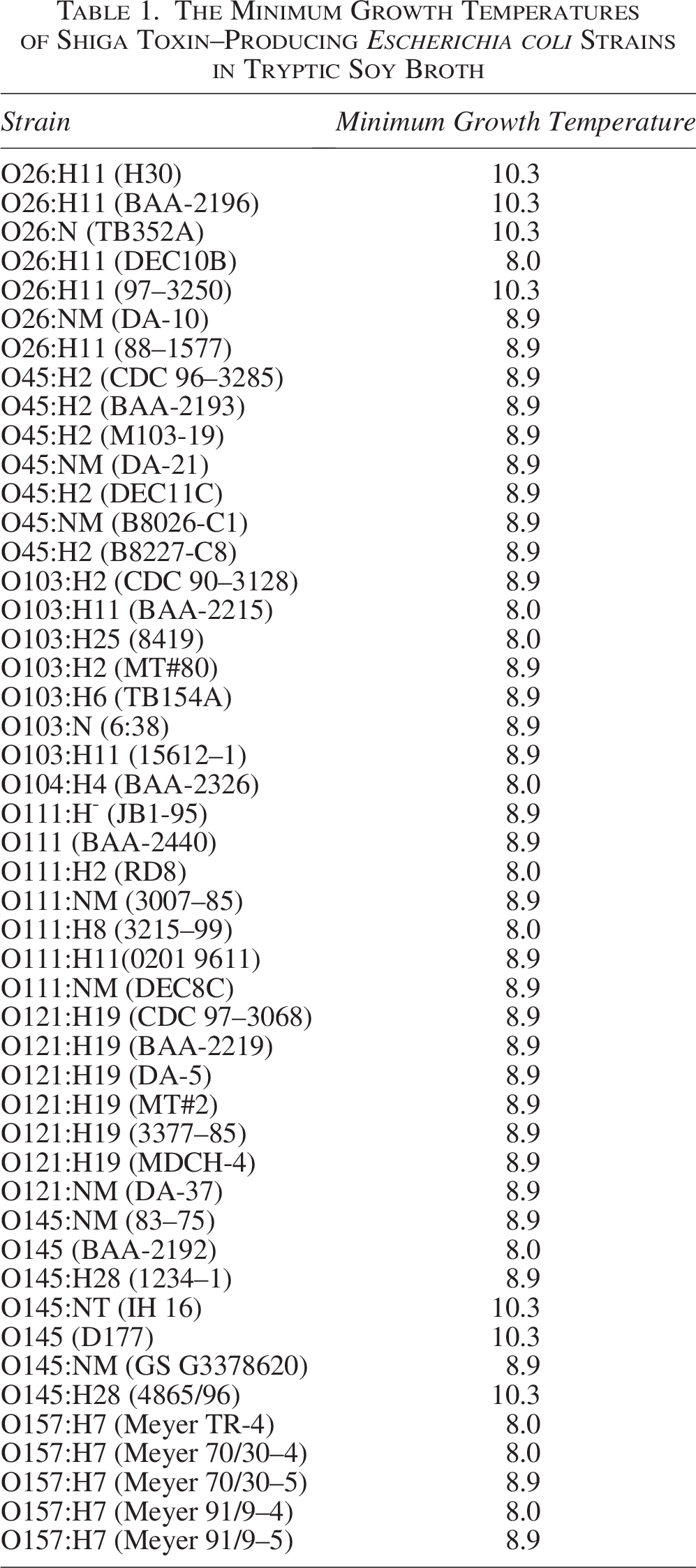
The minimum growth temperatures of the STEC strains ranged between 8.0 ± 0.3°C and 10.3 ± 0.2°C (Tables 1 and 2). Ridell and Korkeala (1997) reported that the minimum growth temperatures of the same Enterobacteriaceae species were similar, and the range was within 2°C or 3°C.
The Minimum Growth Temperatures of Shiga Toxin–Producing Escherichia coli Strains in Tryptic Soy Broth
The Mean Values and Ranges of Minimum Growth Temperatures of Shiga Toxin–Producing Escherichia coli in Tryptic Soy Broth
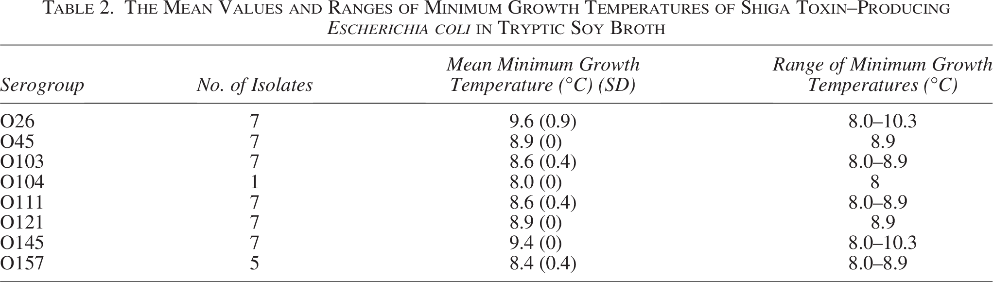
Figure 1 offers a visual representation of the distribution of minimum growth temperatures for the STEC serogroups, which highlights the variability within each serogroup more effectively. There was no variation observed in minimum growth temperatures between replicates of each tested strain. Most STEC strains (31/48; 65%) had a minimum growth temperature of 8.9°C, and 21% of the strains could grow at 8.0°C. The serogroup O26 had a higher minimum growth temperature with an average of 9.6°C, whereas E. coli O104:H4 had a lower minimum growth temperature of 8.0°C and 3 out of 5 of the E. coli O157:H7 could also grow at 8.0°C. All the strains that we tested for E. coli O45 and O121 had the same minimum growth temperature, and we did not see any variation across the strains. The combination of Table 2 and Figure 1 offers a thorough analysis of the minimum growth temperatures of STEC, providing essential insights for improving food safety protocols and guiding future research efforts in this field. Many studies reported similar minimum growth temperatures for E. coli, with examples as follows. Hoffman reported that the minimum temperature of growth for E. coli was about 8°C in a minimal glucose medium. A study on the Enterobacteriaceae isolates reported the range of minimum growth temperature of E. coli between 8.4°C and 8.9°C (Ridell and Korkeala, 1997). Shaw et al. (1971) reported that the minimum growth temperature of an E. coli strain (E. coli ML30) in glucose minimal medium was between 7.5°C and 7.8°C. Similarly, Palumbo et al. (1995) reported that the minimum growth temperature of 16 STEC strains in brain–heart infusion broth was 10°C, with only three strains of E. coli O157:H7 able to grow at 8°C, which is noteworthy considering that the minimum growth temperatures studied by Palumbo and others (1994) reported in 2°C increments were due to the low precision of the methodology used. Different from the rest of studies, Olsvik and Kapperud (1982) reported a much lower temperature (4°C) at which E. coli was able to grow and produce heat-labile enterotoxin in TSB and milk. Possible explanations for this difference may include the following: (1) aeration and agitation were applied to the culture during the incubation and (2) all the strains were stored on deep agar at 4°C before being prepared for inoculum, which might introduce the selection of cold-resistant cells during the storage period.

Distribution of minimum growth temperatures for STEC serogroups. *Each data point represents the minimum growth temperature observed for two replicates per strain. Error bars are not shown as there was no variation between replicates. STEC, Shiga toxin–producing Escherichia coli.
Temperature shift of bacterial cells from optimum to low temperature could result in a prolonged lag time of growth (Shaw et al., 1971) as a result of an accumulation of 70S ribosomal particles and a block in the initiation of protein synthesis by this temperature shift (Anderson, 1975; Broeze et al., 1978; Das and Goldstein, 1968). Therefore, longer incubation is needed to avoid the bias in the determination of the minimum growth temperature caused by temperature shift. In this study, the inoculated samples were incubated for up to 60 days, which was necessary as growth for some strains was observed after the first month of incubation. Thus, it is necessary to observe growth over a longer time when determining the minimum growth temperature of the organisms in specific matrices. In addition, STEC has a low infectious dose, and even minimal increases in bacterial populations can pose significant public health risks (Teunis et al., 2004). Therefore, our definition of growth may not capture slight bacterial increases that are still relevant for food safety. This is a limitation of our study, and future research should use more sensitive detection methods to assess the potential for low-level growth of STEC at temperatures below those reported here.
Recent studies show that while most mesophilic Enterobacteriaceae have limited growth potential at low temperatures, some strains exhibit a degree of cold adaptation. However, STEC’s inability to grow below 8°C is unusual even within the context of E. coli, which generally display better tolerance to cold environments. Recent studies have identified physiological mechanisms that may explain the differences in cold tolerance, suggesting that specific environmental stressors, such as cold temperatures, could uniquely affect the growth dynamics of STEC (Jung and Harris, 2023; Osaili et al., 2020). These findings emphasize the importance of considering the cold tolerance of STEC relative to other bacteria, contributing to a more comprehensive understanding of mesophilic bacterial behavior at low temperatures. Furthermore, stress factors such as acidic environments or low water activity may also play a critical role in shaping the growth competency of these bacteria under cold conditions. The minimum growth temperature is dependent on pH, water activity, and other factors of the food matrix, and it increases under stringent environmental conditions such as low pH and low water activity. The minimum growth temperature of each STEC strain in this study was measured in a microbiological medium that provided optimum growth conditions except for the temperature; thus, the minimum growth temperatures obtained from this study could be considered conservative estimates of the minimum growth temperatures for the organisms compared with what would otherwise be determined in food matrices. This needs to be taken into consideration when applying the results of this study to food preservation. It should also be noted that bacterial responses to temperature stress can be significantly influenced by their culture history and prior exposure to cold environments (Wouters et al., 2000). Cold adaptation, involving the induction of cold-shock proteins (CSPs) and other physiological changes, can enable bacteria to grow at lower temperatures than those observed under nonadapted conditions (Phadtare, 2004). In our study, the cultures were not subjected to cold adaptation before determining the minimum growth temperature. Therefore, the minimum growth temperatures reported here may be higher than those that could be observed in cold-adapted cells. Future research should investigate the impact of cold adaptation on the minimum growth temperature of STEC.
A primary component of bacterial adaptation to cold stress is the cytoplasmic membrane (Mykytczuk et al., 2010). Bacteria can adjust membrane lipid composition and modify the permeability of the membrane to minimize energy expenditure and optimize growth at low temperatures. Bacteria sense the low temperature by modulating membrane fluidity, which induces increases in gene transcription for membrane proteins and enzymes (primarily desaturases) involved in fatty acid biosynthesis (Mykytczuk et al., 2010). The fatty acid chains in membrane phospholipids define the viscosity of the membrane, which in turn affects the permeability and other crucial membrane-associated functions of the bacterium. One of the approaches bacteria use to adjust membrane viscosity is modification of the structure of existing fatty acids, which is necessary for bacterial rapid adaptation to the new environment (Zhang and Rock, 2008). The conversion of saturated and trans fatty acids into unsaturated and cis fatty acids by the enzymes desaturases and cis–trans isomerase is known to increase membrane fluidity at low temperatures (Chattopadhyay and Jagannadham, 2001). The other approach for bacteria to adjust membrane viscosity is the formation of new fatty acids (Zhang and Rock, 2008). Synthesis of short-chain fatty acids, branched-chain fatty acids, and anteiso-fatty acids to maintain an optimum fluidity of bacterial cell membrane at low temperatures has been reported (Saunders et al., 2016). The synthesis of these fatty acids for the maintenance of membrane fluidity is not only through anabolic pathways but also by making use of catabolism (Chattopadhyay and Jagannadham, 2001). The fatty acyl chain composition of E. coli membrane lipids was reported to vary with the growth temperature as follows: the fraction of cis-vaccenoyl chains (18:1c11) decreased, and the fraction of palmitoyl chains (16:0) increased, when the incubation temperature was increased from 17°C to 37°C (Morein et al., 1996). The authors suggested that regulating the membrane lipid composition was necessary for E. coli to grow between a lamellar gel (lipid bilayer) phase and nonlamellar (nonbilayer) phases. It is worth noting that the regulation of membrane fluidity and permeability not only facilitates bacteria to adapt to temperature change but also helps to respond to other environmental changes and exposure to antimicrobials (Zhang and Rock, 2008).
Besides the maintenance of membrane fluidity, bacteria overcome cold stress by other mechanisms, such as CSPs, that are recognized as transcriptional activators or as mRNA chaperones to facilitate transcription and translation at low temperatures (Chattopadhyay and Jagannadham, 2001). A major CSP, CspA, was first reported in E. coli and then its homologs were detected in a variety of bacteria, including thermophiles, mesophiles, and psychrophiles (Graumann and Marahiel, 1998). Although belonging to the same family (Enterobacteriaceae), E. coli and Yersinia enterocolitica present apparently different responses to temperature; the former is a mesophile, whereas the latter is a psychrotroph. Compared with mesophilic bacteria, psychrotrophic bacteria not only transiently induce CSPs following a cold shock but also overexpress cold acclimation proteins (Caps) during prolonged growth at low temperatures (Hébraud and Potier, 1999). The presence of Caps plays a fundamental role in cold-adapted microorganisms and differentiates them from mesophiles. These proteins are reportedly involved in maintaining membrane fluidity and replacing cold-denatured peptides (Hébraud and Potier, 1999). Accordingly, the bacterial mechanisms in response to cold stress are interlinked with the intricacy of cellular machinery (Chattopadhyay and Jagannadham, 2001). Another limitation of our study is the potential presence of viable but nonculturable (VBNC) cells, which may not have been detected using our culture-based methods. Bacteria can enter the VBNC state under stressful conditions, such as low temperatures, and while they are not detectable by standard culturing techniques, they can retain pathogenicity and resuscitate under favorable conditions (Li et al., 2014). Future studies should explore methods to detect and recover VBNC cells to provide a more comprehensive understanding of STEC survival and growth at low temperatures.
Conclusion
The minimum growth temperatures for 48 STEC strains, including E. coli O157:H7, O104:H4, O26, O45, O103, O111, O121, and O145, were measured. The minimum growth temperature was strain dependent. All of the STEC strains were able to grow at ≥10.3°C. Most of the STEC strains (31/48) were able to grow at 8.9°C, with some strains (10/48) of serogroup O26, O103, O111, O145, O157:H7, and O104:H4 able to grow at as low as 8.0°C. Growth was not observed at ≤7.4°C. The results of this study provide basic but important information on the minimum growth temperature of STEC and could be used in either fundamental research or food preservation to control STEC growth. One limitation of our study is the use of laboratory-based conditions, specifically TSB, rather than food matrices. While this approach allowed for controlled comparisons, it may not fully reflect the behavior of the bacteria in real-world food environments. In addition, cold adaptation was not accounted for, which could impact the minimum growth temperatures observed. Strain diversity is another factor that could influence the generalizability of our results and the possibility that our growth detection methods may overlook slight increases significant for food safety. Future studies should address these factors to enhance understanding STEC growth and survival in both research and practical applications.
Authors’ Contributions
L.W.: Conceptualization, data curation, formal analysis, investigation, methodology, project administration, funding, software, formal analysis, and writing—original draft. S.S.: Conceptualization, methodology, resources, project administration, funding, software, review, and editing. H.T.: Conceptualization, formal analysis, methodology, project administration, resources, supervision, visualization, and writing—review and editing.
Footnotes
Acknowledgments
The authors would like to thank the Scientific Research and Innovation Fund of Yantai Institute of Technology for High-level Talents (RC20SP001) and the Food Safety and the Sensory Quality Academic Team (2023XSTD03) for their support and contribution.
Funding Information
This work was supported by the USDA-NIFA STEC Coordinated Agricultural Project (2012-68003-30155).
Disclosure Statement
The authors have no conflict of interest.