




Abstract
The six major non-O157 serogroups of Shiga toxin–producing Escherichia coli (STEC) are responsible for serious foodborne outbreaks worldwide. This research aimed to detect the six major non-O157 STEC in ground beef, artisanal dairy products, lettuce, spinach, turkey, and chicken sold in northern Morocco. Real-time polymerase chain reaction was utilized to identify the presence of the stx1, stx2, eae, wzx O26, wzx O45, wzx O103, wbdl O111, wzx O121, and ihp1 O145 genes. Out of 310 samples analyzed, Shiga toxin (stx) was detected in 55 enrichments (17.74%), stx, and eae were detected in 54/310 enrichments (17.42%), stx, eae, and genes of at least one of the six serogroups were detected in 34/310 enrichments (10.97%). Among the food matrices analyzed, ground beef showed the highest contamination rate with stx, eae, and O serogroups 13/70 (18.6%), followed by dairy 17/100 (17.00%), turkey 3/40 (7.5%), and chicken 1/40 (2.5%). No O serogroups were detected in lettuce and spinach. The most frequent serogroup was O26 (22/34; 64.7%), followed by O145 (12/34; 35.3%), O45 (12/34; 35.3%), O121 (8/34; 23.5%), O103 (8/34; 23.5%), and O111 (6/34; 17.6%). A set of 32 STEC strains were isolated from nine positive samples (9/34; 26.5%). A high rate of food contamination with STEC may indicate firstly a high public health risk due to this pathogen in beef and dairy products and secondly a lack of compliance with standard hygiene practices. Consequently, it emphasizes the urgent need for rigorous monitoring and intervention measures aimed at mitigating the incidence of STEC contamination.
Introduction
Implicated in many major foodborne outbreaks, Shiga toxin–producing Escherichia coli (STEC) is responsible for 2.8 million cases of acute illnesses worldwide on an annual basis between 1990 and 2012 and causes 3890 instances of hemolytic uremic syndrome (HUS), 270 cases of end-stage renal disease, and 230 fatalities (Majowicz et al., 2014). It was recently concluded that all strains of STEC, whatever their serotype, are pathogenic for humans, capable of causing at least diarrhea (EFSA BIOHAZ Panel et al., 2020). However, there are seven serogroups (O157, O26, O45, O103, O111, O121, and O145), which are considered highly pathogenic and cause serious illness, ranging from hemorrhagic colitis to HUS and thrombotic thrombocytopenic purpura (TTP) (Brooks et al., 2005; Brugère et al., 2011). O157 is the most virulent serogroup of STEC, followed by the six major non-O157 serogroups (O26, O45, O103, O111, O121, and O145), which generally cause milder diseases than those induced by O157 (EFSA BIOHAZ Panel et al., 2020; Mathusa et al., 2010; Smith et al., 2014).
The most virulent factors responsible for the pathologies caused by STEC are Shiga toxin (stx) and the adhesion factor “intimin” encoded by the eae gene (Melton-Celsa, 2014; Yang et al., 2020). Once ingested, STEC adheres tightly to intestinal cells, thanks to intimin, which causes attachment-effacement lesions of enterocytes that can lead to hemorrhagic colitis (Brugère et al., 2011). Shiga toxin produced at this level crosses the epithelium of the intestines toward the bloodstream to reach the target cells (vascular, kidney, and brain cells) where it will attach and cause the cessation of protein synthesis, cell apoptosis, and the formation of microthrombi which are the cause of HUS and TTP (Brugère et al., 2011; Melton-Celsa, 2014).
The intestines of warm-blooded animals are the reservoir for STEC, specifically cattle, sheep, and goats which are healthy carriers (Forano et al., 2013). Consumption of raw or undercooked food contaminated with a low dose of STEC is sufficient to cause food poisoning that can develop into HUS and TTP (Eißenberger et al., 2018). Ruminant meat, vegetables, fruits, and dairy products are often the foods most attributed to STEC poisoning (FAO and WHO, 2018; Ouarroud et al., 2024).
Unfortunately, there are no data on human illnesses caused by STEC in Morocco and no surveillance program for STEC in food. Therefore, the prevalence of STEC in food and the sources of contamination are unknown. Monitoring food products at risk of containing STEC remains a necessary step to avoid poisoning with these pathogens in Morocco. This study is the first in Morocco to investigate the prevalence of the six major non-O157 STEC serogroups in ground beef, dairy, lettuce, spinach, turkey, and chicken. This study should contribute to improving Moroccan risk management.
Materials and Methods
Detection of STEC in food was done in accordance with the (ISO/TS 13136, 2012) standard and (LMAP/DGAL/Screening PCR STEC-al.2, 2015) method. These methods include the following consecutive steps: microbial enrichment, DNA extraction, detection of virulence genes (stx and eae), detection of genes associated with serogroups, and isolation of strains from potentially positive samples. The sample collection step is not included in this standard.
Sample collection
A total of 310 samples were taken over a period of time extended from January 2022 to March 2024. The samples taken are: ground beef (n = 70), artisanal dairy products (raw milk [n = 25], fermented milk [spontaneous fermentation of raw milk] [n = 25], raw milk cheese [n = 25], and butter [n = 25]), leafy vegetables (lettuce [n = 30] and spinach [n = 30]) and poultry meat (chicken [n = 40] and turkey [n = 40]). The choice of food matrices analyzed was based on foods implicated in STEC outbreaks reported worldwide. The samples were taken from 56 areas in Tangier city. Ground beef and poultry were taken from butcher shops, dairy products were collected from vendors of artisanal dairy, and leafy vegetables were collected from vegetable vendors. The samples were collected in sterile bags and transported immediately to the laboratory in a cooler, at a temperature between 4°C to 8°C. The collection of different samples was carried out every 2 months.
Sample enrichment
A quantity of 25 g of sample was suspended in 225 mL of buffered peptone water contained in a stomacher bag, homogenized in a stomacher, and incubated at 37°C for 18 to 24 h.
DNA extraction
1 mL of the enrichment was taken into an Eppendorf tube and centrifuged at 12,000 × g for 4 min. The pellet was used for DNA extraction using the “NucleoMag® DNA Bacteria” kit (Macherey-Nagel), following the protocol of manufacturer. This DNA extraction method is based on magnetic beads.
Gene detection by PCR
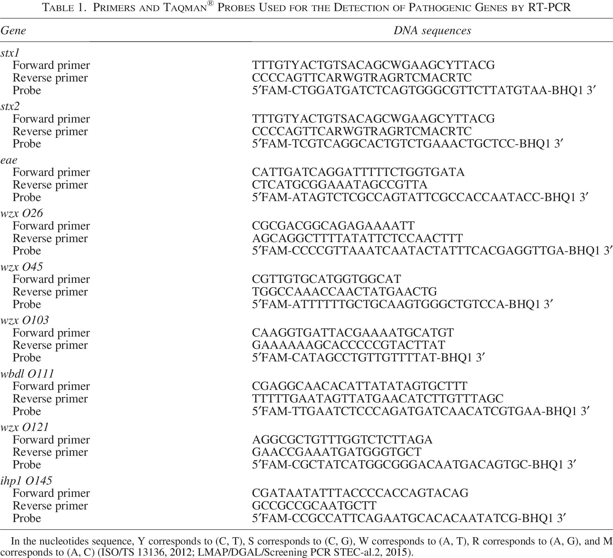
Simple real-time polymerase chain reaction (RT-PCR) was used for the detection of pathogenic genes stx1, stx2, eae, wzx O26, wzx O45, wzx O103, wbdl O111, wzx O121, and ihp1 O145. PCR was executed in a 20 µL reaction mixture composed of 10 µL of master mix (TaqMan™ Fast Universal PCR Master Mix [2X]), 4.5 µL of DNA-free water, 0.6 µL of forward primer [10 µM], 0.6 µL of reverse primer [10 µM], 0.3 µL of probe [10 µM], and 4 µL of DNA.
The sequences of primers and Tasman® probes used are presented in (Table 1). They are cited in (ISO/TS 13136, 2012) and (LMAP/DGAL/Screening PCR STEC-al.2, 2015). All primers and probes were provided by “BIO BASIC CANADA INC.” Forward and reverse primers used for the detection of sox allow the detection of all variants of stx1 and stx2 except the stx2f variant.
Primers and Taqman® Probes Used for the Detection of Pathogenic Genes by RT-PCR
E.coli (O26:H11) CDC 03-3014 (STEC), E.coli (O45:H2) CDC 00-3039 (STEC), E.coli (O103:H11) CDC 06-3008 (STEC), E.coli (O111:H8) CDC 2010C-3114 (STEC), E.coli (O121:H19) CDC 02-3211 (STEC), and E.coli (O145:NM) CDC 99-3311 (STEC) (Microbiologist®, UKCA) were the strains used as positive controls for the PCR amplifications.
The thermal cycler used for DNA amplification is QuantStudio 1 (by Applied Biosystems). The program of temperatures used is step1: 95°C for 10 min, step 2: 94°C for 10 s, and step 3: 62°C for 45 s.
Isolation of STEC strains from positive samples
The isolation of STEC strains was done in accordance with the LNR (National Reference Laboratory) method (LMAP/DGAL/confirmation isolation STEC-al.1, 2015). This method derived from (ISO/TS 13136, 2012), is intended to isolate STEC strains from enrichment broths detected as positive after an initial screening of target genes by RT-PCR. The isolation steps are as follows.
Step 1: Inoculation of the enrichment broth onto TBX agar: After homogenization by vortexing, the enrichment broth was inoculated using a 10 µL loop onto TBX agar (Tryptone Bile X-glucuronide) (Biolife, Italy) and incubated at 37°C for 18–24 h.
Step 2: Random collection of 50 colonies. An isolated colony was picked using a loop, and a streak was traced on the TSA agar (Tryptic Soy Agar) (Biolife, Italy). Each 10 colonies were inoculated in the same petri dish (5 pools of 10 colonies were made) and then incubated at 37°C for 18–24 h. The 10 colonies from each pool were dissolved in a tube containing 1 mL of sterile distilled water.
Step 3: Bacterial DNA was extracted according to the protocol cited above (2.3).
Step 4: Detection of STEC by RT-PCR: stx1, stx2, eae, wzx O26, wzx O45, wzx O103, wbdl O111, wzx O121, and ihp1 O145 genes were amplified by RT-PCR, according to the protocol cited above (2.4).
Step 5: When an STEC was detected in a pool, we returned to the petri dish to isolate the positive colony. Colonies from the positive pool were individually extracted and tested by PCR until the pathogenic strain was found.
Step 6: Positive strains were conserved.
Results
Among 310 samples analyzed, stx (stx1 or/and stx2) was detected in 55 enrichments (17.74%). stx and eae were detected both in 54/310 enrichments (17.42%). Positive enrichments for stx and eae were screened for the presence of the six non-O157 serogroups (O26, O45, O103, O111, O121, and O145) genes. stx, eae, and genes of at least one of the six serogroups were detected in 34/310 samples (10.97%) (Table 2). Ground beef was the food matrix most contaminated with stx, eae, and O serogroups 13/70 (18.6%), followed by dairy 17/100 (17.00%), turkey 3/40 (7.5%), and chicken 1/40 (2.5%) (Table 2).
Prevalence of stx, eae, and non-O157 Serogroups Genes in Samples Analyzed
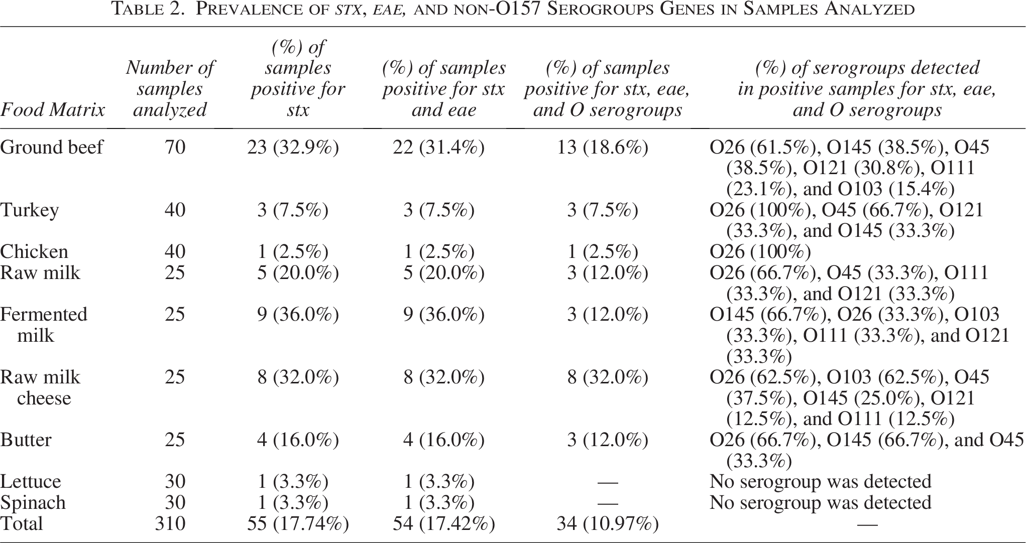
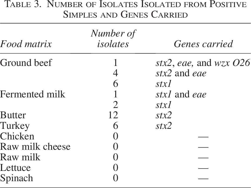
Among the 34 samples with O serogroups identified, the most frequent serogroup was O26 (64.7%), followed by O145 (35.3%), O45 (35.3%), O121 (23.5%), O103 (23.5%), and O111 (17.6%) (Table 2). It should be noted that in certain enrichments, more than one O serogroup was detected. The isolates isolated from positive samples are presented in (Table 3).
Number of Isolates Isolated from Positive Simples and Genes Carried
Discussion
This study detected STEC and non-O157 serogroups in foods consumed by Moroccans, specifically ground beef and dairy products. This discovery highlights the substantial risk faced by consumers, particularly those who consume raw foods such as dairy products. The present study confirms that Moroccan ground beef and artisanal dairy products are vehicles of STEC and are foods at high risk of causing food poisoning. This may be due to the fact that these products are derived from beef and dairy cattle, which are considered major carriers of STEC. Poultry can also be vehicles for STEC, especially turkey. On the contrary, the low prevalence of STEC in lettuce and spinach means that the risk linked to vegetables is minimal compared to other food categories.
The USDA’s (U.S. Department of Agriculture) Food Safety and Inspection Service has declared seven STEC serogroups (O26, O45, O103, O111, O121, O145, and O157) as adulterants in raw beef products (USDA and FSIS, 2012). Ground beef is known to be a vehicle for non-O157 STEC. Etcheverría et al. (2010) found STEC in 40.74% of ground beef samples in Argentina (Etcheverría et al., 2010). Mohammed et al. (2014) detected non-O157 STEC in 16.7% (5/30) of ground beef and 33.3% (10/30) of beef burger samples in Egypt (Mohammed et al., 2014). Toro et al. (2018) detected non-O157 STEC in 43/430 (10%) of samples in Chile (Toro et al., 2018), and Lucatelli et al. (2024) reveled STEC in 1/248 (0.4%) of ground beef samples in Brazil (Lucatelli et al., 2024). According to Bosilevac and Koohmaraie (2011), non-O157 serogroups in ground beef were classified as follows: O103 (7.7%), O26, and O121 (5.5% for each), O45 (4.7%), O111, and O145 (0.05% for each) (Bosilevac and Koohmaraie, 2011). The prevalence pattern of serogroups reported in the previous study was different from that observed in the present study. The high prevalence of STEC detected in ground beef in this study can be explained by the contamination of carcasses at the slaughterhouse, during storage, transport, distribution, and during processing at butcher shops (Etcheverría et al., 2010; Onyeka et al., 2024).
Consumption of artisanal dairy products is common in Morocco, and most of these products are made from raw milk. The contamination of milk and dairy products by STEC revealed in this study shows that this type of product can be a risk for consumers. Studies by other researchers have revealed contamination of dairy by STEC. Perelle et al. (2007) detected STEC in 43/205 (21%) of raw milk samples in France (Perelle et al., 2007). This prevalence is close to the prevalence obtained in our study (20.0%). A smaller prevalence was detected by Arikè Salifou et al. (2020) (2/25; 8%) in Benin (Arikè Salifou et al., 2020). Madic et al. (2011) reported that 29.8% (119/400) of raw milk cheese samples were contaminated with STEC in France. The O26 serogroup was the most detected (4.8%), followed by O103 (1.3%) and O145 (0.8%) (Madic et al., 2011). Guzman-Hernandez et al. (2016) found that 17% (9/52) of traditional cheese samples were contaminated with non-O157 STEC in Mexico (Guzman-Hernandez et al., 2016).
Dairy cattle are known reservoirs of non-O157 STEC (Rapp et al., 2021). Contamination of milk by STEC can occur during milking either by dairy cows (excreting STEC in milk or carrying STEC on their udders, skin, and hair), contact with feces, contaminated milking equipment, the environment or by milking workers. Milk can also be contaminated during storage and transport (EFSA BIOHAZ Panel et al., 2020; FAO and WHO, 2020). Contamination of dairy products with STEC can come from already contaminated milk and can also occur during processing operations through contaminated equipment, handlers, or by transformation environment (FAO and WHO, 2020).
Fresh leafy vegetables are considered a source of pathogenic bacteria, including STEC (Berger et al., 2010). This is consistent with the results obtained in this study. Mazaheri et al. (2014) found that 8/100 of lettuce samples were contaminated with STEC in Iran (Mazaheri et al., 2014). Arrais et al. (2020) detected STEC in 3.33% (1/30) of lettuce samples in Brazil (Arrais et al., 2020). This is the same prevalence obtained in our study. Regarding spinach, Gökmen et al. (2024) detected STEC O145 in 3.7% of spinach samples in Turkey (Gökmen et al., 2024). This prevalence is close to the prevalence obtained in our study. Contamination of lettuce and spinach by STEC can occur in the field (contaminated soil, irrigation water, air, or direct contact with workers and animals), during harvesting, transportation, and processing (EFSA BIOHAZ Panel et al., 2020; Ma et al., 2014; Tyagi et al., 2019).
Chicken is rarely attributed to foodborne STEC outbreaks, with a percentage less than 0.1% (FAO and WHO, 2019). This study revealed a low prevalence of STEC in chicken, but the risk of contamination of chicken meat by these pathogens is present. Other researchers have reached the same conclusion (Elsayed et al., 2021; Sokolovic et al., 2022). Our result was lower than that found by Dishan et al. (2023), who detected STEC in 35/100 of chicken samples in Turkey (Dishan et al., 2023), and Sarwar et al. (2024), who detected non-O157 STEC in 12% (3/25) of chicken meat samples in Saudi Arabia (Sarwar et al., 2024). Other studies conducted on the detection of STEC in chicken did not detect any strains of STEC (Lucatelli et al., 2024). The mode of transmission of STEC to chickens is poorly understood. Ayoade et al. (2022) revealed that chicken slaughterhouses are low reservoirs of STEC (Ayoade et al., 2022). Cross-contamination in butcher shops with other types of meat can occur. Studies on the detection of STEC in turkey are very rare, perhaps because turkey has never been cited as a source of STEC outbreaks between 1998 and 2017 in the Americas, Europe, and Western Pacific regions (Pires et al., 2019). It is therefore difficult to understand the source of contamination of turkey by STEC.
Conclusion
The present study is the first in Morocco to investigate the prevalence of the six major non-O157 STEC in various food matrices. The findings of this study revealed the contamination of different food categories, particularly ground beef and dairy. These results suggest the importance of adherence to standard hygiene practices. Furthermore, it emphasizes the urgent need for rigorous monitoring and intervention measures aimed at mitigating the incidence of STEC contamination.
Research Limitations
This research is a basic study and serves as a reference to develop other more in-depth studies, allowing a better understanding of food contamination by STEC in Morocco. As with the majority of studies, the current study is subject to limitations that could be addressed in future research. The first is the geographical limitation. The second limitation concerns the sampling bias; other food matrices must be analyzed. The third is the limited temporal scope and the fourth is the limited number of samples analyzed.
Authors’ Contributions
B.O.: Conceptualization, data curation, formal analysis, investigation, methodology, writing—original draft. M.E.M.: Conceptualization, project administration, visualization, writing—review and editing. A.K.: Writing—review and editing. L.E.M.: Methodology. A.B.: Supervision, validation, writing—review and editing.
Footnotes
Acknowledgments
B.O. thanks all coauthors for their valuable advice, their valuable guidance, their help, their support, and their availability. The authors would like to thank also the staff of the “Regional laboratory for analysis and research, national office for food safety (ONSSA)” for their technical support, knowledge sharing, advice, and help.
Funding Information
This research did not receive any specific grant from funding agencies in the public, commercial, or not-for-profit sectors.
Disclosure Statement
No competing financial interests exist.