
Abstract
Turmeric (Curcuma longa) is a spice widely used worldwide. It has been used in the treatment and prevention of diseases since ancient times. During harvesting and storage, turmeric is exposed to contamination by different fungi that are responsible for mycotoxins production. Mycobiota and mycotoxins associated with turmeric spices samples were screened in this study. Aspergillus was the dominant genus, it recorded with an average total counts of 4.76 log colony-forming unit/g, and appeared in all 40 tested samples. Molecular identification with internal transcribed spacer sequence was used to identify the species. The common species were Aspergillus flavus (with 80% frequency of occurrence) and Aspergillus niger (100%). Twelve and 20 samples were contaminated with total aflatoxins (AFs) and ochratoxins A (OTA) with concentrations 1.8–2.8 and 3.3–4.6 Parts Per Billioni (PPB), respectively. The search for suitable alternative antimicrobial drug resistance has increased, and this led researchers to explore the use of plant extract in the treatment of infections in both humans and animals. The antimicrobial activity of turmeric with two samples (sample no. 13 positive AFs and OTA, sample no. 2 with no toxins) on selected Candida albicans, Escherichia coli, and Staphylococcus aureus was evaluated. Different types of turmeric extracts were prepared using three solvents namely water, methanol, and ethanol using the disc diffusion method. The two tested turmeric samples extracts showed inhibition activity against all tested microorganisms. The zones of inhibition exhibited by extract of turmeric in methanol solvent from sample no. 13 against test organisms more effective and ranged from 10.6 to 15.4 mm. This is due to the confusion between antimicrobial activity of extract and its positively to mycotoxins content.
Introduction
Turmeric is widely used in the food industry for its natural coloring, flavor enhancement, and health benefits. Its vibrant yellow hue, derived from curcumin, is a popular natural alternative to synthetic dyes. Turmeric is used in a variety of products, including mustards, snack bars, baked goods, soups, and beverages. Its earthy flavor complements many dishes, and its antioxidant and anti-inflammatory properties make it a favored ingredient in health-focused products like teas, smoothies, and supplements (Gul and Bakht, 2015; Jikah and Edo, 2024; Verma et al., 2021). It is being researched with the possibility of using it to prevent and cure metabolic illnesses because of its safe and effective effects as an antioxidant, anti-inflammatory, antibacterial, antitumor, antidiabetic, and anti-obesogenic agent (Servida et al., 2024).
Turmeric is often grown in tropical regions with high humidity, which can promote the growth of molds. Poor handling, inadequate drying, and improper storage can exacerbate this issue. Fungi produce mycotoxins, which are harmful secondary metabolites, on a variety of edible substrates. Mycotoxin-producing fungi that are most frequently found are Aspergillus, Penicillium, and Fusarium species. Certain species within these genera possess the capacity to generate various mycotoxins, including citrinin, ochratoxin, and aflatoxins (AFs). Certain Aspergillus species naturally produce secondary metabolites called AFs, which can cause cancer (Qureshi et al., 2014). Most Aspergillus and Penicillium species also produce ochratoxin A (OTA) and Citrinun (CTN). When these two mycotoxins coexist, hepatorenal carcinogenesis results because OTA is hepatotoxic and CTN is nephrotoxic (Jeswal, 1995; Wichmann et al., 2002). (Thirumala-Devi et al., 2001) assessed the contamination levels of Ochrat toxin A (OTA) in various spices, including turmeric. His results showed that the highest concentration of OTA was found in paprika, while turmeric also showed significant contamination levels.
According to Chainani-Wu et al. (2007), curcuminoids are components of turmeric that mostly consist of curcumin (diferuloylmethane), demethoxycurcumin, and bisdemethoxycurcumin. The main significant component that gives turmeric its biological properties is curcumin (Sasikumar, 2005). Curcumin exhibits antimicrobial activity against a wide range of pathogens, including bacteria, viruses, fungi, and parasites (El-Saadony et al., 2022; Praditya et al., 2019; Prajapati et al., 2021; Pundir and Jain, 2010).
The present study aimed to characterize the mycobiota associated with turmeric spices by molecular identification, natural occurrence of AFs and ochratoxins, and antimicrobial activity of turmeric.
Materials and Methods
Sampling
Forty samples of turmeric powder were acquired at random from retail marketplaces in Jeddah (western Saudi Arabia). The retail shop conditions were ambient temperature (30–35°C), relative humidity (55–70%), and a sample storage time of 4–6 months. Turmeric samples were 150 g per sample. The samples were stored in sterile plastic bags at 4°C until further examination.
Mycobiota determination
Dichloran 18% Glycerol (DG18) agar, obtained from Himedia Laboratories Private Limited in Mumbai, India, consists of the following: glucose (10 g), peptone (5 g), KH2PO4 (1 g), MgSO4·7H2O (0.5 g), glycerol (220 g), agar (15 g), dichloran (2 mg), chloramphenicol (100 mg), and H2O (1 L). This medium, with a lower water activity (aw = 0.955) and preferential growth of xerophilic fungi, was used for total fungal counts (Pitt et al., 1999). Using the surface-spread method, quantitative enumeration was carried out. Each sample weighed 10 g and was homogenized in 90 mL of peptone water. A 0.1 mL sample was inoculated in duplicate onto the culture media after serial dilutions were prepared. For 5–7 days, all the plates were incubated at 25°C in the dark. Counting was performed only on plates containing 15–150 colony-forming units (CFUs), and the findings were represented as log CFU per gram of sample. All colonies were subcultured to malt extract agar (MEA) plates. Macroscopic and microscopic criteria, as well as relevant keys, were used to identify the various isolated genera and species (Klich, 2002; Nelson et al., 1983; Samson et al., 2014).
Molecular identification of fungal isolates
All the fungi (27 species) that were isolated were confirmed through molecular analysis. Using the method described by Gashgari and Gherbawy (2013), the genomic DNA was extracted. PCR primers ITS1-F (5′-CTTGGTCATTTAGAGGAAGTAA-3′) and ITS4-R (5′-TCCTCCGCTTATTGATATGC-3′) were used to amplify the internal transcribed spacer (ITS) region of ribosomal DNA (rDNA) (Gardes and Bruns, 1993; White, 1990). A final volume of 50 μL was used for PCR amplifications, which included 2 μL of DNA, 150 μM of dNTP, 1 U of Taq DNA polymerase (Promega, Corporation, Madison, USA), reaction buffer, and 0.5 μM of each primer. Amplification was carried out in a thermal cycler, beginning with a 3 min denaturation at 94°C, followed by 35 cycles of 1 min at 94°C, 1 min at 50°C, 1 min at 72°C, and a final extension of 10 min at 72°C. Ethidium bromide staining, UV transillumination, and electrophoresis of aliquots of PCR results on a 1% agarose gel were performed. Under license from GE Healthcare, ExoSAP-IT (USB Corporation, Cleveland, USA) purified the PCR products in accordance with instructions from one manufacturer. The BigDye Deoxy Terminator cycle-sequencing kit (Applied Biosystems, Foster City, USA) and an automated DNA sequencer (ABI PRISM 3700, Applied Biosystems) were used to sequence the purified products in accordance with the manufacturer’s instructions. Through The National Center for Biotechnology Information (NCBI) website (http://www.ncbi.nlm.nih.gov), sequences were uploaded to GenBank. To compare the sequences obtained in this study with the GenBank database, the NCBI website’s BLAST software (http://www.ncbi.nlm.nih.gov/BLAST) was utilized.
Detection of natural occurrence of total AFs and OTA in turmeric samples
Using a slightly modified approach based on the Association of Official Analytic Chemists (AOAC) method published by Trucksess et al. (1991), the natural incidence of total AFs and ochratoxins in 40 samples was ascertained. After adding 100 g of each sample to 100 mL of methanol:water (80:20) solvent and 5 g of NaCl, the mixture was mashed in a blender set to maximum speed for 3 minutes. Fluted filter paper (Whatman 2 V; Whatman plc, Maidstone, UK) was used for the filtration procedure. The filtrate was then diluted (1:4) with water and filtered again through glass-fiber filter paper. Two milliliters of the glass-fiber filtrate were put on an AFs or OTs Test RWB SR Column (VICAM, Milford, USA) and left to elute at a rate of one to two drops per second. After washing the columns twice with 5 mL of water, 1 mL of high-performance liquid chromatography (HPLC)-grade methanol was used to elute AF or ochratoxins. After adding 1 mL of bromine developer to the methanol extract, the quantities of total AFs or OTs were measured using a recalibrated VICAM Series-4 fluorometer (Milford, USA) that was set to excite at 360 nm and emit at 450 nm. The recovery rate of the improved fluorometry method was ≥85%, and the detection limit was 1 PPB (Gabal et al., 1994).
Antimicrobial activity of turmeric oil/extract by well diffusion susceptibility method
Using the disc diffusion assay, the antibacterial and antifungal activities of two turmeric samples from solvent (methanol, chloroform, and water) extraction were examined (Bauer et al., 1966). In brief, 8 mm diameter filter paper discs (Whatman no. 1, Cytiva, little Chalfont, Buckinghamshire, UK) were prepared and sterilized for the disc diffusion assay. These discs were aseptically positioned over Nutrient agar plates that had been inoculated with the corresponding test microorganisms (Escherichia coli, Staphylococcus aureus) using sterile forceps. These discs were aseptically loaded with one distinct dose of turmeric extracts (12 μg in 1 mL of Dimethyl sulfoxide “DMSO”). For 24 h, the plates were incubated upright at 37°C. The diameters of the inhibitory zones were subsequently measured in millimeters. Candida albicans was cultured in Czapeck dox agar medium (Himedia Laboratories Private Limited, Mumbai, India), and plates were incubated at 37°C to test for antifungal activity of turmeric extracts. As inoculum for the current investigation, adherent agar and mycelial discs with a diameter of 5 mm were cut from a 7-day-old culture. On the fifth day, radial fungal growths were measured in terms of average diameter (mm). The following formula was applied to the data to determine the percentage inhibition of mycelial growth. The percentage of mycelial zone of inhibition is equal to
Results
Molecular identification of isolated fungi
Twelve fungal genera comprising 27 species were isolated and identified from the collected turmeric spices samples. ITS region sequencing was used to identify the isolates. To confirm the identification, the sequences data obtained in this study were used to compare the GenBank database, the NCBI website’s BLAST software (http://www.ncbi.nlm.nih.gov/BLAST) was utilized. The strains in this study showed identity 100% with strains deposited in the GenBank. The sequences result was submitted to the GenBank, and the accession numbers were obtained and inserted in Table 1.
Isolates Codes, Accession Numbers, and Closely Related Fungal Species Sequence of Isolated Strains from Turmeric Spice Samples in Jeddah, Saudi Arabia
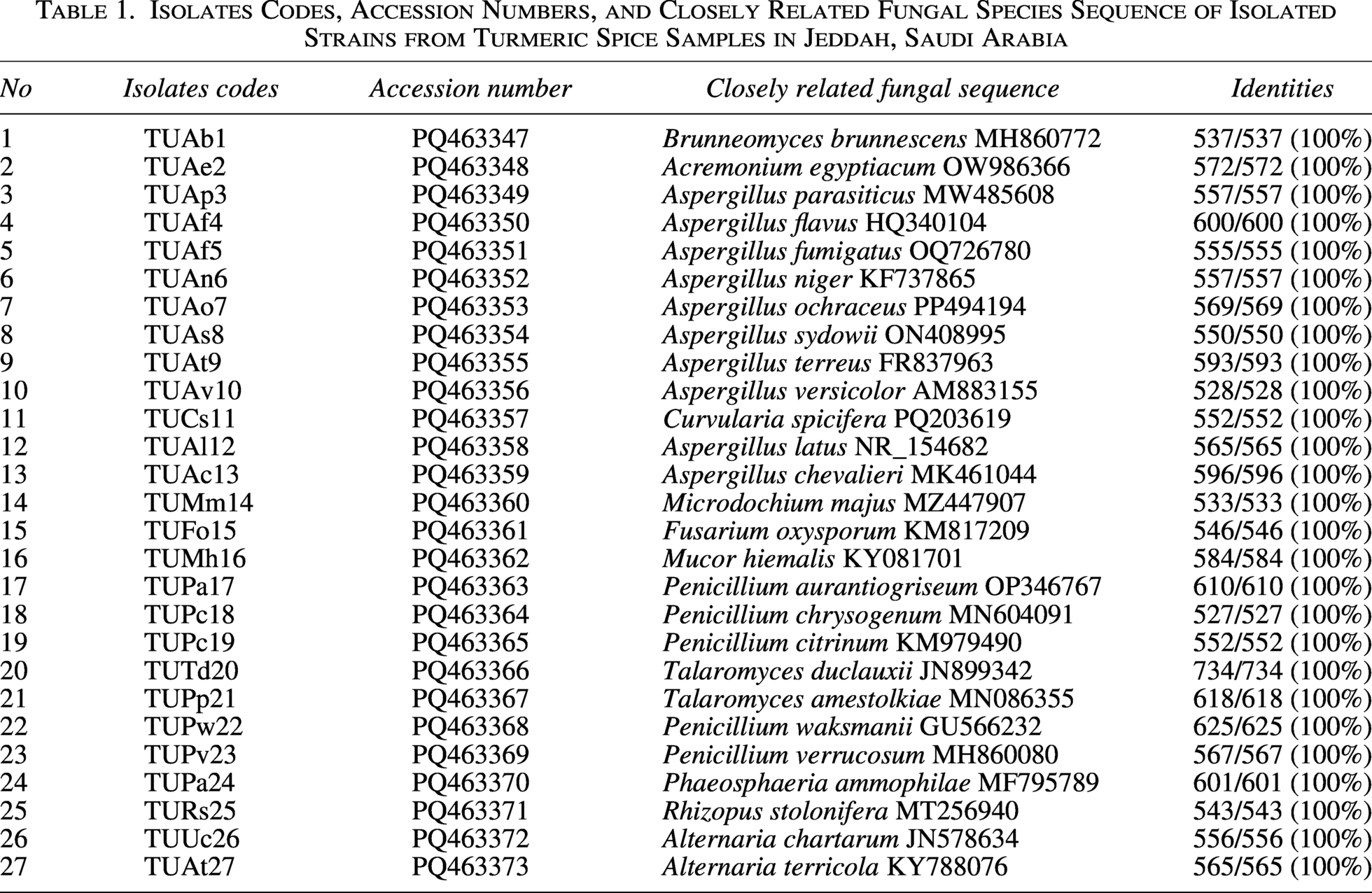
The isolated fungal strains were under five orders. The order Eurotisales comprised three genera of Aspergillus, Penicillium, and Talaromyces. The species were: Aspergillus chevalieri, Aspergillus flavus, Aspergillus fumigatus, Aspergillus latus, Aspergillus niger, Aspergillus ochraceus, Aspergillus parasiticus, Aspergillus sydowii, Aspergillus terreus, Aspergillus versicolor, Penicillium aurantiogriseum, Penicillium chrysogenum, Penicillium citrinum, Penicillium waksmanii, Penicilliu verrucosum, Talaromyces duclauxii, and Talaromyces purpureogenus. The order Glomerellales included Brunneomyces brunnescens. The order Hypocreales contained Acremonium egyptiacum, Fusarium oxysporum, Microdochium majus. The order Plerosporales was represented by the species of Alternaria chartarum, Alternaria terricola, Curvularia spicifera, and Phaeosphaeria ammophilae. The order Mucorales comprised two species of Mucor hiemalis and Rhizopus stolonifer (Table 1 and Fig. 1).
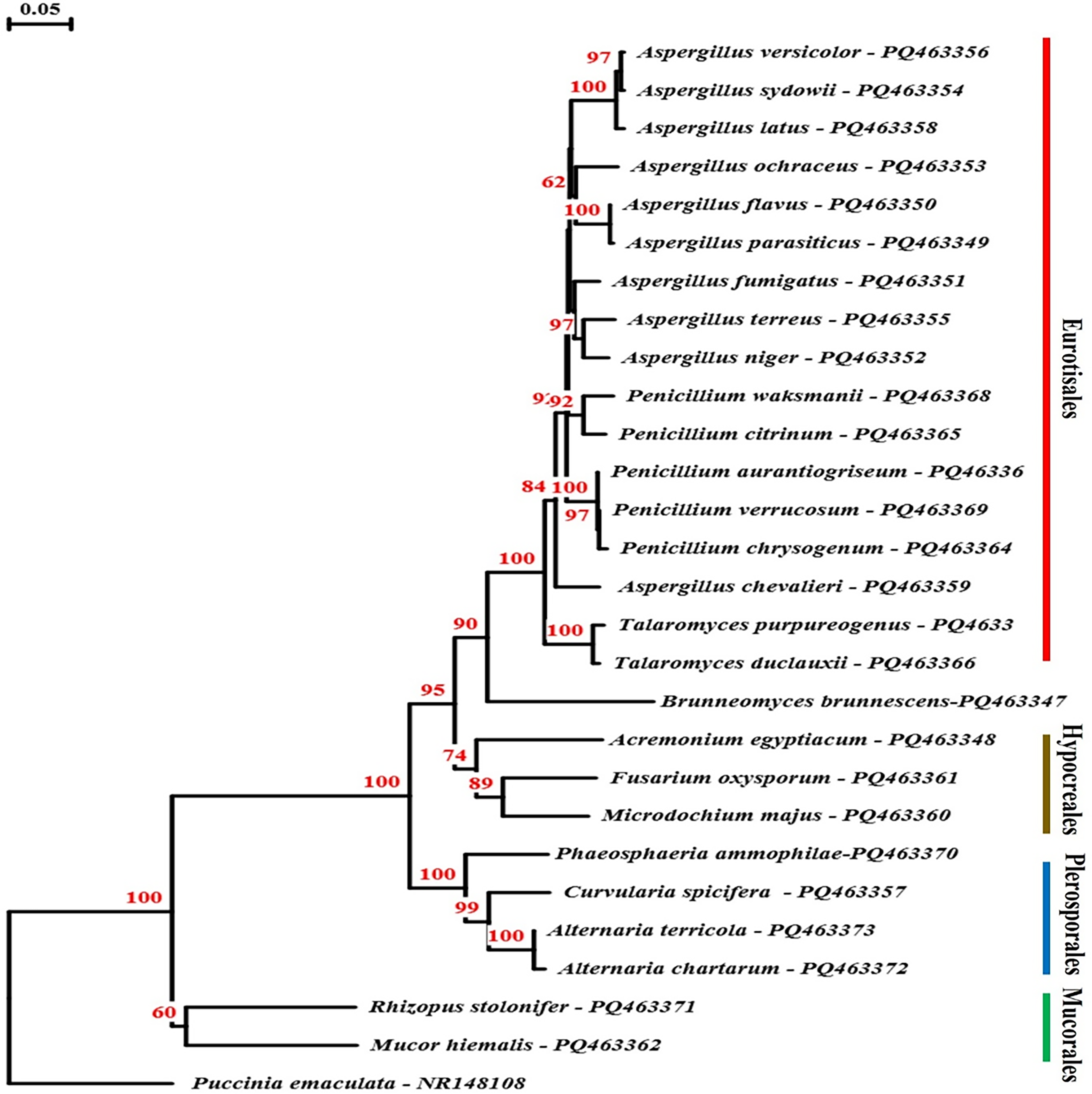
Phylogenetic tree of isolated strains obtained from turmeric powder samples based on ITS region sequence results. The numbers above branches indicate bootstrap values. Only values >70% were shown. ITS, internal transcribed spacer.
Occurrence of turmeric mycobiota
Turmeric mycoflora was isolated from 40 rhizome powder samples by using dilution-plate method on dichloran 18% glycerol (DG18) agar. The fungal colony was isolated and purified on potato dextrose agar (PDA) medium. Twenty-seven fungal species representing 12 genera were recorded from the collected turmeric spices samples. The dominant genus was Aspergillus, which was recorded with average total counts of 4.76 log CFU/g. It appeared in all 40 tested samples. Nine samples were heavily contaminated with Aspergillus, samples no. 13 and 33 were the highest ones, appeared with 3.396 and 3.390 log CFU/g, respectively. Seven samples of 7, 11, 12, 27, 29, 32, and 34 were noticed to be contaminated with Aspergillus, with ranges 3.31–3.36 log CFU/g. Sample no. 6 was the lowest contaminated sample with Aspergillus, it showed 2.900 log CFU/g (Fig. 2). A. niger and A. flavus were the highest contributed species, estimated ATC with 4.436 and 4.303 log CFU/g, they appeared with number of cases of isolation in 40 (100%) and 32 (80%) of the tested samples. A. chevalieri, A. latus, and A. ochraceus were noticed with ATC ranges 3.301–3.465 log CFU/g, contributed number of cases of isolation (NCI) with 20, 11, and 12 of the samples and recorded frequency with 50, 27, and 12%, respectively. A. sydowii and A. fumigatus were estimated with ATC 3.018–3.172 log CFU/g, showing NCI of 20 and 6 with F% 50 and 15. A. parasiticus, A. terreus, and A. versicolor contributed ATC 1.921, 2.619, and 2.523 log CFU/g, NCI 1, 8, and 2 of the samples, and frequencies (F%) 2.5, 20, and 5, respectively.
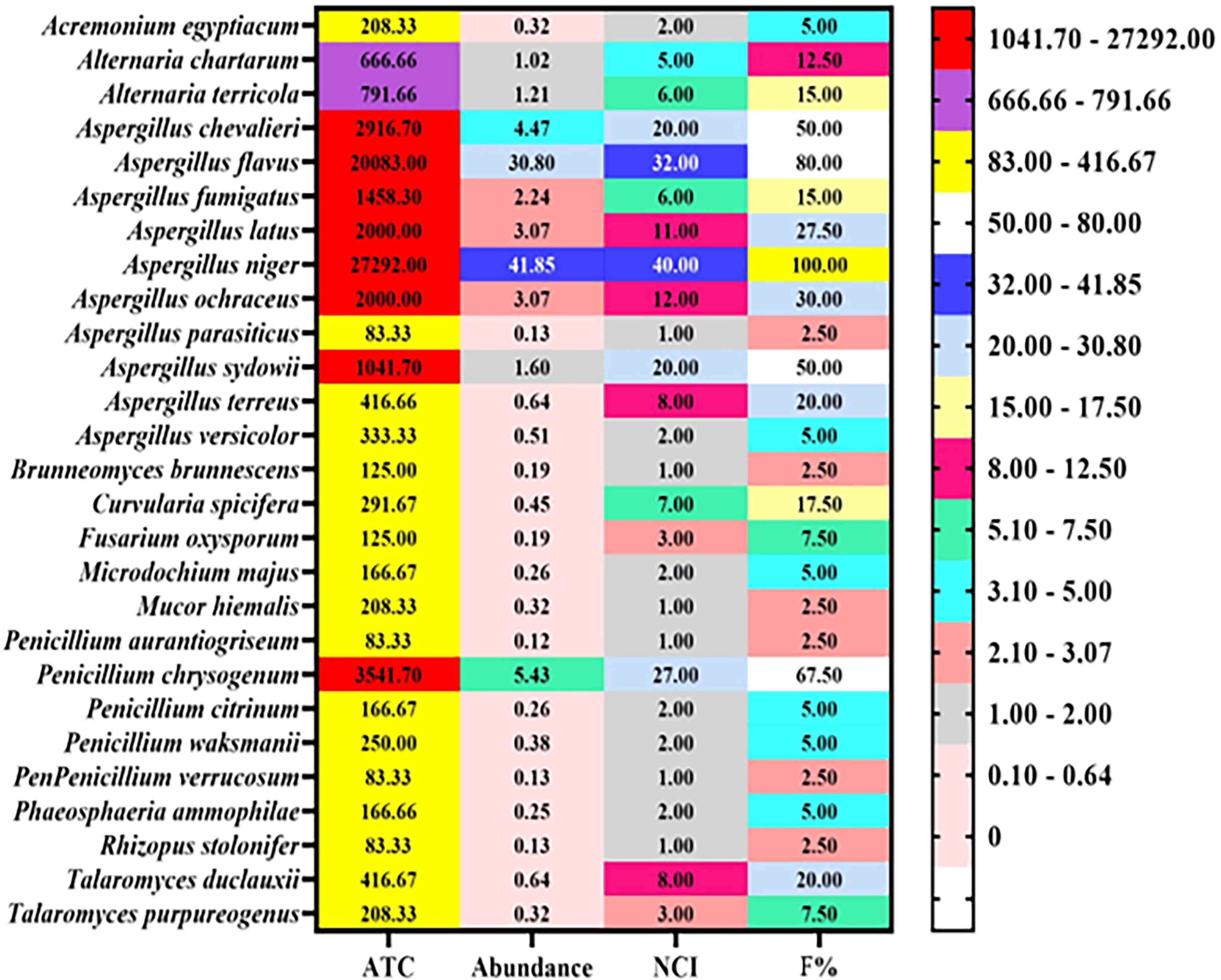
Average total counts (ATC), abundance (percentage of C%), frequency (F%) of fungal species isolated from 40 samples of Curcuma on dichloran 18% glycerol (DG18) agar and incubation at 25°C for 1 week.
P. chrysogenum ranked the second most isolated species after Aspergillus spp. It recovered from 27 (67.5%) samples with 3.550 log CFU/g. Other species of Penicillium, P. aurantiogriseum, P. citrinum, P. waksmanii, and P. verrucosum showed ATC with ranges 1.921–2.398 log CFU/g, NCI 1–2, and F% 2.5–5%.
A. egyptiacum, A. chartarum, and A. terricola appeared in 2, 5, and 6 of the samples with ATC 2.319, 2.824, and 2.898 log CFU/g.
B. brunnescens, M. hiemalis, and R. stolonifera were recorded in one sample for each with ATC 2.097, 2.319, and 1.921 log CFU/g.
C. spicifera, F. oxysporum, and M. majus estimated in 7, 3, and 2 of the samples, exhibited ATC with 2.465, 2.097, and 2.222 log CFU/g.
Three fungal species of P. ammophilae, T. duclauxii, and T. purpureogenus were noticed in 2, 8, and 3 of tested samples with ATC 2.222, 2.619, and 2.319 log CFU/g (Fig. 2).
Natural distribution of total AFs and OTA
The 40 powder samples of turmeric were tested for natural contamination of total AFs and ochratoxins. It was found that 12 samples (out of 40) were contaminated with the two types of mycotoxins. Principal component analysis in Figure 3, showed the correlation between the contaminated samples with the two types of mycotoxins (AFs and OTA) appeared with the same percent of PC. Twelve samples of tested turmeric were free from the two types of mycotoxins (Fig. 3). Twelve turmeric samples were positive for AFs. The concentration of AFs ranged 1.8–2.8 PPB. The highly contaminated samples were samples no. 7 and 29, and the lowest one was sample no. 12. Samples no. 11, 27, and 34 exhibited the same value of AFs with 2.5 PPB. Twenty samples (out of 40) were positive for the occurrence of ochratoxins. The amount of occurrence ranged 3.3–4.6 PPB. The highest contaminated sample with ochratoxins was sample no. 37, and the lowest one was sample no. 11. Six samples showed the same concentration of OTA with a value of 4.2 PPB (Table 2; Fig. 3).

Principal component analysis (PCA) analysis of aflatoxins and ochratoxins based on the natural occurrence, isolates inside the green circle were negative for the two toxins; the blue circle comprised positive isolates, and the red circle included positive isolates for OTA. OTA, ochratoxins A; S, sample of turmeric.
Detection of Total Aflatoxins (AFs) and Ochratoxins (OTA) from Turmeric Powder Samples
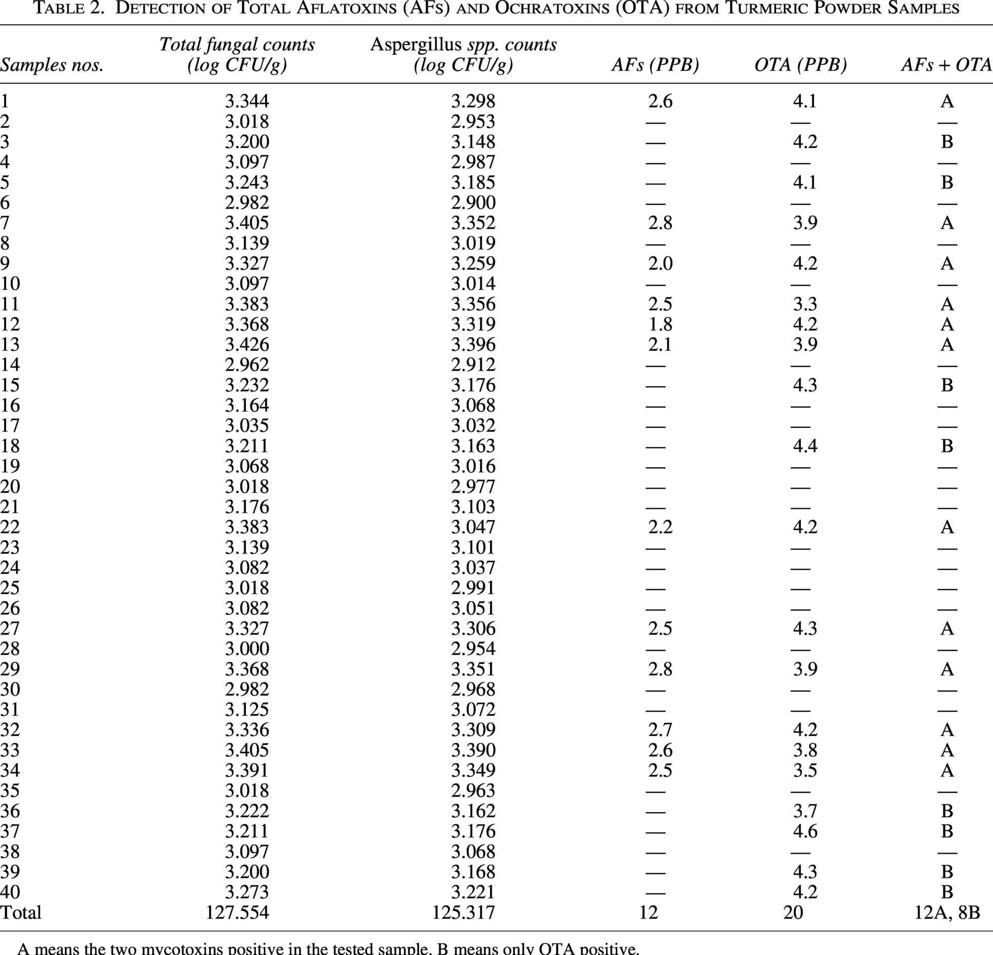
A means the two mycotoxins positive in the tested sample. B means only OTA positive.
Antimicrobial effect of turmeric extracts
Three extracts from turmeric were tested against C. albicans, E. coli, S. aureus with the disc diffusion assay. The choice of sample nos. 2 and 13 to compare the sample exhibited no AFs and OTA (2) and the other one (13) positive with the two types of mycotoxins. Sample no. 13 showed more effectiveness against the three tested strains. It exhibited an inhibition diameter of 3.2–15.4 mm. The methanol extract appeared diameter with 15.4 mm against S. aureus (Table 3).
Antimicrobial Activity of Turmeric Powder Extracts in Water, Methanol, and Chloroform against Escherichia coli, Staphylococcus aureus, and Candida albicans
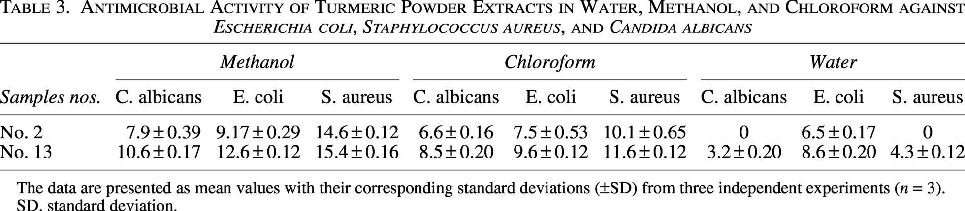
The data are presented as mean values with their corresponding standard deviations (±SD) from three independent experiments (n = 3).
SD, standard deviation.
Discussion
This study aimed to detect the fungal species contaminated with turmeric powder spices samples collected from Jeddah (Western Saudi Arabia). Fungi belonged to four orders Eurotisales, Glomerellales, Hypocreales, and Plerosporales were isolated. A. niger and A. flavus were the dominant species, showed Average Total Counts (ATC) with 27292 and 20083 CFU/g, NCI in 40 (100%) and 32 (80%) of the tested samples. In China, Hu et al. (2015) reported that A. flavus was the common contaminant in turmeric. Jeswal and Kumar (2015) found that A. flavus and A. niger naturally contaminated nine different Indian spices turmeric, black pepper, red chili, coriander, cumin, ginger, fenugreek, fennel, and caraway. Also, AFs, OTA, and citrinin were detected in tested spices. El‐Dawy et al. (2019) stated that 18 fungal genera were obtained from 50 spices samples (Cuminum cyminum, Curcuma longa, Nigella sativa, Salvia officinalis, and Zingiber officinale) collected from the stored markets in Upper Egypt. The predominant genus in the count and frequency was Aspergillus, 71.32% and 100%. Rani and Saxena (2022) claimed that A. flavus and A. niger are common contaminants in turmeric.
Spices can be naturally contaminated with harmful substances such as AFs and ochratoxins, which are produced by specific fungi during various stages such as cultivation, harvesting, or storage. Over time, exposure to these toxins may have adverse effects on human health due to their potential toxic and carcinogenic properties. In this study, we found that 12 samples (out of 40) were contaminated with the two types of mycotoxins. In India, (Thirumala-Devi et al., 2001) reported that OTA contamination in turmeric samples ranged from 11 to 102 µg/kg. Sadef et al. (2023) analyzed the AFs contamination of 474 samples of turmeric and red chili gathered from Lahore city by thin-layer chromatography (TLC). The turmeric samples and red chili with ≥10 μg/kg of AFs were 33% and 70%, which are considered unsuitable for human consumption according to the European Union standard limit. The mean estimated daily intake of turmeric among humans was 0.0006–0.0008 μg/kg bw/day. Turmeric is often added to food during the cooking process or food preparation. The effectiveness of heat in inactivating mycotoxins depends on factors such as the type of mycotoxin, the temperature, and the duration of cooking. While cooking can help reduce the risk, it’s important to use good quality spices and proper storage methods to minimize contamination in the first place.
The study assessed the antimicrobial properties of turmeric extracts. Findings indicated that turmeric sample no. 13 exhibited inhibitory effects against the tested yeast and bacteria, whereas sample no. 2 demonstrated the least activity. Mycotoxin analysis revealed the presence of AFs and OTA in sample no. 13, while sample no. 2 tested negative. This suggests that mycotoxin contamination in turmeric may be mistaken for its antimicrobial effectiveness. In a study conducted by Gulel et al. (2024), the concentration of Salmonella typhimurium in chicken decreased by 2.37, 2.71, and 2.84 log levels when stored in an extract of turmeric after 6 days. This result was obtained with 1%, 2%, and 3% curcumin treatment, respectively. Fatimah et al. (2022) found that turmeric ethanolic extract had positive effect against S. aureus. The results showed inhibition zones at concentrations of 20%, 40%, 80%, and 100% (11.86, 12.04, 12.53, and 10.97 mm). The use of spices extracts and their compounds as antimicrobial in traditional medicine showed significant concerns about the toxicity, safety, and quality of these spices. There are several methods to decontaminate turmeric and remove microbial contaminants while preserving its antimicrobial properties for use as a food additive: soaking in ethanol, boiling in water, boiling in acetic acid, steaming, and autoclaving. These methods help ensure that turmeric retains its bioactive compounds, such as curcuminoids, which are responsible for its antimicrobial properties (Chusri et al., 2012).
Studies have reported the contamination of plant materials with medicinal properties by mycotoxins (Makhuvele et al., 2020; Thipe et al., 2020). Many researchers concluded that many plant extracts used as food ingredients are toxic and mutagenic, so screening of toxicity is necessary. Extracts exhibiting no signs of toxicological effect are used safely (Sajid et al., 2019).
In conclusion, turmeric is extensively utilized in the food industry for its color, flavor, and nutritional benefits. This study revealed that turmeric can be contaminated with fungi, leading to the natural formation of mycotoxins. Additionally, turmeric shows promise as an antimicrobial alternative. However, it is vital to ensure that turmeric samples are free from mycotoxins, as their presence can compromise the spice’s antimicrobial properties. Therefore, it is essential to monitor and control mycotoxin contamination to ensure food safety and maintain the beneficial properties of turmeric.
Authors’ Contributions
H.F.A.-H.: Conceptualization, methodology, validation, formal analysis, investigation, data curation, writing—review and editing, funding were performed by the author. I.P.: Writing—review and editing. E.E.-D.: Writing—original draft preparation, writing—review and editing. Y.G.: Conceptualization, writing—original draft preparation, writing—review and editing.
Footnotes
Acknowledgment
The authors extend their appreciation to Taif University, Saudi Arabia, for supporting this work through project number (TU DSPP-2025-12).
Author Disclosure Statement
No potential conflicts of interest were reported by the author(s).
Funding Information
This research was funded by Taif University, Saudi Arabia, Project No. (TU-DSPP-2025-12).
Data Availability Statement
All data generated or analyzed during this study were included in this article.