




Abstract
HER2 has been thought to play a critical role in both breast cancer development and progression. Any functional polymorphisms can potentially affect breast cancer risk as well as cancer phenotype and outcome. In our study, we analyzed three polymorphisms in the HER2 gene: the single-nucleotide polymorphism (SNP) HER2 Ile655Val as well as another SNP (rs903506) close to it and a new screened dinucleotide repeat H(AC)I4 in intron 4, in a sample of 148 cases and 290 controls from the Tunisian population and investigated their association with breast cancer risk. For the HER2 Ile655Val, we found similar allele frequencies between cases and controls (frequency of I allele was 0.92 and 0.91, respectively). The same was observed for the noncoding SNP (rs903506). These two SNPs also showed no association with any clinical parameters, except the association of HER2 Ile655Val with tumor size (p = 0.002). But, a significant association was found between the short tandem repeat (STR) [H(AC)I4] and breast cancer risk at both genotypic and allelic levels (p = 0.0004 and p = 0.0001, respectively). Multivariate analysis with binary logistic regression of disease status on genotypes of the three polymorphisms confirmed the association of STR with breast cancer risk (p = 0.016). Therefore, this STR seems to be a promising biomarker in breast cancer and deserves further investigation.
Introduction
H
Further, it has been shown that HER2 gene amplification/overexpression is correlated with worse clinicopathological parameters such as steroid hormone receptor-negative tumors, tumor size, lymph node involvement, high nuclear and histological grade, aneuploidy, proliferation index, and reduced response to chemotherapy and hormone therapy (Slamon et al., 1987; Pegram et al., 1997; Gregory et al., 2000; Witton et al., 2003; WisemAn et al., 2005). In addition to high expression level, the active form of HER2 could be induced by changes in transmembrane amino acid composition (Stern et al., 1988; Shigematsu et al., 2005).
Mutations in the transmembrane region of the rat homologue of HER2 occur in chemically induced rat neuroblastomas and lead to increased tyrosine phosphorylation (Prenzel et al., 2001). Sequence analysis of the cDNA identified an A/G transition polymorphism in the first position of codon 655 in the DNA region encoding the transmembrane domain (Papewalis et al., 1991). This variant corresponds to Ile655Val nonsynonymous substitution. Many investigators addressed the relationship between the HER2 Ile655Val polymorphism and breast cancer (Papewalis et al., 1991; Xie et al., 2000; Fleishman et al., 2002; Millikan et al., 2003; Montgomery et al., 2003). The initial report of positive association between this variation and breast cancer (Xie et al., 2000) has not been successfully replicated till now (Montgomery et al., 2003; Kamali-Sarvestani et al., 2004; An et al., 2005; Benusiglio et al., 2006).
However, subgroup analysis in several studies suggested that among women who were younger (Xie et al., 2000; Millikan et al., 2003; Montgomery et al., 2003), were physically inactive (Xie et al., 2000), had larger body mass (Xie et al., 2000; Millikan et al., 2003), or had a positive family history of breast cancer, the Val/Val genotype was associated with increased risk of breast cancer in comparison with the Ile/Ile genotype.
As the HER2 gene is also an important biomarker, particularly in breast cancer, in our study we analyzed the HER2 Ile655Val polymorphism (rs1801200) as well as another single-nucleotide polymorphism (SNP) (rs903506) close to it and also a conserved dinucleotide repeat H(AC)I4 in intron 4 which we recently described (Kharrat et al., 2008) in a case-control study. We also investigated the association of these polymorphisms with HER2 protein expression and other pathological and clinical parameters.
Materials and Methods
Subject
A retrospective study was conducted on a group of 148 Tunisian women with breast cancer selected from the register of Sfax University Hospital Habib Bourguiba between the years 2001 and 2007, with a mean age of 51 ± 11.43 years (27-79). The control group contains 290 women with a mean age of 54 ± 17.24 years (20-87). All were healthy and clinically asymptomatic women randomly selected from blood donors of the Blood Transfusion Center of Sfax (Tunisia) and matched with patients for age and social conditions.
Immunohistochemistry
The status of HER2, Bcl2 protein, estrogen, and progesterone receptor was assessed by immunohistochemistry on frozen tissue. Primary antibodies used were BM5084 (HER2; ACRIS, Hiddenhausen, Germany), clone 124 (bcl2; DAKO, Carpinteria, CA), clone 1D5 (ER1; DAKO), and clone PgR636 (PR; DAKO), which were incubated for 30 min at room temperature. A biotin-labeled secondary antibody (goat anti-rabbit, 1:500; DAKO) was incubated for 15 min at room temperature. Then the avidin-biotin-horseradish peroxidase (HRP) complex was applied for 15 min as directed by (DAKO), and finally, the immunoprecipitate was visualized by treating with diaminobenzidine tetrahydrochloride (DAKO) for 30 min. The sections were counterstained with hematoxylin.
Immunostaining was considered positive if more than 5% of tumor cells were stained, and it was scored on the basis of the approximate percentage of positive tumor cells and the relative immunostaining intensity (Khabir et al., 2003).
Phenotyping
One hundred thirty-one patients had invasive ductal carcinoma, 7 patients had invasive lobular carcinoma, and 10 patients had medullar or tubular cancers. Histological grade was not available for lobular carcinomas because tubule formation is not a characteristic of the tumor. The microscopic grading of Scarff-Bloom-Richardson was used (Bloom and Richardson, 1957); among the patients with ductal carcinoma, 9 (6.8%) had grade I, 77 (58.8%) had grade II, and 45 (34.3%) had grade III tumors. The nodal status was known for 144 patients; 52 (36.1%) had no nodal involvement. Lymph node invasion was detectable in 92 cases (63.8%), of which 51 had one to three nodes involved and 41 had more than three nodes involved.
DNA extraction and polymerase chain reaction
DNA was extracted from freshly frozen breast cancer tissue of 148 patients, using a Wizard Genomic DNA purification kit (Promega, Madison, WI), and also from blood samples of all controls and patients using standard phenol-chloroform protocol.
The genotypes of the polymorphisms were determined with polymerase chain reaction (PCR) for the dinucleotide repeats H(AC)I4 in intron 4, using labeled primers, as reported by Kharrat et al. (2008), and with PCR-restriction fragment length polymorphism-based method (Pinto et al., 2004) for the two SNPs (rs1801200 and rs903506).
The primers for PCR amplification of the studied polymorphisms are given in Table 1 and Figure 1.

The position of polymorphisms studied in the HER2 gene.
SNP genotyping
For Ile655Val, PCR products were digested by restriction enzyme Alw26 for 2 h at 37°C and subsequently separated by electrophoresis in 3% Nuseiv/agarose (2:1, wt:wt) (digestion of the G allele at codon 655 results in two fragments of 116 and 32 bp, whereas the A allele is undigested and gives a fragment of 148 bp). For the SNP rs903506, PCR products were digested by restriction enzyme NciI for 2 h at 37°C and also separated by electrophoresis in 3% Nuseiv/agarose (2:1, wt:wt) (digestion of the G allele results in two fragments of 106 and 91 bp).
The two SNPs are located at 175 bp apart; Ile655Val (rs1801200) is located in exon 17 of the gene HER2 (encoding the transmembrane domain), whereas the A23271G polymorphism (rs903506) is within intron 17.
The dinucleotide repeat H(AC)I4 genotyping
Assessment of the dinucleotide repeat H(AC)I4 in intron 4 was performed using Super Mixte Applied Biosystems (2 × ) in a 6.25 μL reaction volume containing 100 ng DNA and 10 μM of each primer. The PCR reactions were performed using a GenAmp PCR system 9600 thermocycler (PerkinElmer, Massy, France). Cycling conditions for screening the microsatellite were as follows: initial heating at 95°C for 5 min, followed by 30 cycles of denaturation at 94°C for 11 min, annealing at 56°C for 1 min, and extension at 72°C for 1 min. About 0.5 μL of the PCR products was mixed with 9.5 μL of formamide containing 0.3 μL of ROX400 molecular weight standard (Applied Biosystems, Foster City, CA). The denatured PCR products were separated on the ABI PRISM 310 DNA Analyzer (Applied Biosystems). Evaluation of the collected data was done with the GeneScan Analysis software (Applied Biosystems) and the fragment lengths were determined. To confirm the number of repeats, direct sequencing of the PCR products was carried out.
Statistical analysis
Genotypic and allelic frequencies were calculated by simple counting using Microsoft Excel. The exact test for the Hardy-Weinberg equilibrium (HWE) was performed using the Genetic Data Analyses program (version 1.1) (Hauptmann et al., 2003). Association of the studied polymorphisms with breast cancer susceptibility was assessed using the case-control chi-square test with p ≤ 0.05 taken as the cutoff for statistical significance. Odds ratios (OR) and their 95% confidence intervals (CIs) were estimated using programs from the linkage utility package (http://linkage.rockefeller.edu).
The association of the polymorphisms with pathological and clinical parameters was evaluated by two-way analysis of variance, Spearman correlation, and Student's t-test. A multivariate logistic regression of the disease status on the polymorphisms and age was performed to test associations with risk corrected for age. These analyses were computed using appropriate functions from the R language (www.r-project.org).
Results
HER2 expression and clinical parameters
In this sample, overexpression was observed for HER2, bcl2, ER1, and PR in 62%, 69%, 65%, and 58% of tumors, respectively. HER2 was positively correlated with tumor size (rs = 0.318; p = 0.003) and bcl2 status (rs = 0.270; p = 0.013) but not with Scarff-Bloom-Richardson grade (p = 0.25). There was no overall significant association between the expression of HER2 and that of hormonal receptors.
HER2 polymorphisms
Genotype frequencies in control samples were in HWE for both SNPs (p = 0.14 and p = 0.33). For the noncoding SNP (rs903506), the frequencies of the wild-type (G) allele were 75% and 66% in cases and controls, respectively, but this difference is not statistically significant (Table 2). No association was found between SNP rs903506 and clinical parameters in our study.
For SNP rs1801200, no significant difference was found in allele frequencies between cases and controls (frequency of I allele was 0.92 and 0.91, respectively). In contrast, the homozygous V genotype was present in 2.7% of patients but absent in the control group. When genotype frequencies are considered, we found a significant difference (Fisher's exact test, p = 0.0017) between controls and patients (Table 2). However, this association was not detected when we compared the [Val/Val + Ile/Val] versus [Ile/Ile] (p = 0.16).
Moreover, we looked for a potential association between the rs1800200 and clinicopathological parameters in the patient's sample. Associations between genotype and major parameters studied were nonsignificant (Table 3). However, when we compare genotype frequencies within cases stratified by age group (> or ≤ 45 years) and hormonal status (ER+) for the rs1801200, a weak association was found (p [ ≤ 45] = 0.048). Also, association of the rs1801200 polymorphism with clinical parameters assessed by two-way analysis of variance showed a significant result with tumor size (p = 0.002).
p-value by Fisher's one-way analysis of variance test.
STR, short tandem repeat; SBR, Scarff-Bloom-Richardson.
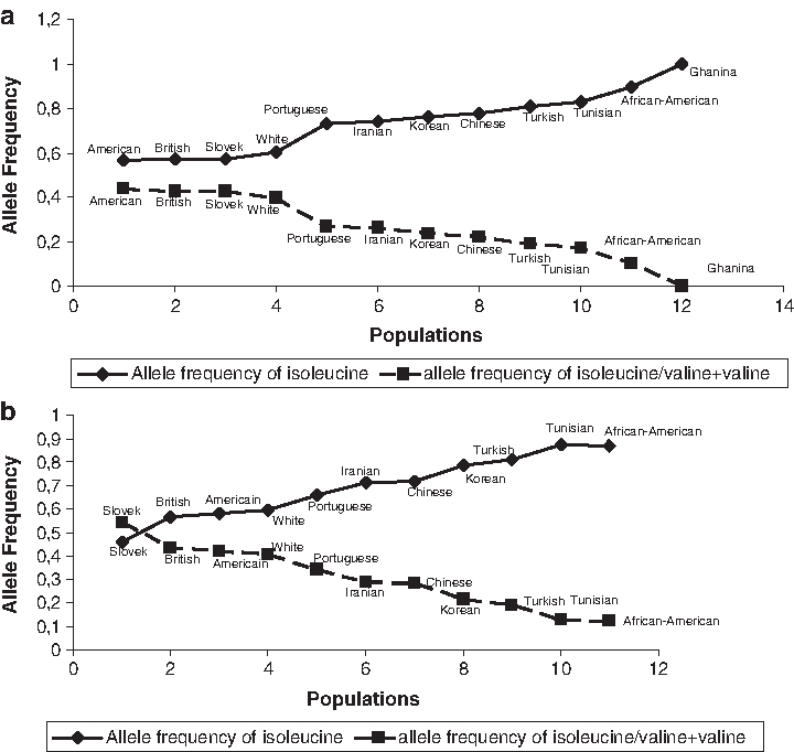
To investigate ethnic specificities, we compared the genotype frequencies of the rs1801200 in our population with published data available for Chinese (Xie et al., 2000), African (Ghanaian) (Ameyaw et al., 2000), Afro-American (Millikan et al., 2003), British (Benusiglio et al., 2005), Portuguese (Pinto et al., 2004), Turkey (Akisiki and Dalay, 2004), Iranian (Kamali-Sarvestani et al., 2004), Slovak (Zubor et al., 2006), Korean (An et al., 2005), and American (Nelson et al., 2005).
Figure 2 shows the frequency of the Ile/Ile and Val/Ile + Val/Val genotype; we observed that we can distinguish three ethnic groups in the control sample (Fig. 2a): Caucasian, Asiatic, and African. The same pattern was found for the patient subset (Fig. 2b).
Allele frequencies of the Ile655Val polymorphism in (
The dinucleotide repeats H(AC)I4 and breast cancer
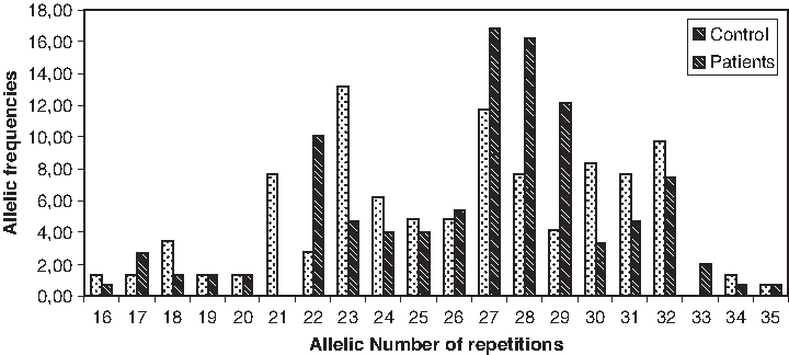
The number of AC repeats ranged from 16 to 35 among the Tunisian samples (Fig. 3). Genotype frequencies in control samples were in HWE for the [H(AC)I4] (corrected p = 0.44).
Allelic distribution of the H(AC)I4 repeats.
We subdivided the alleles into two classes: short (S) (≤25 repeats) and long (L) (>25 repeats). A highly significant association was found between the short tandem repeat [H(AC)I4] screened and breast cancer risk at genotypic and allelic levels (p = 0.0004 and p = 0.0001, respectively) (Table 4). Particularly, we found that the subjects having two copies of the H(AC)I4 allele with >25 repeats showed 2.44-fold increased risk for breast cancer (OR = 2.44; p = 0.00017; 95% CI: 1.53, 3.91) compared with subjects having one or both alleles with ≤25 repeats. The risk also tended to increase when we compared the subject having two copies of the H(AC)I4 allele with >25 repeats and those having one copy of this polymorphism (OR = 2.93; p = 0.00013; 95% CI: 1.67, 5.14) (Table 4).
[LL] versus [SL + SS].
[LL] versus [SL].
[LL + SL] versus [SS].
[L] versus [S].
OR, odds ratio; CI, confidence interval.
Further, binary logistic regression of disease status on these three polymorphisms confirmed the association of the microsatellite polymorphism with the risk of breast cancer (p = 0.016). However, no association was found between H(AC)I4 and any of the clinical parameters considered in our study (after correction for multiple testing) (Table 3).
Discussion
HER2 is the second member of the EGFR family which has emerged over the past 20 years to be one of the most important proto-oncogenes in invasive breast cancer. In fact, breast cancer is the most widespread malignant disease affecting women. Its incidence has a slight growing potential. Given the determining role of HER2 overexpression in breast cancer pathogenesis and potential tumorigenicity of point mutation in the transmembrane-encoding region of HER2 gene in animal model, any polymorphism in this gene region might conceivably be a candidate gene for human breast cancer.
The most studied polymorphism of such type has been the Ile655Val polymorphism (rs1801200). However, until now no convincing evidence for any association between this polymorphism in HER2 and breast cancer risk has been reported.
A study by Fleishman et al. (2002) stated that the presence of isoleucine in codon 655 destabilizes the formation of active HER2 heterodimers, even in the presence of receptor overexpression. In contrast, the presence of valine can render the receptor in an active state, with slower endocytosis and rapid receptor recycling. But, Pinto et al. (2005) hypothesized that GG (Val/Val) genotype encodes receptors with higher dimerization capacities and possibly constitutive activation. However, use of bioinformatics tools for prediction of SNP effect on protein function failed to find any functional impact of this polymorphism (Choura and Rebai, 2009).
Several investigators have examined the relationship of this polymorphism with breast cancer risk (Xie et al., 2000; Montgomery et al., 2003; Kamali-Sarvestani et al., 2004; An et al., 2005; Benusiglio et al., 2005; Kalemi et al., 2004; Nelson et al., 2005; Benusiglio et al., 2006; Zubor et al., 2006). There have been inconsistent findings from one study to another. One population-based case-control study among Chinese women suggested that Val/Val homozygosity at erbB-2 codon 655 was associated with increased risk of breast cancer among younger women less than 45 years of age (Xie et al., 2000). Other studies (British women, Caucasian subjects, African-American, Korean, and Iranian), however, stated that the valine allele was not a breast cancer risk factor (Montgomery et al., 2003; Kamali-Sarvestani et al., 2004; An et al., 2005; Benusiglio et al., 2006). Our study also showed no association between HER2 Ile655Val polymorphism and risk of breast cancer development. This is in agreement with most published studies (Montgomery et al., 2003; Kamali-Sarvestani et al., 2004; An et al., 2005; Benusiglio et al., 2005; Benusiglio et al., 2006; Zubor et al., 2006). However, our study showed that the frequency of Val/Ile in Tunisian controls and patients is similar to that in African-American women and Turkish, but higher than that in Caucasian (Slovak, American, British, White, and Portuguese). Therefore, this difference in distribution can be explained by a different genetic background.
No correlation was found between HER2 polymorphisms analyzed in our study with histological grade or lymph node status. By contrast, Papadopoulou et al. (2007) reported a significant association in Greek samples with these parameters. However, the tumor size was found significantly associated with this polymorphism in our study. The same result was observed in gastric cancer Japanese patients (Kuraoka et al., 2003).
No significant association was found between HER2, Bcl2 protein, and estrogen/progesterone receptor and the SNPs studied in our populations. This polymorphism does not seem to alert or increase the expression of proteins implicated in breast cancer mechanisms. The same results were reported by Kamali-Sarvestani et al. (2004) in an Iranian population. Conversely, in the African-American population, Millikan et al. (2003) observed an insignificant trend for HER2 overexpression in women with Val/Val genotype. Puputti et al. (2006) suggested that in some HER2-positive breast tumors, the Val allele is overrepresented and could be potentially advantageous in the progression of breast cancer. These inconsistent results cannot be clearly explained, but it may be possibly due to a difference in the extent of the disequilibrium among different ethnic groups (Hauptmann et al., 2003).
It has been shown that the first intron in several genes, including EGFR, plays an important regulatory role (Bornstein et al., 1988; Sica et al., 1992), particularly the AC repeat (Maekawa et al., 1989). In fact, polymorphic dinucleotide repeat are rarely found in the coding regions of genes and are very often located in intergenic noncoding regions or in introns (Akai et al., 1999). Further, it was suggested that these repeats have some physiological functions, such as regulation of gene expression (Kashi et al., 1997; Sharma et al., 2007). The dinucleotide repeats H(AC)I4 is the only AC repeat found by homology in the HER2 gene in intron 4 (Kharrat et al., 2008). In our study, this polymorphism proved a significant association with breast cancer. This polymorphic region may be involved in conformational changes after transcription factor binding, as reported by Kharrat et al. (2008). In fact, it was observed that (TG/CA) n repeats of length n ≥12 units exert a downregulatory effect on transcription. Moreover, this negative modulatory effect increases with increasing length of repeat. Indeed, we observed that genes with long introns harbor more repeats compared with genes with short introns, suggesting a strong role of intron length as controlling factor for abundance of repeats (Sharma et al., 2007). These preliminary results deserve further confirmation.
Conclusion
Our results suggest that the well-studied Ile655Val genetic polymorphism of HER2 is not associated with an increased risk of breast cancer in Tunisian women. Also, we did not find an association between this polymorphism and major pathological and clinical features such as tumor grades, node involved, ER, PR, and bcl-2, and HER2 status of the disease. However, a new microsatellite in intron 4 showed promising results with a high association with breast cancer risk. Further investigations are needed to replicate this initial finding and assess its potential as a risk predictor.
Footnotes
Acknowledgments
This work was supported by the Ministry of Higher education, Scientific Research and Technology, Tunisia, and by the Kuwait University/Research Administration (grant no. YN01/05).
Disclosure Statement
No competing financial interests exist.