
Abstract
Aims: Molecular biology techniques based on the detection of genomic sequences by reverse transcription combined with polymerase chain reaction (PCR) have enabled the detection of different RNA viruses in serum or plasma samples. Since the dengue epidemic outbreak declared in Argentina in 2009, numerous patients' samples were analyzed for the acute phase of infection. One of the main methodological drawbacks is the lack of internal control to measure the effectiveness of the viral extraction and reverse transcription process. In this article, we propose to standardize a molecular method to detect beta actin (β-Act) and glucose 6 phosphate dehydrogenase (G6PDH) complementary DNAs (cDNAs) present in patient's plasma/serum, as a control process. Results: RNA extraction, reverse transcription, and PCRs for human G6PDH, β-Act, and the dengue virus genome were performed. cDNA fragments for β-Act and G6PDH were amplified for all samples, regardless of the presence or absence of viral RNA. Conclusions: Amplification of β-Act and G6PDH cDNAs can be used as a control for the extraction and reverse transcription processes during dengue virus detection. This could also be a useful method for controlling the above steps when infections caused by other RNA viruses are studied, even if another methodology is employed, such as real-time PCR.
Introduction
S
For diagnosis, clinical laboratories use tissue culture for virus isolation and serological methods to confirm the viral serotype (Shu and Huang, 2004). The reverse transcriptase-polymerase chain reaction (PCR) amplifies nucleic acid sequences and has provided a fast, sensitive, early technique for dengue virus detection. One problem remains unresolved—the inability to have a method that ensures that the early stages of viral RNA detection have been properly done. This problem also occurs in the detection of other types of RNA viruses. Nevertheless, nucleic acids (DNA and RNA) are present in blood samples and used in fetal medicine, oncology, transplant, etc. (Holford et al., 2008). Based on these findings, our work aims to contribute with a method to ensure that the entire processes of RNA extraction and reverse transcription have been correctly made, thus avoiding false-negative results due to errors during these steps.
Materials and Methods
Virus
Sera pool from patients infected with dengue virus donated by Dr. Sandra Gallego was used (Central Laboratory of the Province of Córdoba, Córdoba, Argentina).
Clinical samples
Freshly frozen plasma and/or serum samples were collected from patients at the LACE. The study was approved by the Committee of Ethics and Research from the Oulton-Romagosa and Ministerio de Salud de Córdoba and is in accordance with the Declaration of Helsinki. In all cases, the subjects were informed and their consent was obtained.
RNA extraction
Total RNA was extracted manually using a Viral Mini kit system (Qiagen, Inc.), following the manufacturer's instructions. RNA was eluted in 60 μL of nuclease-free water.
Reverse transcription
The complementary DNA (cDNA) was synthesized using Superscript III (Invitrogen, Inc.). Briefly, a mixture containing 6 μL of MilliQ water (DNase and RNase free), 300 ng of random primers, 5 μL of viral RNA, and 0.5 mM of each of dGTP, dATP, dCTP, and dTTP was incubated at 65°C for 5 min. Then, the mixture was left 1 min at 4°C. This mix was combined with 4 μL of 5×First Strand Buffer, 5 mM DTT, 4 U RNAse OUT, and 5 U Superscript III and incubated at 25°C for 5 min and 55°C for 60 min. Finally, the enzyme was inactivated for 15 min at 70°C.
Design of oligonucleotides and PCR
The primer designs for β-Act and glucose 6 phosphate dehydrogenase (G6PDH) amplification were performed with Primer-Blast software (www.ncbi.nlm.nih.gov/tools/primer-blast/), taking into account the position of those genes in the genome, to amplify and discriminate both cDNA and genomic DNA. This was necessary because in the amplification process, genomic DNA contamination could be observed in the RNA extraction step. The oligonucleotide sequences are listed in Table 1, as the size of the fragments expected. Oligonucleotides and PCR conditions for dengue detection were performed according to the procedure of Lanciotti et al. (1992).
PCRs were carried out using an MJ Research thermal cycler. The reaction volume of 15 μL consisted of 1 μL cDNA, 3 μL of 5×buffer, 200 μM each of dGTP, dATP, dCTP, and dTTP, 0.6 nM forward primer, 0.6 nM reverse primer; 0.6 U GoTaq polymerase (Promega), and water qsp 15 μL. Amplification conditions were 94°C for 5 min, 35 cycles of 94°C for 40 s, 59°C for 40 s, and 72°C for 75 s, and a final extension at 72°C for 7 min.
The amplified fragments were separated by electrophoresis at 80 V for 30 min on a 1% agarose gel in TAE 1×, previously stained with ethidium bromide (10 mg/mL).
Results
RNA extractions and reverse transcriptions were performed using serum and/or plasma from patients suspected to have dengue infection. Freshly frozen samples were analyzed. Figure 1 shows the PCR products obtained for different samples amplified for G6PDH (A) and β-Act gene (B). All plasma/serum samples gave positive amplification. In all cases wherein amplification is found, it corresponds to cDNA and not to DNA. This indicates that our method allowed us to know whether the RNA extraction and reverse transcription were properly done, regardless of the presence or absence of viral RNA (Fig. 2).
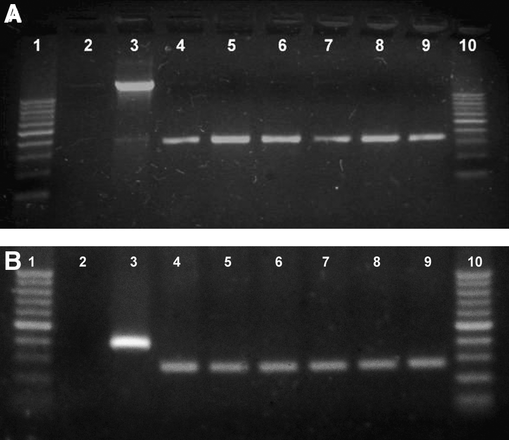
Results of the PCR assay for G6PDH
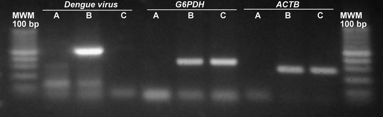
Results of the PCR assay for dengue virus; G6PDH and β-Act genomes after separation in agarose gel by electrophoresis.
Discussion
The results obtained in this work show that PCR employed to detect G6PDH or β-Act cDNAs can be used to control RNA processing in the dengue virus detection in both human serum/plasma. Moreover, according to the design of oligonucleotide, we can easily know whether the amplified products correspond to cDNA or genomic DNA. This consideration is important because, to the moment, there was no way to determine whether the RNA extraction and reverse transcription processes had been successful done. Without these controls, in some cases, samples reported negative for dengue virus could be wrong because of errors in the first steps of RNA extraction and reverse transcription processes. In short, the addition of either of these controls can offer greater security to inform negative results. Moreover, this procedure can be also applied to retroviruses present in human specimens.
Footnotes
Acknowledgments
The authors acknowledge the funding provided by the LACE laboratory and the Central Laboratory of the Province of Córdoba for providing positive control of dengue virus.
Disclosure Statement
The authors declare that they have no conflict of interests.