




Abstract
Background: The brother of the regulator of imprinted sites (BORIS) is a novel member of the cancer testis antigen gene family, which are normally expressed only in spermatocytes, but abnormally activated in different malignancies. Aim: The aim of this study was to explore the expression of BORIS in hepatocellular carcinoma (HCC) and its correlation with the clinicopathologic features and prognosis of HCC. Methods: We investigated BORIS expression in HCC cell lines and 105 primary HCC clinical surgical specimens using real-time polymerase chain reaction and Western blot analysis. We further examined the correlation of BORIS with a liver stem cell marker (CD90) in HCC tissues by histochemical double staining. The correlation of BORIS with clinicopathologic features and prognosis of HCC was analyzed using patient data. Results: The expression of BORIS was found in SMMC-7721, BEL-7402, and Huh-7, but not in hep-G2 cells. The expression rate of BORIS was significantly higher in the HCC tissues than in the adjacent noncancerous tissues (p=0.000). BORIS expression was correlated with the tumor size (p=0.000), CD90 expression (p=0.000), and satellite nodule (p=0.000). Kaplan-Meier survival curves showed that patients with positive expression of BORIS had lower overall survival rate (p=0.003). Conclusions: Our data indicate that BORIS may be an auxiliary diagnosis index and a novel favorable prognostic indicator of HCC.
Background
H
The BORIS gene was first described as a DNA-binding protein that shares 11 zinc-finger (ZF) domains with CCCTC-binding factor (CTCF) (Filippova et al., 1996; Qi et al., 2003), but differs from this molecule at the N- and C-termini (Loukinov et al., 2002). Lobanenkov et al. originally identified the CTCF as a transcription factor regulating cmyc expression (Lobanenkov et al., 1990; Filippova et al., 1996), which was subsequently found to act as a tumor suppressor (Filippova et al., 1998; Rasko et al., 2001; Filippova et al., 2002). BORIS belongs to the same protein family as CTCF and has gained increasing attention due to its decreasing methylation of DNA. BORIS protein is absent in nonmalignant male tissues with the exception of testis, and is totally absent in females. However, BORIS transcripts were detected in more than half of the cancer cell lines (Klenova et al., 2002). Several collaborating laboratories have initiated studies of BORIS expression in a variety of primary cancers (Hong et al., 2005; Vatolin et al., 2005). As yet, there have been no reports about the expression of BORIS in HCC.
Based on the above discoveries, in the present study, we investigated BORIS expression in a total of 105 tissue samples of HCC and several liver cancer cell lines by immunohistochemical, polymerase chain reaction (PCR), and Western blot analysis, and demonstrated that BORIS expression was a reliable indicator for disease diagnosis and its poor prognosis. The expression of CSC marker CD90 is related to the expression of BORIS in HCC tissue.
Materials and Methods
Ethics
This work has been carried out in accordance with the Declaration of Helsinki (2000) of the World Medical Association. This study was approved by the medical ethics committee of the West China hospital, Sichuan University. All patients (n=141) involved in the study voluntarily offered written informed consent when approached by research personnel.
Tissue samples
HCC tissue samples (n=105) were obtained from patients who underwent surgical treatment without prior radiotherapy or chemotherapy treatment at the West China hospital between 2005 and 2006 (Chengdu, China). Normal liver samples (n=20) and cirrhosis samples (n=16) were collected from individuals without cancer and were used as controls. Clinical information on gender, age, serum hepatitis B surface antigen (HBsAg), serum alpha fetoprotein (AFP), cirrhosis, satellite nodule, and tumor capsule of patients were received from the West China Hospital. All samples were obtained with previous written consent. The histological cell types were assigned according to the criteria of the WHO classification.
Cell lines and culture
Different human liver cancer cell lines, Hep-G2, and Huh-7 were obtained from the American Type Culture Collection (ATCC). BEL-7402 and SMMC-7721 cell lines were obtained from the Committee of Type Culture Collection of the Chinese Academy of Sciences (Shanghai, China). Cells were cultured in the DMEM or RPMI-1640 supplemented with 10% fetal bovine serum and 1% penicillin-streptomycin at 37°C with 5% CO2.
Immunohistochemical and histochemical double staining
The embedded tissues were cut into 4-μm-thick sections for histological studies and stained using hematoxylin-eosin. BORIS protein was detected with a specific rabbit anti-human polyclonal antibody (Abcam). Paraffin-embedded tumor sections were boiled in a cooker for 4 min in citrate buffer. Then, slides were treated with 3% hydrogen peroxide 20-30 min before blocking with 3% bovine serum albumin. The tumor sections were incubated with the primary antibody (dilution, 1:50) overnight at 4°C. For negative control, the primary antibody incubation was replaced with 3% blocking solution. Sections were washed in PBS three times and incubated with goat-anti-rabbit IgG (Jackson ImmunoResearch) for 1 h at room temperature. After further washing, the samples were covered with a Vectashield mounting medium (Vector Laboratories) and stored at −20°C. Negative control slides were probed with normal goat serum under the same experimental conditions.
To determine the relationship between the expression of BORIS and CSC marker CD90, we performed histochemical double staining for BORIS (Abcam) and CD90 (Dako) on the same sections using Envision Doublestain System (Dako). Deparaffinization and antigen retrieval were performed as described above. After quenching the endogenous peroxidase activity with a peroxidase-blocking reagent (Dako), tissue sections were incubated with mouse monoclonal anti-CD90 antibodies for 1 h at room temperature, followed by rinsing with a washing buffer. Then, the tissue sections were subjected to secondary staining for BORIS with the sequential steps of quenching, incubation with anti-BORIS antibodies, incubation with anti-goat IgG antibodies, and detection of the phosphatase activity in accordance with the manufacturer's instructions (Envision Doublestain System; Dako).
Immunofluorescence detection of BORIS and CD90 derived from frozen liver cancer tissue sections were performed using Alexa fluorescent dye-conjugated antibodies (Invitrogen). Fluorescent images were acquired at 2-mm Z-axis intervals using a confocal microscope (Leica TCS SP5 II).
Immunohistochemical staining and assessment of BORIS levels in HCC and paracancer tissues
Immunohistochemical analysis was done by staining normal (n=20), cirrhosis (n=16), paracancer tissues (n=34), and HCC (n=58) using the Vecta stain Elite ABC standard kit (Vector Laboratories) as suggested by the manufacturer. Immunohistochemical staining was evaluated by using the immunoreactivity score (IRS) as previously described (Gion et al., 2001). In brief, the percentage of BORIS-positive cells was divided into four categories (<10%, 11%-50%, 51%-80%, and >80%, with corresponding scores of 1, 2, 3, and 4, respectively), whereas the staining intensity was given a value between 0 (no detectable immunostaining) and 3 (strong immunostaining). The IRS (0-12) was then calculated by multiplying the score values. Scoring was done in a blinded fashion by two independent scorers, with each slide read twice.
Reverse transcription-PCR
To determine the BORIS transcription in human HCC cell lines, total RNA was extracted from cells with TRIzol reagent (Invitrogen) according to the manufacturer's instructions. Total RNA was reverse-transcribed with 25 units of MMLV reverse transcriptase (Promega) and oligo-dT as primer. The resulting cDNAs were amplified with the following oligonucleotide sequences: BORIS, 5-CCCATTGTGCCACCATCA-3 (forward), BORIS, 5-AGCATGCAAGTTGCGCA-3 (reverse). Actin transcription was used as an invariant endogenous control. The primer sequences were as follows: actin F: 5-TCATCACCATTGGCAATGAG-3 (forward), actin R: 5-CACTGTGTTGGCGTACAGGT-3 (reverse).
Western blot analysis
For Western blot analysis, lysates from cells were prepared according to Klenova et al. (1993) with modifications. Lysates from testis tissues were prepared as follows; tissues were homogenized in the lysis buffer at the ratio 50 mm3 tissue/100 mL of buffer. The homogenate was kept on ice for 30 min, filtered through gauze, and centrifuged for 15 min, at 4°C and 13,000 rpm. Samples containing high concentration of lipids were additionally precipitated with acetone. The supernatant was discarded, the pellet dried at room temperature, and resuspended in sodium dodecyl sulfate loading/lysis buffer. Western blot assay was conducted as described previously, and membranes were probed with anti-BORIS antibody (Abcam; dilution 1:100) or anti-α-tubulin antibody (Sigma; dilution 1:200). Detection was performed using enhanced chemiluminescence reagent (Amersham Biosciences, now GE Healthcare) according to the manufacturer's instructions.
Statistical analysis
Data were expressed as mean±standard deviation or median with range. SPSS program for Windows (version 15.0; SPSS, Inc.) was used for statistical analysis. Comparisons of BORIS tumor expression with clinical and pathologic features were evaluated by using chi-square tests or two-tailed Fisher's exact test. Overall survival analyses were estimated by using the Kaplan-Meier method. The Student's t-test was used to detect differences between groups. All tests performed were two-tailed. A difference was considered significant if p<0.05.
Results
BORIS expression in HCC cell lines
BORIS expression was investigated in different human liver cell lines by means of reverse transcription-polymerase chain reaction (RT-PCR) and Western blot method. As shown in (Fig. 1), we found that normal testis (positive control), BEL-7402, SMMC-7721, and Huh-7 cells constitutively express BORIS mRNA and protein. However, BORIS could not be detected in Hep-G2 cells or normal liver tissue.

Detection of the brother of the regulator of imprinted sites (BORIS) mRNA and protein in four human liver cancer cell lines by reverse transription-polymerase chain reaction
BORIS expression in HCC clinical samples
To screen the state of BORIS expression in vivo, we investigated BORIS protein expression by immunohistochemistry, in a total of 105 primary HCC, 20 normal liver, and 16 cirrhosis tissues. Fifty-eight cases of BORIS expression (55.2%) localized in the cytoplasm and the nucleolus were found in HCC surgical resection samples, whereas the remaining 47 cases (44.8%) displayed almost undetectable BORIS expression. BORIS expression was not detected in cirrhosis or normal liver tissues. Analyses of the relationship between BORIS expression and various clinicopathological parameters are listed in Table 1. Expression of BORIS was significantly correlated with tumor size (p=0.000) and satellite nodule (p=0.000), but not with age, gender, serum HBSAg, cirrhosis, or tumor capsule (Table 1). On the other hand, we calculated that the IRS values for 34 BORIS-positive samples (2.43±0.51) in adjacent noncancerous tissues were lower than 58 BORIS-positive samples (5.22±0.53) in HCC tissues (p=0.01) (Fig. 2).
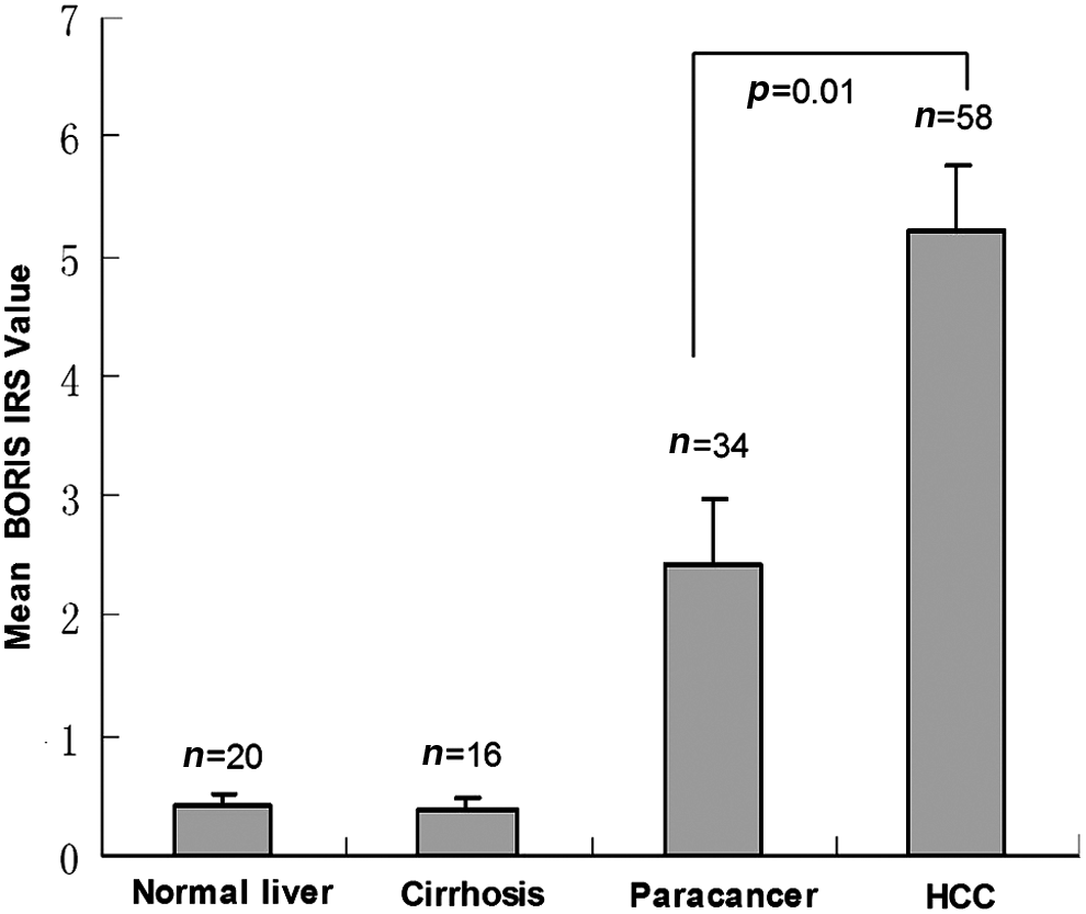
The immunoreactivity score (IRS) values for 34 BORIS-positive samples (2.43±0.51) in paracancer tissues were lower than 58 BORIS-positive samples (5.22±0.53) in hepatocellular carcinoma (HCC) tissues (p=0.01).
p-Value<0.05 is bolded.
BORIS, brother of the regulator of imprinted sites; HBsAg, hepatitis B surface antigen; AFP, alpha fetoprotein.
BORIS present in HCC associated with CD90 expression
We performed histochemical double staining for BORIS and CD90 in 105 primary HCC specimens. CD90 expression was observed in 70 cases (66.7%), localized in the plasma membrane and the cytoplasm, and BORIS expression was observed in 58 cases (55.2%) localized in the nucleolus and the cytoplasm (Fig. 3). Forty-nine of the 105 cases (46.7%) showed double staining, and the expression of BORIS was correlated with CD90 (p=0.000, Table 1). We also found BORIS expression in HCC tissue associated with the expression of CD90 using multiple fluorescence staining technique (Fig. 3).
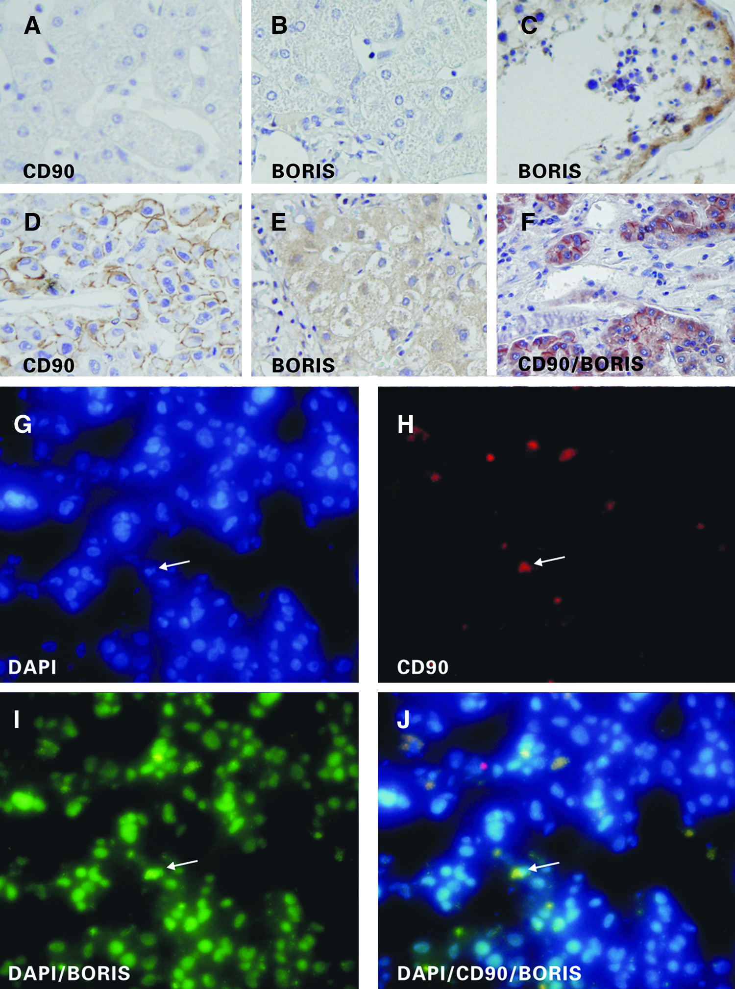
Detection of BORIS and CD90 protein in normal human liver
Prognostic significance of BORIS expression
To elucidate the prognostic role of BORIS in HCC, overall survival rates were estimated by Kaplan-Meier survival curves (Fig. 4). The expression of BORIS was associated with poor prognosis of patients with HCC (p=0.003, log-rank test). Overall survival rate and 5-year survival rate of patients with BORIS expression were lower than those that did not express BORIS (46.8% vs. 20.7% [overall survival rate] and 38.3% vs. 15.6% [5-year survival rate], respectively, p<0.05, Fig. 4). On the other hand, the presence of satellite nodule tends to be higher in the BORIS-positive group (34/58 cases, 58.6%) when compared to the BORIS-negative group (10/47 cases, 21.3%, p=0.000).
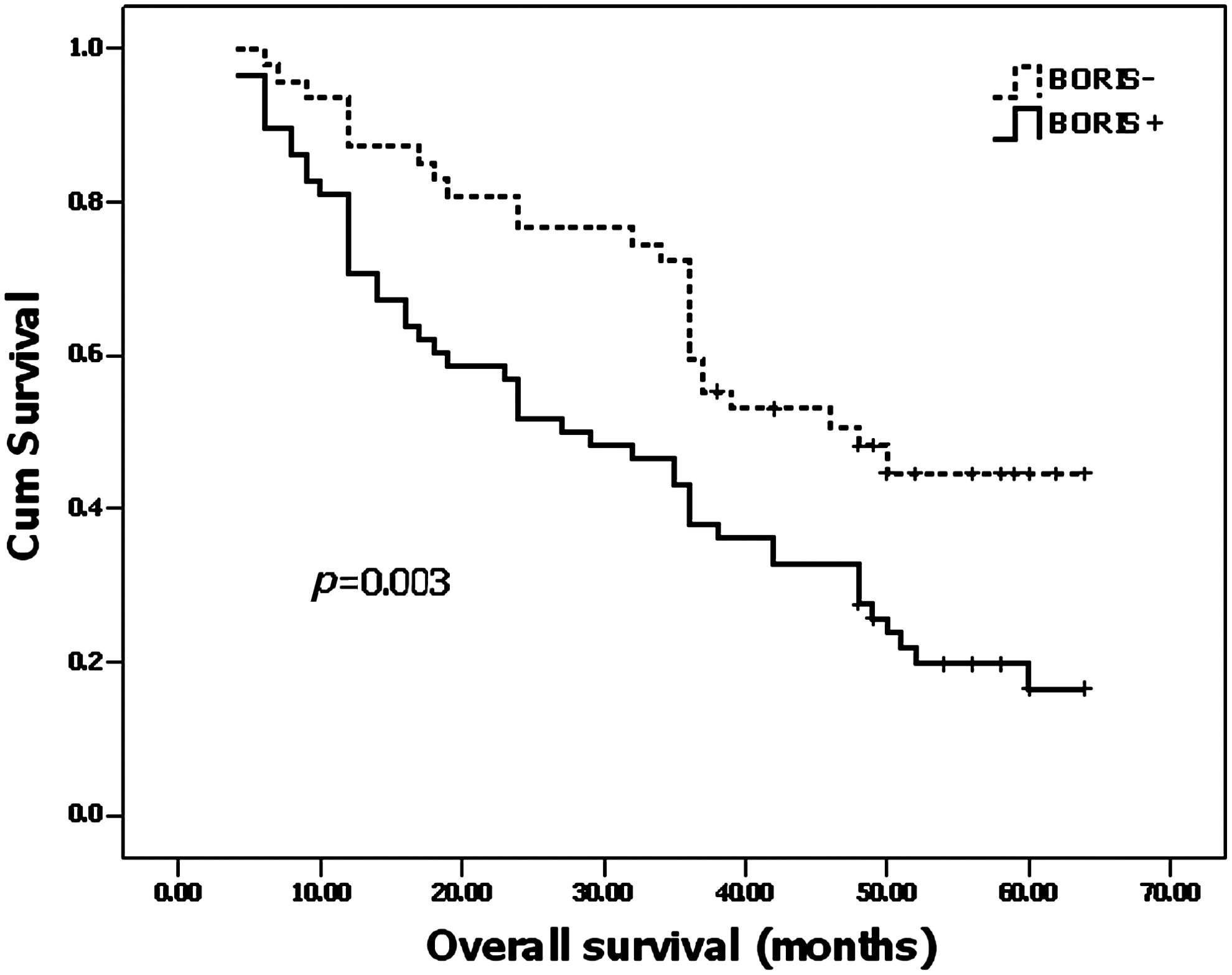
Kaplan-Meier survival curves for BORIS-positive group versus BORIS-negative group in 105 patients with HCC showed a highly significant separation (p=0.003, log-rank test).
Discussion
A number of recent studies have found that epigenetic changes play an important role in the progression of cancer. In epithelial ovarian cancer, the BORIS/CTCF expression ratio is also associated with a higher stage of tumor and decreased rate of survival (Woloszynska-Read et al., 2011). It has been suggested that the expression of BORIS displaces CTCF in the genome and leads to proliferation of cancer cells. Ghochikyan et al. (2007) demonstrated that BORIS protein was always expressed in pancreatic carcinoma, but there was no significant expression in normal tissues. D'Arcy et al. (2008) showed that normal glands of the breast were negative for BORIS, whereas all types of breast carcinoma expressed this protein with very high incidence. BORIS has also been shown to be expressed in many other cancers, such as head and neck squamous cell carcinoma, prostate cancer, endometrial cancer, colon carcinoma, and melanoma (Vatolin et al., 2005; Risinger et al., 2007; Kholmanskikh et al., 2008; Cuffel et al., 2011; Makovski et al., 2012). The results of these studies, including ours, are similar in that they show BORIS expression to be more intense in the carcinoma nest than in normal or benign lesions. However, BORIS, as a regulatory protein of DNA demethylation, has not been reported to play a role in liver tumors. This is the first study showing the expression of BORIS in HCC and its role in disease prognosis. We studied protein levels of BORIS in a large number of cases of HCC and compared them to noncancerous tissues and observed that BORIS was expressed in 55.2% of the HCC cases. Our study also showed that the expression of BORIS in HCC is significantly related to the CSC marker CD90 in HCC tissues.
In this study, we also investigated BORIS mRNA and protein expression in HCC cell lines or primary HCC tissues, and evaluated the correlation of BORIS expression to clinical outcome of HCC patients, and we found that BORIS expression in HCC tissue was significantly higher than that in normal tissue, which suggests that BORIS may be involved in the genesis and development of liver cancer, and it could be used as an indicator of liver cancer. These results combine with the findings that BORIS was expressed in different human liver carcinoma cell lines suggest that BORIS is likely to be associated with malignant liver cells.
Our results demonstrated that BORIS was very frequently expressed in HCC when compared to noncancerous tissue. We obtained more informative results probably because of the larger number of cases examined by immunohistochemistry analysis. We found that the expression of BORIS is also correlated with high proliferation activity and poor overall survival rate. Our study also revealed that the expression of BORIS in HCC is significantly related to the CSC marker CD90 in HCC tissue. The research efforts on BORIS and CD90 have shed light on new directions for the eradication of CSCs. The expression of BORIS in CSCs provides evidence that BORIS could be a therapeutic target in treating liver cancer. Kaplan-Meier survival analysis showed that the expression of BORIS significantly correlated with a shorter survival time of HCC patients (p=0.003). Moreover, the expression of BORIS also showed a tendency to correlate with high tumor metastasis rate in the liver (Table 1). These results have revealed that the expression of BORIS strongly reflects the biological aggressiveness of HCC and plays an important role in its progression. These results also suggest that HCC patients with BORIS expression could probably have a relapse with poor prognosis. BORIS could be used as a prognostic indicator for the HCC, and inhibiting BORIS expression could prove to be an adjuvant therapeutic approach in treating HCC.
In summary, our study has shown for the first time that BORIS is expressed in HCC tissues, and its expression significantly correlates with poor prognosis. Furthermore, we demonstrated that the expression of CSC marker CD90 is related to the expression of BORIS in HCC tissue and HCC cell lines. The expression of BORIS in CSCs could provide experimental basis for relevant studies.
Footnotes
Acknowledgments
This research was supported by grants from the National Natural Science Foundation of China (No. 81172372) and Sichuan University Young Teachers in start-up funding (No. 2011SCU11046). We thank Shu Wang Duan and Ling Wei for his critical review and language editing of this manuscript as well as Yan He and Qun Ying Li for his technical support.
Authors' Contributions
B.L, Y.Q., and K.F.C. designed and directed the study. Y.Q., B.L., K.F.C., W.Q.H., B.H., and Y.G.W. performed the majority of the experiments and composition of the manuscript. Y.G., W.Q.H., and Y.G.W. were responsible for data collection and analysis, and reviewing and scoring the degree of immunostaining of sections. All authors have read and approved the final manuscript.
Author Disclosure Statement
The authors declare that they have no competing interests.