




Abstract
Aims: The prognostic role of dickkopf-1 (DKK1) in gastric cancer (GC) remains poorly characterized. We performed a meta-analysis to evaluate correlations between DKK1 overexpression and the prognosis of patients with GC. Materials and Methods: We included five published studies to assess the relationship between DKK1 and the clinicopathological characteristics and overall survival of GC patients. Literature searches, article selection, data collection, and statistical analysis were performed using RevMan 5.3 software. Results: Our analyses revealed that DKK1 overexpression was significantly associated with vascular invasion (odds ratio [OR] = 2.43, 95% confidence interval [CI] = [1.21, 4.89], p = 0.01, random effect), lymphatic invasion (OR = 2.61, 95% CI = [1.30, 5.24], p = 0.007, random effect), and distant metastasis (OR = 2.99, 95% CI = [1.95, 4.59], p < 0.00001, fixed effect). Moreover, we also found that DKK1 overexpression was significantly associated with poor overall survival in GC patients (risk ratio = 2.67, 95% CI = [2.24, 3.48], p < 0.00001, fixed effect). Conclusion: This meta-analysis demonstrated that DKK1 may be a useful prognostic marker for GC.
Introduction
D
As a negative regulator of the Wnt signaling pathway, dickkopf-1 (DKK1) plays a crucial role in tumor carcinogenesis and has known roles in cell proliferation, apoptosis inhibition, angiogenesis, invasiveness, immunosuppression, and cell motility (Sato et al., 2010). Overexpression of DKK1 has been reported in many tumors, including multiple myeloma, hepatoblastoma, Wilms' tumor, prostate cancer, kidney cancer, and breast cancer, as well as lung and esophageal cancers (Liu et al., 2014; Huo et al., 2015). Moreover, recent reports have indicated the oncogenic potential of DKK1 activation and its utility as a cancer biomarker; however, these proposals remain controversial (Shen et al., 2012). By contrast, in GC, DKK1 expression and its prognostic value also remain inconsistent (Gomceli et al., 2011; Lee et al., 2012; Cai et al., 2014). Therefore, a systematical and comprehensive meta-analysis to identify the prognostic significance of DKK1 in GC is urgently needed. This study is aimed to more precisely estimate the prognostic value of DKK1 overexpression in GC.
Materials and Methods
Search strategy
Published studies were identified by searching the PubMed, EMBASE, Cochrane Library, Web of Science, Chinese Biomedical Literature Database, China National Knowledge Infrastructure (CNKI), and Wanfang databases (the last search was updated to May 2015). The search strategy used “DKK1 OR dickkopf-1” AND “gastric cancer” or “gastric carcinoma.” No language limitation was applied. Additional articles or abstracts were retrieved using hyperlinks and by manually scrutinizing the reference lists of relevant publications.
Eligibility criteria
Studies were included if they met all of the following inclusion criteria: (1) clinical research that directly examined DKK1 expression in GC; (2) results that included clinicopathological characteristics or overall survival; and (3) studies that reported sufficient data to allow an estimation of the hazard ratio and 95% confidence intervals (CIs) according to DKK1 expression or that could be obtained through contacting the study author(s). Noneligible trials included ecological studies, case reports, reviews, editorials, and animal trials. When multiple publications of the same study population were identified or when study populations overlapped, only the most recent or complete study was included in this present analysis. Duplicate reports were only included in specific analyses if they used different antibodies or performed different subgroup analyses.
Quality assessment and data extraction
The quality of each study was assessed independently by two researchers according to the REporting recommendations for tumor MARKer prognostic studies (REMARK) guidelines (Altman, et al., 2012). Data extracted from eligible studies included the first author's name, study year, region of origin, number of patients, tumor stage, patient characteristics, first-line therapies, follow-up period, detection methods, cut-off level to classify DKK1 as overexpressed, number of patients with high DKK1 expression, and risk ratio (RR) with 95% CIs for overall survival. If a study reported both the results of univariate and multivariate analyses, the latter was selected as its consideration of confounding factors makes it more precise.
Data analysis
Meta-analyses were performed using RevMan 5.3 software. We used the Q-test and I2 test to assess heterogeneity between studies. We used odds ratio (OR) value to evaluate the correlation of DKK1 with clinicopathological parameters, and RR value to evaluate the association between DKK1 expression and overall survival in patients with GC. To test for publication bias, we utilized RevMan 5.3 software to generate a funnel plot; p < 0.05 was considered to indicate a significant difference.
Results
Study characteristics
A total of 22 studies were retrieved from the PubMed database and 29 studies were identified in other electronic databases. The process used for retrieving and eliminating studies is shown in Figure 1. Ultimately, five eligible studies (Gomceli et al., 2011; Gao et al., 2012; Lee et al., 2012; Pan et al., 2013; Cai et al., 2014) published from 2012 to 2014 were deemed to be eligible for our present study, including four reports that provided sufficient original data regarding DKK1 expression and clinicopathological characteristics and another study for which data were obtained by contacting its author. There were three studies that assessed the prognostic value of DKK1 expression for overall survival in GC patients using the Kaplan-Meier method.

The flow diagram shows criteria used for the inclusion and exclusion of studies.
We obtained relevant information by directly extracting original data or by obtaining original data by emailing the author. The basic characteristics of the five eligible articles are listed in Table 1. A total of 1286 GC patients were included in this meta-analysis, including 471 who were DKK1 positive and 815 who were DKK1 negative. All patients came from the following three countries: China, South Korea, and Turkey. DKK1 expression in GC patients was assessed by immunohistochemistry analysis of tissues and ELISA analysis of sera.
DKK1, dickkopf-1; ELISA, enzyme-linked immunosorbent assay; IHC, immunohistochemistry.
Correlations between DKK1 expression and clinicopathological characteristics in GC patients
As shown in Figures 2B, C, and E, our findings reveal that elevated levels of DKK1 expression were significantly associated with the following clinicopathological characteristics: vascular invasion (OR = 2.43, 95% CI = [1.21, 4.89], p = 0.01, random effect), lymphatic invasion (OR = 2.61, 95% CI = [1.30, 5.24], p = 0.007, random effect), and distant metastasis (OR = 2.99, 95% CI = [1.95, 4.59], p < 0.00001, fixed effect).
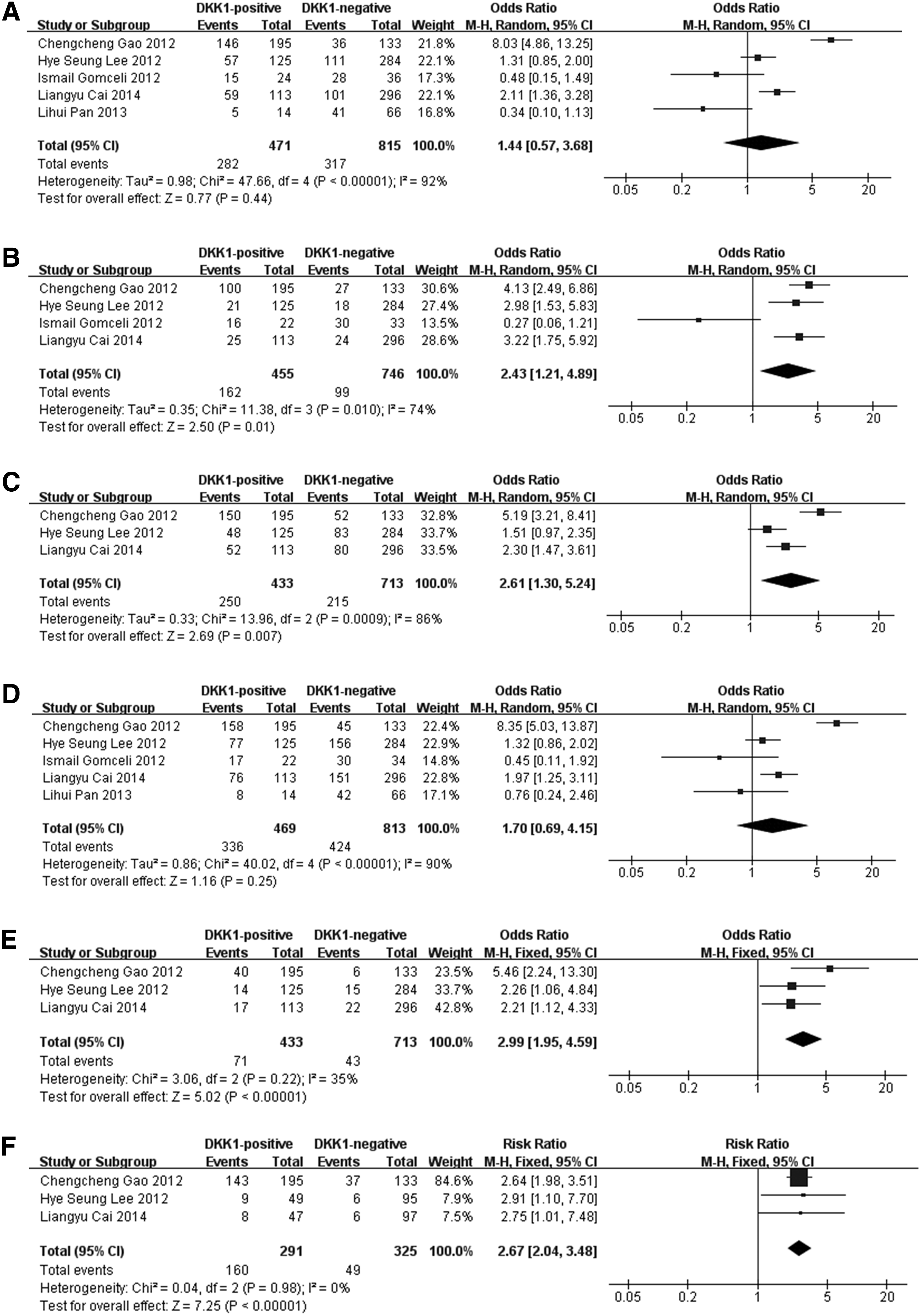
Forest plots show the correlation of DKK1 with clinical characteristics or overall survival.
However, as shown in Figures 2A and D, DKK1 overexpression was not significantly associated with the tumor, nodes, metastases (TNM) stage (OR = 1.44, 95% CI = [0.57, 3.68], p = 0.44, random effect) or lymph node metastasis (OR = 1.70, 95% CI = [0.69, 4.15], p = 0.25, random effect).
The impact of DKK1 overexpression on the overall survival of GC patients
Because the RR of overall survival (OS) was not directly presented, we calculated the RR of OS by extracting or emailing the author to obtain original data from these three studies. The pooled RRs of three studies, which contained 616 patients, were analyzed. Our data (Fig. 2F) indicated that DKK1 overexpression was significantly associated with poor overall survival in GC patients (RR = 2.67, 95% CI = [2.24, 3.48], p < 0.00001, fixed-effect). Moreover, no significant heterogeneity was found among the three included studies for GC patients (χ2 = 0.04, I2 = 0%, p = 0.98).
Publication bias
We tested for publication bias using RevMan 5.3 software. As shown in Figure 3, all funnel plots, except for Figure 3B, indicate that the points were evenly distributed and symmetrical, and Figures 3E and F show that most points were also within the 95% CI. These findings indicate that no publication bias existed for the TNM stage, lymphatic invasion, lymph node metastasis, distant metastasis, or overall survival; furthermore, the results of our present study are credible. Additionally, as shown in Figure 3B, publication bias existed for vascular invasion, which indicates that more studies need to be included for further research.

Funnel plot analysis of publication bias.
Discussion
In GC, prognostic factors, such as the patient age, tumor location, tumor size, tumor differentiation degree, and TNM stage, are commonly used to evaluate the prognosis of GC patients (D'Angelo et al., 2014; Li et al., 2014; Lin et al., 2014). However, none of these parameters can totally and effectively reflect the tumor biological characteristics or therapeutic responses after treatment. Therefore, explorations of additional novel and potential biological markers for GC are necessary.
Increasingly, many experimental studies have linked DKK1 overexpression to worse survival in various cancers, such as hepatocellular carcinoma (Tao et al., 2013), breast carcinoma (Forget et al., 2007), lung cancer (Kim et al., 2014), and GC (Sato et al., 2007). However, in GC patients, the prognostic value of DKK1 is not consistent or convincing. Therefore, a meta-analysis of the prognostic significance of DKK1 may be helpful and is urgently needed.
DKK1 functions as a negative regulator of the canonical Wnt signaling pathway. Specifically, Wnt proteins bind to the frizzled receptor (Fz) and low-density lipoprotein receptor-related protein-5/6 (LRP5/6), resulting in β-catenin accumulation in the cytoplasm followed by its migration to the nucleus. In the nucleus, β-catenin interacts with the transcription factor TCF/LEF and drives the expression of c-myc, cyclin D1, and MMP7, which are important for cell proliferation and migration. DKK1 binds to LRP5/6 and blocks its interaction with Wnt molecules, resulting in β-catenin degradation and the reduction of cell proliferation and migration. Many studies have shown that DKK1 is involved in carcinogenesis for many types of cancer. A previous study (Xu et al., 2014) found that DKK-1 expression is reduced in human lung cancer, which suggests that DKK-1 acts as a tumor suppressor gene in this type of neoplasm. However, other studies (Forget et al., 2007; Fukuzawa et al., 2008; Tao et al., 2013; Kristensen et al., 2014) detected the overexpression of DKK1 in many malignant tissues, including breast cancer, multiple myeloma, Wilms' tumor, and hepatocellular carcinoma. Thus, the expression and role of DKK1 may vary between different types of cancer. So, what occurs in GC?
In this present analysis, we included five studies of the prognostic value of DKK1 in GC. Among these studies, three (Gao et al., 2012; Lee et al., 2012; Cai et al., 2014) showed that DKK1 was present at a higher level in cancerous tissues compared with adjacent noncancerous tissues. Nevertheless, Lihui Pan (Pan et al., 2013) reported that DKK1 had a low-level expression in GC tissues, whereas it was higher in normal gastric tissues. Furthermore, Ismail Gomceli (Gomceli et al., 2011) assessed the expression of DKK1 in the serum of GC patients and found that serum levels DKK1 were elevated in GC patients and might represent a potentially useful novel serologic marker for GC. However, overall, no consistent conclusions could be reached and a larger meta-analysis would appear to be warranted.
In our present meta-analysis, DKK1 overexpression was not significantly associated with the TNM stage (OR = 1.44, 95% CI = [0.57, 3.68], p = 0.44, random effect) or lymph node metastasis (OR = 1.70, 95% CI = [0.69, 4.15], p = 0.25, random effect). However, we found that high levels of DKK1 expression were significantly associated with vascular invasion (OR = 2.43, 95% CI = [1.21, 4.89], p = 0.01, random effect), lymphatic invasion (OR = 2.61, 95% CI = [1.30, 5.24], p = 0.007, random effect), and distant metastasis (OR = 2.99, 95% CI = [1.95, 4.59], p < 0.00001, fixed effect). Intriguingly, we also found that DKK1 overexpression was significantly associated with poor overall survival in GC patients (RR = 2.67, 95% CI = [2.24, 3.48], p < 0.00001, fixed effect). In assessments of publication bias, all findings, except for vascular invasion, exhibited no publication bias. As few articles were available for meta-analysis, and to overcome the publication bias regarding vascular invasion, additional studies will be needed to arrive at an accurate conclusion regarding the relationship between DKK1 and vascular invasion. Nevertheless, a prospective study with a larger sample size and multifactor analysis would be helpful. After taking publication bias into consideration, we conclude that DKK1 overexpression is significantly associated with lymphatic invasion and distant metastasis, and may represent a potential prognostic marker for GC.
Alternatively, although biomarkers can be detected in cancerous tissue using immunohistochemistry (IHC)—which is the preferred method for the evaluation of tumor markers—blood or body fluid sample assays based on non-IHC methods are more readily available and involve relatively noninvasive procedures (Mallett et al., 2010). Thus, in future studies, detecting DKK1 in serum would be useful. Moreover, additional studies of DKK1 expression levels in the sera of GC patients and healthy individuals are urgently needed, which may establish serum DKK1 to be a useful marker for monitoring GC in a convenient, noninvasive, and low-cost way.
Although larger well-designed studies that include additional ethnic groups as well as larger population studies are required, our present meta-analysis indicated that elevated DKK1 expression was associated with lymphatic invasion and distant metastasis, and also led to poorer survival of GC patients. To the best of our knowledge, this is the first meta-analysis to evaluate the prognostic role of DKK1 expression in GC. These findings suggested that DKK1 may represent a novel biomarker to predict the prognosis of GC, and could be a potential target for the development of diagnostic and therapeutic approaches in the treatment of GC.
Footnotes
Acknowledgment
The authors would like to acknowledge the anonymous reviewers for providing helpful comments on this article.
Author Disclosure Statement
No competing financial interests exist.