




Abstract
Aim:
To identify whether miR-148a-3p interacts with DNA (cytosine-5)-methyltransferase 1 (DNMT1) in esophageal cancer.
Methods:
A luciferase assay and immunoblotting were performed to detect the relationship between miR-148a-3p and DNMT1. The MTT method, Annexin V/propidium iodide staining, and Transwell assays were adopted to assess the biological behaviors in EC109 cells. The association between the expression level of miR-148a-3p, clinical features, and prognosis were evaluated by chi-square test and univariate survival analysis.
Results:
In this study, DNMT1 was identified as a direct target of miR-148a-3p by luciferase assay and Western blot. Real-time quantitative PCR analyses showed that the relative expression levels of miR-148a-3p and DNMT1 were reduced in esophageal cancer samples compared with adjacent tissues; and a negative relationship between both was indicated. Upon overexpression of miR-148a-3p in esophageal cancer cells, proliferation and invasion were significantly suppressed, and apoptosis was promoted. A higher level of miR-148a-3p was correlated with better patient outcomes.
Conclusions:
Our study indicated that miR-148a-3p, by targeting DNMT1, likely regulates cell proliferation and invasion in esophageal cancer. miR-148a-3p might also be used prognostically in esophageal cancer and serve as a therapeutic target in the future.
Introduction
Esophageal cancer is one of the most prevalent malignancies, ranking sixth among cancers across the world (Ferlay et al., 2015). Despite therapeutic advancements in recent years, including surgery, chemotherapy, and radiotherapy, the 5-year survival rate of esophageal cancer remains <20%, making it a globally fatal malignancy (DeSantis et al., 2014; Rustgi and El-Serag, 2014). In light of the sophisticated factors that account for the initiation and progression of esophageal cancer, there is an urgent need to elucidate the dominant underlying molecular mechanisms that are responsible for cancer development (McCabe and Dlamini, 2005). This would be highly beneficial toward the discovery of novel therapeutic targets of esophageal cancer.
miRNA is one type of small noncoding RNA, which is generally composed of 18-25 nucleotides (Yu and Li, 2015). Mechanistically, miRNA can regulate the expression of its target genes by binding to the 3′ untranslated region (3′-UTR) of the messenger RNAs (mRNAs), and thus inhibit translation and promote the degradation of target mRNAs (Jili et al., 2016). In recent decades, accumulating evidence has suggested that the functional role of miRNA in the progression of esophageal cancer includes modulation of tumor proliferation, invasion, migration, and epithelial-mesenchymal transition (EMT). Based on previous findings, both up- and downregulation of miRNAs are implicated in the tumorigenesis and progression of esophageal cancer (He et al., 2016; Zhou et al., 2016; Qi et al., 2017; Lindner et al., 2018). It has been reported that miR-675-5p, an oncogenic miRNA, could promote tumor development and metastasis of esophageal cancer by binding to RALBP1-associated Eps domain containing 2 (REPS2), and thus activating the RalBP1/RAC1/CDC42 signaling pathway (Zhou et al., 2016). In contrast, after identifying the lower expression levels of miR-30a-3p/5p in esophageal cancer tissues, Qi et al. (2017) proposed that miR-30a-3p/5p might repress the Wnt signaling pathway by directly binding to wingless-type MMTV integration site family, member 2 (Wnt2), and Frizzled class receptor 2 (Fzd2), thereby inhibiting cell proliferation of esophageal cancer. miR-143-3p also might exert its tumor-suppressing effect by targeting quaking 5 (QKI-5) in esophageal cancer, and presenting as inhibition of proliferation, invasion, and EMT of the tumor cells (He et al., 2016). In addition, Lindner et al. (2018) showed that when selecting chemotherapy resistant-related miRNAs, miR-148a-3p might inhibit cell migration of esophageal cancer; the underlying mechanisms, however, remain insufficiently illustrated.
DNA (cytosine-5)-methyltransferase 1 (DNMT1) is the most abundant form of DNMT, and is chiefly responsible for maintaining and establishing DNA methylation patterns (Romanek-Piva et al., 2016). DNMT1 could epigenetically contribute to the hypermethylation of tumor suppressor genes, by inducing the progression of cancer (Raggi et al., 2014; Bashtrykov and Jeltsch, 2015). Researchers have increasingly appreciated the important role that DNMT1 plays in the development of esophageal cancer. A study by Bai et al. indicated that, upon inhibition of DNMT1, proliferation and invasion as well as metastasis of esophageal cancer were repressed, primarily due to the reduced methylation level of the promoters of Ras association domain-containing protein 1 (RASSF1A) and death-associated protein kinase (DAPK) (Bai et al., 2016). In addition, an epigenetic circuit demonstrating the interplay between DNMT1 and miRNA126 has been identified in esophageal cancer, implying the potential effect of miRNAs on DNMT1 (Liu et al., 2015). Recently, several studies have suggested that DNMT1 is the target gene of miR-148a-3p in both bladder cancer and laryngeal cancer (Jili et al., 2016; Wang et al., 2016; Wu et al., 2016), which contributes to cancer progression. However, the possible relationship and biological function of miR-148a-3p and DNMT1 remains to be illuminated in esophageal cancer.
In this study, we identified DNMT1 as a direct target of miR-148a-3p in esophageal cancer, which is consistent with findings in other cancers. Next, it indicated a lower expression of miR-148-3p, whereas a higher expression of DNMT1 in esophageal cancer tissues compared with the adjacent tissues, and an inverse correlation between these two molecules was presented. Within the in vitro study, by overexpressing miR-148a-3p in the EC109 cells, the protein level of DNMT1 was significantly reduced, along with the decreased capability of cell proliferation and invasion. The expression level of miR-148a-3p was shown to be positively correlated with the overall survival probability and progression-free survival probability of esophageal cancer patients. This study confirmed the interplay between miR-148a-3p and DNMT1 in esophageal cancer. It also suggests the potential clinical uses of miR-148a-3p in the near future.
Materials and Methods
Cell line and cell culture
Human esophageal cancer cell line EC109 cells were purchased from the cell bank of Chinese Academy of Science (Shanghai, China). The cells were grown in RPMI 1640 medium supplemented with 10% fetal bovine serum (FBS) and 1% penicillin/streptomycin. The cells were maintained in a 37°C humidified incubator supplemented with 5% CO2. All the culture materials were from Gibco.
Tissue sample
Formalin-fixed paraffin-embedded samples and the matched adjacent tissues of 126 patients suffering from esophageal cancer were collected from the archive of the department of Henan Province Hospital of TCM from 2008 to 2018, along with the corresponding clinical data. Hematoxylin and eosin-stained sections were reviewed and rediagnosed by one senior pathologist working at the Henan Province Hospital of TCM. The patients ranged in age from 21 to 83 years (mean, 58.61 ± 7.29). The follow-up time ranged from 2014.01 to 2018.06 weeks (mean, 141 weeks). All of the tissues were tested by real-time quantitative PCR (RT-qPCR). This study was approved by the Ethics Committee of Henan Province Hospital of TCM, and informed consent was obtained.
RNA extraction and qPCR
Total RNA was extracted from the cell line EC109 with Trizol reagent (Invitrogen) using traditional methods, and the RNA concentration and quality were determined by UV-2401 spectrophotometer (Shimadzu, Japan). Certain amounts of RNA (1 μg) were used for reverse transcription into cDNA using the PrimerScript™ RT reagent kit (Takara, China), PCR amplification for mRNA, and miRNA were performed by SYBR Premix EX TaQ™ II kit and xx (Takara, China). Primers for the amplification, including GAPDH, DNMT1, U6, and miR-148-3p, were designed by Taraka Company; details are shown in Table 1. miR-NC mimics, miR-NC inhibitor, miR-148-mimics, and miR-148-inhibitor were designed by Jima Tech (Shanghai, China). GAPDH and U6 were used as the internal reference for mRNA and miRNA, respectively. 2−ΔΔCt method was applied to calculate the relative final amounts of target mRNAs and miRNAs.
Primers
Western blot analyses
Western blot assays for human DNMT1 (1:1000; Abcam) were performed on cell line tissues. The samples were lysed by RIPA buffer supplemented with a protease inhibitor mixture for 30 min at 4°C. After the protein concentrations were measured, the cell lysates were separated on SDS-PAGE gels. The proteins were then transferred to polyvinylidene difluoride membranes and blocked for 1 h in 5% skimmed milk at room temperature. Subsequently, the membranes were incubated with primary antibodies, followed by a HRP-conjugated secondary antibody. Anti-GAPDH (1:5000; Kangchen Bio-Tech, China) was used as a loading control. The protein bands were visualized by enhanced chemiluminescence method.
Cell proliferation assay
Two thousand transfected cells were plated in each well of a 96-well plate and cultured for 24-96 h with 100 μL miR-148a, inh-148, and/or inhibitor negative control (iNC). MTT solution was added to the wells and incubated at 37°C with 5% CO2 for 4 h. The medium was then removed and 10 μL of DMSO was added to each well. After which the cells were incubated at 37°C with 5% CO2 for 2 h, and hidden from light. The absorbance was measured at 490 nm using a microplate reader at 490 nm microplate reader (BioRad).
Apoptosis assay
Flow cytometry was used to detect the rate of apoptotic cells after miRNA transfection using the Annexin V-FITC/PI Detection Kit (Abcam). The EC109 cells were digested after miR-148a was transfected for 48 h, and then resuspended with binding buffer. Next, the cells were stained with Annexin V-FITC and propidium iodide. Finally, flow cytometric analyses were performed to detect the apoptosis rate based on the FCS Express software.
Transwell assay
A 24-well Transwell chamber with 8 μm pore size polycarbonate membrane (Corning) was used to evaluate the cells' invasion behaviors. Matrigel matrix (BD, Franklin Lakes) was applied to precoat the membrane after premelting at 4°C. Next, 4 × 10⁵ suspended cells in serum-free medium were added to each upper chamber, and 500 μL of DMEM containing 10% FBS was added to the lower chamber. After 24-48 h incubation period, the cells were whipped off the surface of the upper chamber with cotton swabs, and the invasive cells were fixed on the lower membrane surface with 4% paraformaldehyde, and stained with 0.1% crystal violet. The remaining cells were counted under a light microscope (Olympus, Japan).
Plasmid and cell transfection
The miR-148-3p inhibitor encoded plasmids and their corresponding controls (miR-NC and miR-NC inhibitor) were purchased from ABM (ABM, Canada). The cells were transfected with plasmids together with lipofectamine 2000 (Thermo Fisher Scientific) for 48 h according to the manufacturer's instruction, and then the transfected cells were plated or harvested for further experiments.
Luciferase reporter assay
The luciferase reporter assay was performed per the manufacturer's instructions (Promega). The pGL3-DNMT1 was established by amplifying the 5′-UTR of DNMT1 gene containing the binding site of miR-148a-3p and then cloning it into the Xbal site of the pGL3 control vector (Promega), which is immediately downstream of the luciferase gene. The pGL3-DNMT1-mutant vector was generated as a negative control; it has four mismatches at the miR-148a-3p binding site of the 5′-UTR. For the luciferase assay, HEK293T cells were seeded in 96-well plates and each was co-transfected with pGL3-DNMT1 or pGL3-DNMT1-mutant and miR-148a-3p. The well that was transfected with the pGL3 vector was used as a control. After a 48 h transfection period, the relative luciferase activity was measured using the Dual-Luciferase Reporter Assay System (Promega).
Statistical analysis
Statistical analyses were performed by software of SPSS version 20.0 (Chicago) and GraphPad 6.0 (GraphPad Software, La Jolla). Chi-square test was used to evaluate the correlation between the miR-148-3p expression and clinicopathological parameters. A Pearson correlation analysis was conducted to analyze the correlation between miR-148-3p and DNMT1. A Student's t-test was used to calculate the intergroup difference. Overall survival and progression-free survival were analyzed by Kaplan-Meier method. A p-value <0.05 was considered statistically significant.
Results
DNMT1 is a direct target gene of miR-148a-3p in esophageal cancer
DNMT1 was predicted to be one of the candidate targets of miR-148a-3p using bioinformatic software Target Scan. The findings revealed that the potential binding sites of miR-148a-3p were observed in the 3′-UTR of DNMT1 (Fig. 1A). The 3′-UTR wt-DNMT (wild type) and mut-DNMT (mutant type) luciferase reported vectors were constructed, and the EC109 cells were co-transfected with 3′-UTR miR-NC and pGL3, miR-148a-3p and wt-DNMT, miR-148-3p and mut-DNMT, respectively. The results exhibited that the luciferase activity in the miR-148a-3p along with the wt-DNMT group were significantly decreased compared with the other two groups (p < 0.05, Fig. 1B). The findings of the western blot in vitro were consistent with earlier findings. In the cell line co-transfected with miR-NC inhibitor and miR-148-3p, the protein level of DNMT1 was dramatically induced (Fig. 1C, D). Our results showed that DNMT1 is a direct target of miR-148a-3p in the esophageal cancer cell line EC109.
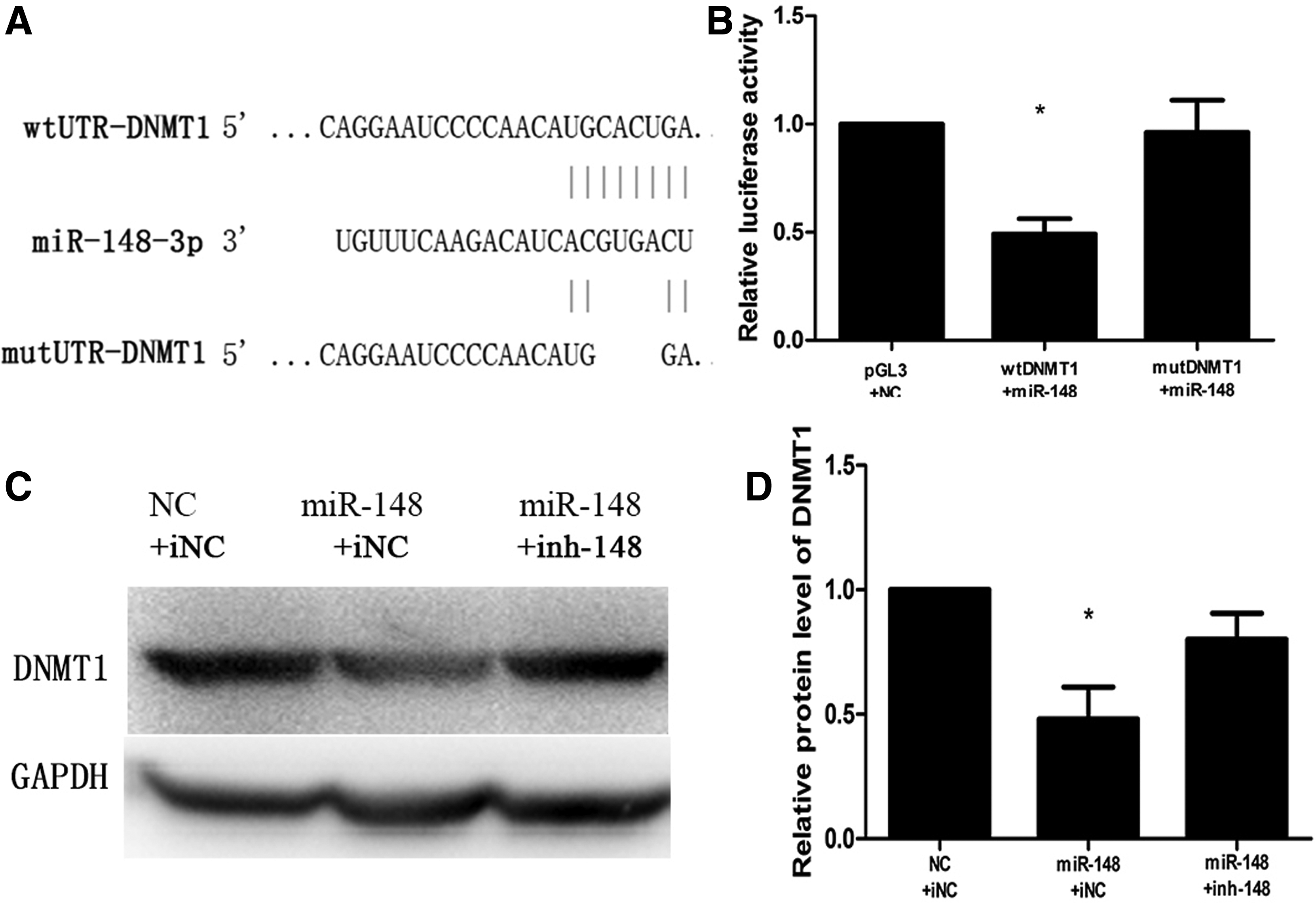
miR-148a-3p's target reaction with DNMT1 in esophageal cancer.
miR-148a-3p is downregulated and DNMT1 is upregulated in esophageal cancer
To further investigate the correlation between miR-148a-3p and DNMT1, a qPCR was performed on the samples of patients with esophageal cancer. The results demonstrated that the relative expression level of DNMT1 was higher in cancerous tissues when compared with the adjacent noncancerous tissues (p < 0.05, Fig. 2A). However, as for the miR-148-3p, the relative expression level was significantly lower in carcinomatous tissues than in their adjacent tissues (p < 0.05, Fig. 2B). In addition, the findings of the Pearson correlation test indicted a negative correlation between the miR-148-3p and DNMT1 in esophageal cancer (p < 0.001, r = −0.448, Fig. 2C). The area under the curve (AUC) value is 0.818, which confirmed the high reliability of miR-148-3p (Fig. 2D). The aforementioned findings indicate that miR-148a-3p might be a potential target for DNMT1.

Relative expression levels of DNMT1 and miR-148-3p in adjacent noncancerous and cancerous tissues, respectively.
miR-148a-3p overexpression might contribute to the inhibition of esophageal cancer cell proliferation and invasion and promote apoptosis
The cell proliferation, Annexin V, and Transwell assays were performed in three groups of cells that were transfected with miR-NC and miR-NC inhibitor, miR-148-3p and miR-NC inhibitor, and miR-148-3p and miR-148-3p inhibitor, respectively. We found that cell growth was suppressed significantly when the miR-NC inhibitor and miR-148-3p mimics were transfected into the cells (Fig. 3A). In addition, the overexpression of miR-148-3p in EC109 cells contributed to an increase of the Annexin V positive cell population, which was twofold higher than the cells transfected with miR-NC, whereas the addition of the miR-inhibitor could reduce cell apoptosis (Fig. 3B, D). The matrigel invasion assay was used to detect that whether miR-148-3p would affect the invasion of EC109 cells. The results demonstrated that overexpression of miR-148-3p reduced the invasive ability of EC109 cells (Fig. 3C, E). These findings demonstrated that miR-148-3p could contribute to the inhibition of cell proliferation and invasion of esophageal cancer.

miR-148-3p mediated biological effect of EC109 cell.
miR-148a-3p serves as a predictor for the prognosis of esophageal cancer
Correlations between the expression level of miR-148-3p and clinicopathological parameters of esophageal cancer
We also investigated correlations between miR-148-3p expression levels and clinicopathological parameters in the 126 patients examined. Several clinicopathological parameters, including gender, age, tumor size, clinical stage, degree of differentiation, and lymph node involvement, were enrolled into our study. After analysis by chi-square test, only clinical stage and differentiation degree were shown to be significantly associated with the expression level of miR-148-3p. No significant correlations were observed for the other clinicopathological parameters (Table 2). There is a lower expression of miR-148 in high clinical stage (III/IV) patients compared with low clinical stage patients (p = 0.016), and so is in poor differentiated patients (p = 0.032).
Correlation of miR-148-3p with Clinical Features of Esophageal Squamous Cell Carcinoma Patients
Correlations between the expression level of miR-148-3p and survival
Overall survival and progression-free survival were assessed by the Kaplan-Meier method in our cohort (Fig. 4). The results revealed that the overall survival (Fig. 4A, p = 0.009) and progression-free survival (Fig. 4B, p = 0.032) are both significantly reduced in patients with high expression levels of miR-148-3p.
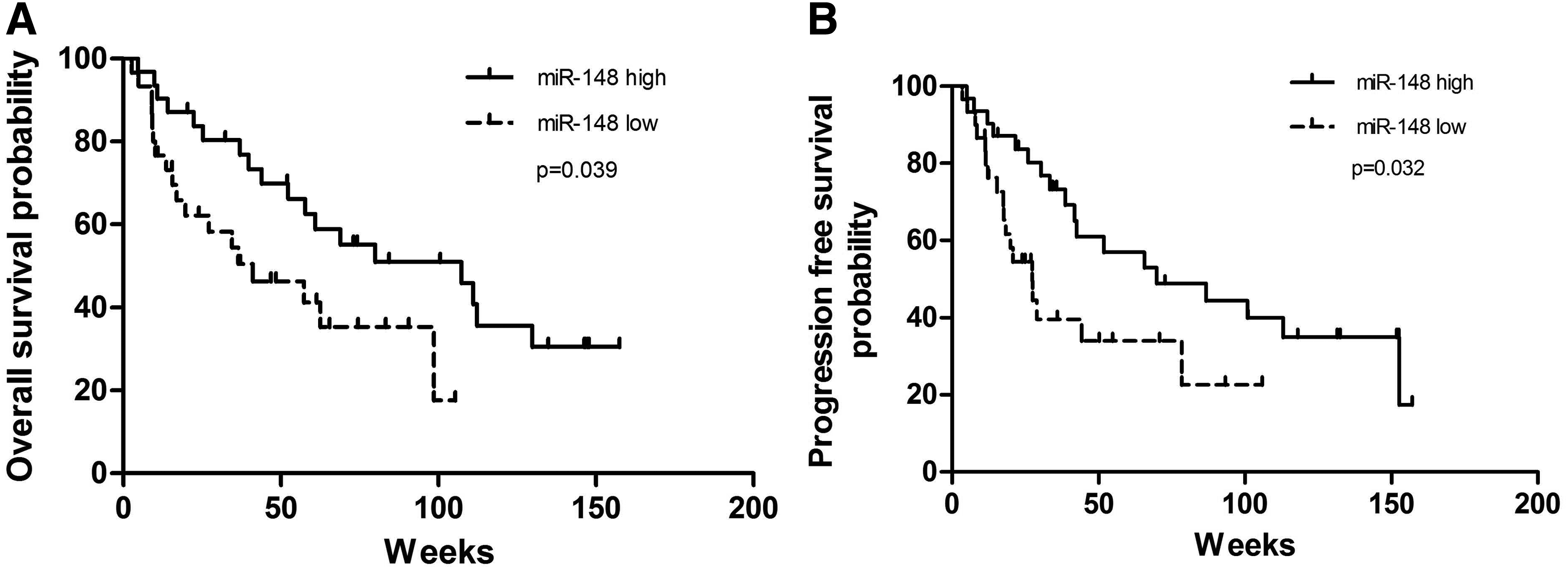
Kaplan-Meier analysis for survival in patients with different level expression of miR-148-3p.
Discussion
In recent decades, abnormal up- or downregulation of miRNAs has been suggested to implicate in the initiation and development of cancers (Zhou et al., 2016). Based on previous findings, miR-148-3p has been revealed to be downregulated and can be used a favorable biomarker for better clinical outcomes in several cancers, namely bladder cancer, laryngeal cancer, colorectal cancer, and esophageal cancer (Dong et al., 2014; Wang et al., 2016; Wu et al., 2016; Lindner et al., 2018). DNMT1 has been reported to play a pivotal role in epigenetically silencing tumor suppressor genes. Several studies have indicated DNMT1 as a target gene of miR-148-3p in mediating cancer progression. Wang et al. (2016) demonstrated that miR-148-3p was generally downregulated in bladder cancer due to the hypermethylation induced by DNMT1, and it is also targeted by miR-148-3p; and miR-148-3p and DNMT1 form a regulatory loop in bladder cancer. It has also been observed that miR-148-3p and DNMT1 worked as an axis for controlling the expression of Runt-related transcription factor 3 (RUNX3), a tumor suppressor gene, implicating it in the development of laryngeal cancer (Jili et al., 2016). Wu et al. (2016) reported that, as lncRNA H19 is upregulated in laryngeal cancer, knockdown of H19 could inhibit cell invasion, migration, and proliferation by upregulating the expression of miR-148-3p and the subsequent decreased levels of DNMT1. As far as we know, there is no study regarding the association between miR-148-3p and DNMT1 in esophageal cancer. Here, we hypothesized that miR-148-3p might interact with DNMT1 and be involved in the progression of esophageal cancer.
In this study, we set out to investigate the expression level of miR-148-3p and DNMT1 in clinical samples from esophageal cancer patients and their adjacent normal tissues by qPCR. The level of miR-148-3p was lower and DNMT1 was higher in tumor tissues than the adjacent normal tissues, with a negative correlation displayed between the levels of both. This verified our presumptions. In addition, the analysis regarding the expression of miR-148-3p and the clinicopathological factors has revealed the possible negative relationship of miR-148-3p in regard to the progression of esophageal cancer. The luciferase assay confirmed DNMT1 as a target gene for miR-148-3p, which was in accordance with the observations from bladder and laryngeal cancer studies. The western blot analyses validated the conclusion and indicated that miR-148-3p was an important factor in modulating the expression level of DNMT1. The in vitro cell assays of overexpression miR-148-3p with or without its inhibitor have revealed the effect that miR-148-3p plays on regulating cell proliferation as well as invasion of esophageal cancer. This implies that miR-148-3p plays a significant role in the development of esophageal cancer. Another recent study suggested that overexpression of miR-148-3p could impair cell migration of esophageal cancer (Lindner et al., 2018), which supports the findings of this study. In an attempt to figure out the prognostic value of miR-148-3p in cancer patients, a survival analysis was conducted. The results showed the higher expression levels of miR-148-3p were an indicator for better outcome and survival probability.
Although we have uncovered the potential involvement of miR-148-3p and DNMT1 in the development of esophageal cancer, the detailed mechanisms and function of miR-148-3p are insufficiently understood. Further studies are required to delineate the mechanism. From the perspective of the molecular level, we found that DNMT1 is a downstream target of miR-148-3p. However, it is still unclear that what is the downstream gene that is methylated by DNMT1. The potential upstream regulator for the miR-148-3p-DNMT1 axis is still unknown. In addition, in light of the versatile function of miR-148-3p in modulating the progression of esophageal cancer, the questions arises as to whether there are any other target genes that are responsible? The function of miR-148-3p might need to be validated in more cell lines of esophageal cancer. Beyond the biological function of miR-148-3p in cancer cells, it is also necessary to explore its possible role in vivo.
Our study has revealed that DNMT1 is a direct target of miR-148-3p in esophageal cancer, and the expression level of miR-148-3p is decreased. DNMT1 is also enhanced in esophageal cancer, exhibiting a negative correlation. The overexpression of miR-148-3p in esophageal cancer cells could inhibit the tumor proliferation and invasion. A higher expression level of miR-148-3p could potentially predict a better probability of overall survival and progression-free survival. In the near future, miR-148-3p might be utilized as a prognostic indicator and an intervention target for esophageal cancer.
Conclusions
Our study indicated that miR-148a-3p, by targeting DNMT1, might regulate cell proliferation and invasion of esophageal cancer. miR-148a-3p also might be used as an indicator for the prognosis of esophageal cancer and a promising therapeutic target in the future.
Footnotes
Acknowledgments
The authors thank the patients and volunteers recruited from Henan Province Hospital of TCM for participating in the research.
Author Disclosure Statement
The authors declare that they have no conflict of interest.