




Abstract
Background:
Differential expressions of cancer-associated genes, including histone deacetylases (HDACs), were identified in distinctive molecular subtypes of breast cancer. Compared with hormone receptor-positive breast cancer, triple-negative (TNBC, ER−PR−HER2−) is the most aggressive form of breast cancer.
Aims:
To determine the association of HDAC7 mRNA expression levels with clinicopathological features and patients' survival with TNBC or ER+PR+HER2− breast cancers.
Methods:
Total RNA was extracted from 61 TNBC and 74 ER+PR+Her2− tumors. Relative gene expression was evaluated by SYBR Green RT-PCR, normalized to glyceraldehyde-3-phosphate dehydrogenase. The HDAC7 mRNA expression was defined as high or low, according to receiver operating characteristic analysis. Kaplan-Meier and Cox regression analyses for overall survival were assessed to evaluate the prognostic relevance of HDAC7 overexpression.
Results:
The HDAC7 overexpression was predominantly found in invasive ductal carcinomas (p = 0.023), high histologic grade (p = 0.007), and high nuclear grade tumors (p = 0.030). TNBC subtypes had a significantly lower mean HDAC7 gene expression compared with ER+PR+HER2− tumors (p = 0.005). However, HDAC7 overexpression predicted unfavorable survival of TNBC patients (p = 0.003). Multivariate Cox regression analysis indicated that recurrences (hazard ratio [HR] = 5.432, p = 0.003), and HDAC7 overexpression (HR = 9.287, p = 0.033) persisted as independent prognostic factors for poor survival of TNBC patients.
Conclusions:
HDAC7 mRNA overexpression is associated with poor survival in patients with TNBC tumors.
Introduction
Breast carcinomas are an intrinsic group of tumors, characterized by molecular heterogeneity of distinctive subtypes that leads to diverse clinical manifestations and prognosis (Park et al., 2012). Subtypes of breast cancer are primarily diagnosed by estrogen-receptor (ER), progesterone-receptor (PR), and the human epidermal growth factor 2 (HER2) expressions. As opposed to prognostically most favorable hormone-receptor-positive and HER2-negative tumors (ER+PR+HER2−), triple-negative breast cancers (TNBC) lack expression of the hormone- and HER2-receptor (ER-PR-HER2−), and are the most aggressive forms, associated with early metastasis, chemotherapeutic resistance, and poor survival (Trivers et al., 2009). TNBC accounts for 10-20% of all breast cancers and is commonly diagnosed in women under 40 years (Trivers et al., 2009), and remains the most challenging molecular subtype.
Epigenetic modifications are heritable alterations of gene expression that occur without changes in the DNA sequence. Histone acetylation is one of the key epigenetic mechanisms that contribute to the transcriptional activation. On the other hand, histone deacetylation by enzyme families' histone deacetylases (HDACs) decreases negative histone charges, which promote compact, transcriptionally inactive chromatin state. An aberrant pattern of histone acetylation is a common hallmark of tumor phenotype, demonstrated in a number of malignancies, including breast carcinoma (Weichert, 2009; Barneda-Zahonero and Parra, 2012).
In humans, HDACs are commonly divided into four classes based on phylogenetics, cellular localization, and biological function. Class II HDACs (HDAC4, 5, 6, 7, 9, and 10), localized in both the nucleus and cytoplasm, is expressed in a tissue-specific manner (Villagra et al., 2009). Histone deacetylase 7 (HDAC7) has been implicated in the epigenetic regulation of the key homeostatic biological processes, such as cell growth, differentiation, and apoptosis (Zhu et al., 2011). HDAC7 has a role in the maintenance of vascular integrity, endothelial cell migration, and angiogenesis (Chang et al., 2006; Mottet et al., 2007; Zhu et al., 2011). Furthermore, HDAC7 associates with a number of diverse transcription factors and corepressors, including STAT3 (signal transducer and activator of transcription 3 activation) (Lei et al., 2017), ERα, FOXP3 (Li et al., 2007), and FOXA1 (Malik et al., 2010). HDAC7 has a crucial role in cancer cell proliferation through inhibition of c-Myc expression and the induction of p21 and p27 tumor suppressor expression (Zhu et al., 2011).
The number of previous studies demonstrated different expression patterns of cancer-associated genes among distinctive subtypes of breast cancer (Wiechmann et al., 2009; Caslini et al., 2019). Furthermore, HDAC7 belongs to intriguing Class IIa HDACs, which members can act either as oncogenes or as tumor suppressors, depending on the cellular context (Weichert, 2009). Also, emerging studies implicate that HDAC7 might be a potential target for new therapy modalities with HDAC inhibitors in specific molecular subtypes of breast cancer, such as TNBC (Caslini et al., 2019). Thus, the aim of this study was to investigate the potential association of HDAC7 gene expression with clinicopathological features and prognosis in two distinct molecular subtypes of breast cancer, TNBC and ER+PR+HER2− tumors.
Materials and Methods
Subjects and tumor samples
The study was carried within the period 2015-2019 and included 135 breast cancer patients, 61 with TNBC and 74 with ER+PR+HER2− tumors. All of the patients were females with a median age 60, range 33-85 years that underwent tumor surgical resection at the Institute for Oncology and Radiology of Serbia, Belgrade, Serbia. None of the patients was subjected to chemotherapy or radiotherapy before surgery.
Tumor tissue samples were fresh frozen and stored at −196°C in the tumor bank of the Institute for Oncology and Radiology of Serbia. All tumors were graded and histologically classified by a breast cancer pathologist. Informed consent was obtained from all participants. All experiments performed in this study involving human samples were in accordance with the ethical standards of the Ethics Committee of the Institute for Oncology and Radiology and with the 1964 Helsinki Declaration and its later amendments or comparable ethical standards.
RNA isolation and quantitative PCR
Total RNA was extracted from fresh-frozen tissue samples by using TRIzol reagent (Invitrogene, Carlsbad, CA), according to the manufacturer's protocol. Briefly, 200 ng of total RNA was used for cDNA synthesis using the Tetro cDNA Synthesis Kit (Bioline, London, United Kingdom) in the final volume of 20 μL. cDNA samples were diluted to the final concentration of 20 ng/μL. Primers used for Real-time PCR were previously described (Lei et al., 2017). Relative expression of HDAC7 was quantified by Maxima SYBR Green PCR Master Mix (Thermo Fisher Scientific, Waltham, MA), according to the manufacturer's recommendations. Quantification was performed in 7500 Real-time PCR System (Applied Biosystems, Foster City, USA). All reactions were carried out in triplicates.
To identify the most stable reference gene, we examined GAPDH (glyceraldehyde-3-phosphate dehydrogenase), ACTB (β-actin), 18S (18S ribosomal RNA), HPRT (hypoxanthine guanine phosphoribosyl transferase), and TBP (TATA box-binding protein) gene expression in the pilot study of this research. A total of 60 tumor samples were examined, 30 TNBC and 30 ER+PR+HER2− carcinomas. The stability of the candidate reference genes was evaluated with delta Ct method and two softwares: NormFinder (Andersen et al., 2004), for evaluations of the intergroup and intragroup variations, and BestKeeper (Pfaffl et al., 2004), as a commonly used software that evaluates the stability reference gene expression. In our analysis, GAPDH was ranked the highest, with the lowest intra- and intergroup variations and therefore used as an endogenous control for normalization.
The fold changes of HDAC7 gene expression were calculated by the 2−ΔΔCt method, and the Pfaffl efficiency correction (Pfaffl, 2001). Calibrator sample consisted of pooled cDNA from all samples. The large pool of standard and calibrator cDNAs was aliquoted and used in every run through the completion of a study, to provide consistent Real-time PCR results. The fold change of HDAC7 gene expression was dichotomized as high or low, according to optimal cutoff based on survival outcome, assessed by receiver operating characteristic (ROC) analysis.
Statistical analyses
The statistical analyses were performed using SPSS 20.0 software (IBM Corporation). The Mann-Whitney U test was used to compare the difference of HDAC7 gene expression between TNBC and ER+PR+HER2− tumors. ROC and AUC (area under the ROC curve) analysis was used to evaluate the HDAC7 gene expression as a potential biomarker for breast cancer diagnosis and prognosis. Furthermore, ROC curves and the Manhattan distance method were assessed to determine the optimal threshold value of gene expression based on survival outcome. The HDAC7 gene expression was defined as high or low, according to optimal cutoff. Associations of HDAC7 gene expression with clinicopathological characteristics of the patients were evaluated by chi-square test (χ2) or Fisher's exact test.
The Kaplan-Meier method and log-rank tests were used to compare survival between two groups. The Cox analysis was assessed to evaluate the hazard ratio (HR) for overall survival (OS), with a 95% confidence interval (95% CI). The variables significant in univariate analysis, including the variables with p < 0.200, were included in the multivariate analyses to determine the independent predictors of OS. p-values of <0.05 were considered statistically significant.
Results
Clinicopathological features are presented in Table 1. The relative expression of HDAC7 mRNA was evaluated in two molecular subtypes of breast cancer, TNBC and ER+PR+HER2−, Figure 1. A comparison of HDAC7 gene expression profiles between two distinct prognostic subgroups of breast cancer revealed that TNBC patients had a significantly lower HDAC7 expression compared with ER+PR+HER2− patients (mean ± standard error of the mean 8.23 ± 1.66, 21.39 ± 5.09, respectively, p = 0.005), Figure 1.
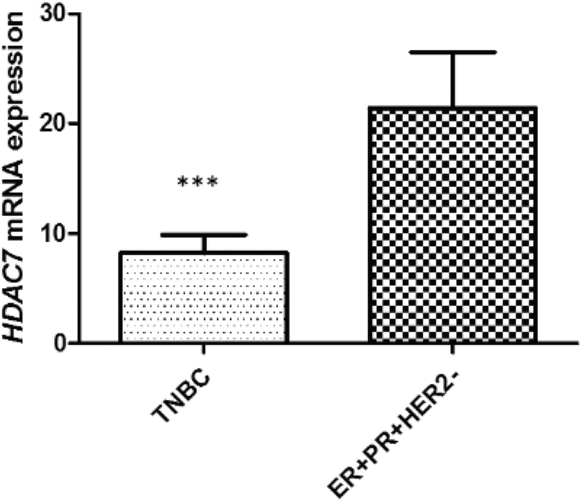
Relative fold change of HDAC7 gene expression in tumor tissues of triple-negative and ER+PR+Her2− breast cancer patients. Relative fold changes are calculated by 2−ΔΔCt method, and data are expressed as the mean fold change ± SEM (standard error of mean). *** Represents Mann-Whitney U test p < 0.001. ER, estrogen-receptor; HDAC7, histone deacetylase 7; Her2, human epidermal growth factor 2; PR, progesterone-receptor; SEM, standard error of the mean.
Patients' Clinicopathological Features and Their Association with Histone Deacetylase 7 Expression
Significant values (p < 0.05) are in bold.
Combined h/nG1 with h/nG2.
ER, estrogen-receptor; HDAC7, histone deacetylase 7; Her2, human epidermal growth factor 2; PR, progesterone-receptor; TNBC, triple-negative breast cancer.
The ROC analysis was used to evaluate the prognostic potential of the HDAC7 mRNA expression for subtype-specific expressions in TNBC and ER+PR+HER2−, as previously suggested (Sorlie et al., 2003; Xiao et al., 2011). The ROC curve analyses using original data of all cases indicated that for total cohort the value of 2.1 is an optimal cutoff for HDAC7 fold change (AUC = 0.57, sensitivity 94.7%, specificity 28.7%, p = 0.045), Figure 2. However, the results of ROC analysis using subtype-specific expression data were better than those using data for all cases. The AUC score increased from 57% for total cohort to 66% and 60% in TNBC and ER+PR+HER2− subtype, while specificity significantly increased from 27% for the total cohort to 47.8% and 49.3% in TNBC and ER+PR+HER2− subtypes, respectively, Figure 2. ROC analysis suggested the value of 2.35 as an optimal cutoff for HDAC7 fold change in TNBC patients (AUC 0.66, sensitivity 92.9%, specificity 47.8%, p = 0.005), and a value of 7.1 as an optimal cutoff for HDAC7 fold change in ER+PR+HER2− patients (AUC 0.60, sensitivity 80%, specificity 49.3%, p = 0.21), Figure 2. Therefore, we dichotomized HDAC7 fold changes of gene expression as low (underexpressed) or high (overexpressed) using these subtype-specific expression cutoffs.
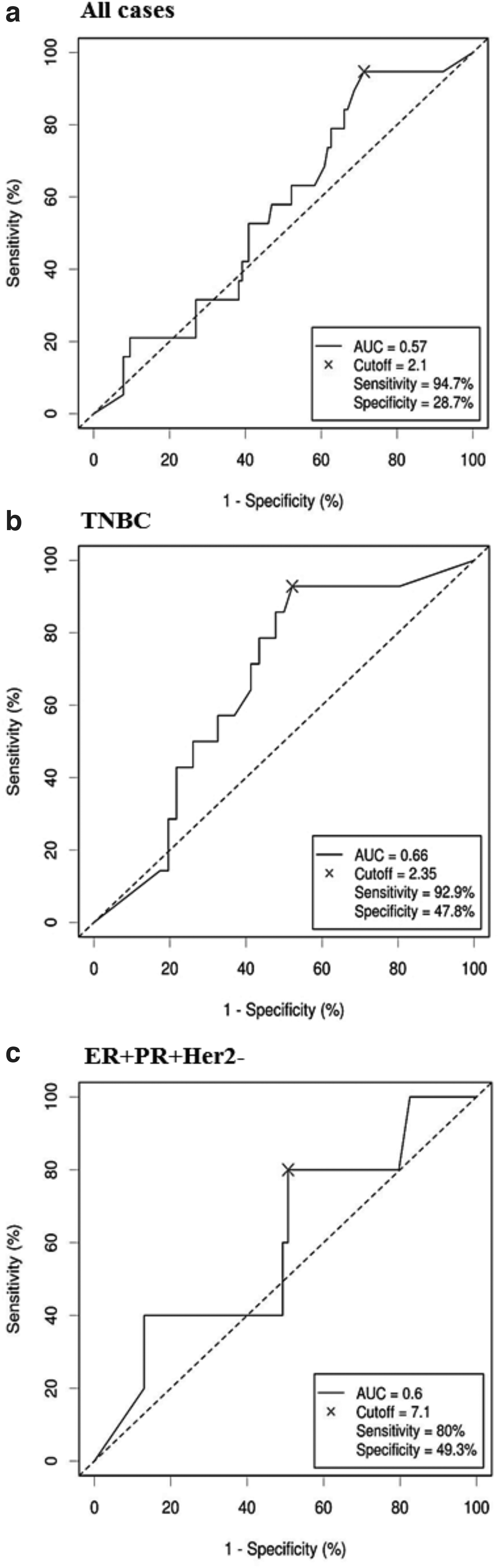
ROC-AUC analyses for all cases
We observed a higher prevalence of HDAC7 overexpression in invasive ductal histological type than lobular breast carcinomas (p = 0.023), in tumors with a high histologic grade than with a low histologic grade (p = 0.007), and with a high nuclear grade compared with low nuclear grade tumors (p = 0.030), Table 1. Higher expression of the HDAC7 was found in 62% of TNBC subtype (38/61), whereas we observed similar percentage between high HDAC7 and low HDAC7 expressing ER+PR+HER2− tumors (53% [39/74] vs. 47% [35/74], respectively), Table 2.
Association of Histone Deacetylase 7 Gene Expression with Clinicopathological Features in Triple-Negative Breast Cancer and ER+PR+Her2− Patients
The associations of HDAC7 fold changes of gene expression with clinicopathological features of TNBC and ER+PR+HER2− patients are shown in Table 2. None of the clinicopathological features was associated with HDAC7 overexpression in both TNBC and hormone-positive subtypes of breast cancer.
To our knowledge, all patients died from breast cancer. However, to prevent a potential misattribution of deaths, we have used OS rather than disease-specific survival, since all-cause mortality is unaffected by bias in classifying the cause of death. According to subtype-specific survival analysis, TNBC patients with HDAC7 overexpression had worse OS compared with those with low HDAC7 expression (p = 0.005), Figure 3. In the subgroup of ER+PR+HER2− patients, no survival difference was detected between high- and low HDAC7-expressing tumors, Figure 3. There was no association between HDAC7 expression and disease-free survival in either of the studied subgroups of breast cancer patients.

Kaplan-Meier survival curves for HDAC7 expression in TNBC
The univariate Cox regression analysis in the TNBC subgroup of breast cancer patients revealed that distant metastasis (HR = 4.687, 95% CI [1.643-13.371], p = 0.004), recurrences (HR = 6.178, 95% CI [2.075-18.393], p = 0.001), and HDAC7 overexpression (HR = 11.460 95% CI [1.502-87.428], p = 0.019) significantly contributed to poor survival, Table 3. Multivariate Cox regression analysis indicated that in TNBC patients, recurrences (HR = 5.432, 95% CI [1.798-16.406], p = 0.003), and HDAC7 overexpression (HR = 9.287 95% CI [1.203-71.677], p = 0.033) persisted as independent prognostic factors for poor survival. In ER+PR+Her2− patients none of the clinicopathological features, including HDAC7 overexpression, did not persist as independent prognostic factors to worse OS, Table 3.
Univariate and Multivariate Cox Regression Analysis in Triple-Negative Breast Cancer and ER+PR+Her2− Breast Cancer Patients, According to Overall Survival
Significant values of confidence interval (p < 0.05) are in bold.
CI, confidence interval; HR, hazard ratio.
Discussion
In recent years, due to its heterogeneity, breast cancer is no longer considered as a single entity, but as a group of tumors composed of distinct molecular subtypes, with diverse clinical and pathological features, different therapeutic responsiveness, and outcomes (Wiechmann et al., 2009; Park et al., 2012). Elucidating their molecular profiles could be essential for determining treatment and surveillance modalities.
A number of recent studies show that histone modifications could have a critical impact on the chromatin landscape in breast cancer. HDAC7 plays an important role in the most relevant aspects of carcinogenesis, including apoptosis, proliferation, invasion, and metastasis (Zhu et al., 2011). The HDAC7 expression has been frequently dysregulated in a number of tumor types (Moreno et al., 2010; Lei et al., 2017; Sang et al., 2019), including breast carcinoma (Weichert, 2009; Witt et al., 2017; Caslini et al., 2019). However, the exact role of HDAC7 in the pathophysiology and its potential impact on survival in distinctive molecular subtypes of breast carcinoma are yet to be fully elucidated. Furthermore, the exact role of HDAC7 in the complex network of other HDACs, as well as the transcriptional response of their downstream targets and hormone receptors in breast cancer is still largely unknown.
There are a limited number of studies investigating the potential prognostic utility of HDAC7 gene expression. To our knowledge, our study is the first one evaluating the prognostic potential of HDAC7 expression in two clinically and prognostically diverse molecular subtypes of breast carcinoma, TNBC and hormone-receptor positive, HER2-negative tumors (ER+PR+HER2−). Our data indicate that TNBC patients with HDAC7 overexpression had significantly worse OS, as opposed to ER+PR+HER2− patients, where HDAC7 expression was not associated with survival.
Our findings of variable HDAC7 gene expression patterns in triple-negative and hormone-receptor-positive breast carcinomas are supported by previous findings of different expression signatures of cancer-associated genes among distinctive subtypes of breast cancer (Wiechmann et al., 2009). As previously suggested (Sorlie et al., 2003; Xiao et al., 2011), subtype-specific expression analysis provided more precise insight of the relationship between HDAC7 and these breast cancer subtypes. Our subtype-specific HDAC7 expression analysis showed significantly better results than those using data sets for all breast cancer cases, suggesting that subtype-specific ROC analysis can further improve the interpretation of gene expression profiling from breast cancer studies. Our results showed that although ER+PR+HER2− patients generally had a significantly higher HDAC7 expression compared with TNBC patients, the prognostic impact of HDAC7 overexpression was observed only for patients with the TNBC subtype, suggesting that HDAC7 could potentially have a differential role depending on the subtype of breast cancer.
Moreover, it was previously demonstrated that various reference genes show differential expression in cell lines of different breast cancer subtypes (Liu et al., 2015). Although GAPDH is the most commonly used reference gene for normalization of quantitative expression, it has been questioned as reference gene in breast cancer since the level of GAPDH expression can be downregulated by chemotherapic drugs (Valenti et al., 2006). None of the patients in our study was subjected to chemotherapy or radiotherapy before surgery. To ensure credible evaluation of the HDAC7 gene expression in our cohort, we identified GAPDH as the optimal reference gene, with the lowest intra- and intergroup variations when comparing TNBC to ER+PR+HER2− subtype of breast carcinomas.
Our findings of significant associations between HDAC7 gene expression and poor survival in TNBC patients are in line with those reported in acute lymphoblastic leukemia (Moreno et al., 2010), ovarian (Yano et al., 2018), gastric (Yu et al., 2017), lung (Lei et al., 2017; Sang et al., 2019), and colorectal cancer (Gao et al., 2018). The exact mechanism of HDAC7 influencing survival in TNBC patients is still not fully understood, but its oncogenic role has been indicated. HDAC7 promotes lung cancerogenesis by repressing STAT3 (Lei et al., 2017). In addition, HDAC7 inhibits tumor suppressor plakoglobin, which subsequently leads to proliferation and invasion potential of lung cancer cells (Sang et al., 2019). In various cancer types, HDAC7 induces c-myc oncogene activation, leading to cancer cell proliferation (Dokmanovic et al., 2007; Zhu et al., 2011).
HDAC7 could exert its oncogenic role through the modulation of endothelial cell migration and angiogenesis. It has been reported that HDAC7 affects the vascular integrity and angiogenesis through inhibition of the matrix metalloproteinase 10 expression (Chang et al., 2006), and platelet-derived growth factor and its receptor, key mediators of the advanced stages of angiogenesis (Mottet et al., 2007). Moreover, among the other HDACs, HDAC7 silencing is sufficient to inhibit angiogenesis in vitro (Mottet et al., 2007). These findings indicate that HDAC7 could be a promising molecular target of antiangiogenic drugs in cancer treatment.
Previous studies demonstrated that HDAC inhibitors (valproic acid, entinostat) can convert ER-negative to ER-positive cell lines (Zhou et al., 2007; Sabnis et al., 2011), which suggests that HDACs could have a crucial role in the regulation of ER expression. It has been demonstrated that HDAC7 could have a unique role in ERα transcriptional repression and regulation of a subset of estrogen-repressed genes, including a cell cycle inhibitor Reprimo (Malik et al., 2010). Tripartite interaction of ERα, HDAC7, and FoxA1 induce transcriptional repression of Reprimo, which and subsequently leads to cell proliferation (Malik et al., 2010).
HDAC7 could play a critical role in driving and maintenance of breast cancer stem cells by controlling a number of cancer-associated genes. A recent study demonstrated cancer stem cell-specific effects of the HDAC1 and HDAC7 in breast and ovarian cancer cells, showing that both HDAC1 and HDAC7 are necessary to maintain cancer stem cell state, whereas HDAC7 overexpression alone is sufficient to enhance the cancer stem cell phenotype (Witt et al., 2017). However, as opposed to the deacetylase-dependent activity of HDAC1, the mechanism of transcriptional regulation by HDAC7 is complex and could be mediated by deacetylase-independent mechanisms (Caslini et al., 2019; Sang et al., 2019). Moreover, while HDAC1 is predominantly associated with transcriptional repression, HDAC7 can be associated with both transcriptional activation and repression. In acute lymphoblastic leukemia and Burkitt lymphoma, HDAC7 acts as a transcriptional repressor of the c-myc oncogene and promotes apoptosis (Barneda-Zahonero et al., 2015). However, in epithelial carcinoma cell lines, such as HeLa, MCF7, and HCT-116, HDAC7 knockdown results in c-MYC repression and consequent increase in p21Cip1 and p27Kip1 protein levels (Zhu et al., 2011). Therefore, it seems that HDAC7 could have a differential effect on activation or repression of gene expression depending on cell lineage (hematopoietic vs. epithelial cells), a molecular subtype of cancer, cellular context (stem vs. nonstem cells), as well as tumor microenvironmental oxygen conditions (hypoxia).
It seems that HDAC7 could have a differential effect on activation or repression of gene expression depending on cell lineage (hematopoietic vs. epithelial cells), a molecular subtype of cancer, cellular context (stem vs. nonstem cells), as well as tumor microenvironmental oxygen conditions (hypoxia). Despite the contribution of both HDAC7 mRNA overexpression and distal metastasis to poor survival, no significant association was found between these parameters in TNBC. HDAC7 mRNA expression could be an independent marker of poor survival. These findings lead to the suggestion that there might be no explicitly causal relation between HDAC7 mRNA expression and metastasis formation, and support the suggestion that the role of HDAC7 appear to vary in different types of cancer. Another potential reason could be inherent heterogeneity in the biology of breast carcinomas, or by the heterogeneity of the patient cohort recruited in this study.
Emerging data are introducing HDAC inhibitors as promising therapy modality of TNBC patients (Huang and Ling, 2017; Su et al., 2018), in advanced stage ER+PR+HER2− breast cancers (Zhang et al., 2018), and as a novel approach in targeted therapy of primary or secondary resistance in breast cancer (Ediriweera et al., 2019). Drugs that specifically inhibit HDAC7 are still not available, however, one study revealed that class-selective HDACi downregulated HDAC7 at the protein level (Witt et al., 2017). Furthermore, it has been reported that pan-HDACi repressed the enzymatic activity of the entire HDAC family, but only downregulated mRNA and protein expression of HDAC7 (Dokmanovic et al., 2007). These findings suggest that HDAC7 might be the point of convergence of HDAC inhibition, introducing HDAC7 as a downstream target of the whole HDAC family (Dokmanovic et al., 2007).
Interestingly, inhibitors targeting HDAC1 and HDAC7 could preferentially target cancer stem cells (Witt et al., 2017). A number of the specific and pan-HDAC inhibitors are inhibiting HDAC1, increasing the global H3K27 acetylation, while selectively decreasing acetylation at super enhancers in breast cancer stem cells (Caslini et al., 2019). This effect probably occurs through an HDAC7, as HDAC7 activity is downstream from HDAC1 and other HDAC molecules.
It was demonstrated that HDAC7 binds to transcription start regions and super enhancers, subsequently contributing to transcriptional regulation of many cancer-associated genes, including c-MYC, CD44, CDKN1B, SMAD3, VEGFA, Hypoxia inducible factor 1α, p21 (Zhu et al., 2011; Caslini et al., 2019). Therefore, an HDAC7-specific inhibitor could potentially be a better therapeutic target than pan-HDAC inhibitors, particularly for cancer stem cells. HDAC7 inhibition could exert a multiplying effect through the simultaneous repression of multiple oncogenes. Moreover, HDAC7 inhibitors could eradicate the treatment-resistant cancer stem cell pool and be a more efficient approach to eliminate long lived cellular reservoirs of cancer stem cells (Caslini et al., 2019).
A potential limitation of our study is the lack of HDAC7 protein and the microRNA expression data. Previous studies indicated that HDAC7 protein overexpression correlates with poor outcome in lung, ovarian, gastric, and nasopharyngeal cancers (Lei et al., 2017; Yu et al., 2017; Yano et al., 2018; Sang et al., 2019; Li et al., 2020). However, it has been demonstrated that HDAC7 overexpression in cancer does not necessarily reflect an increase in mRNA levels (Witt et al., 2017; Caslini et al., 2019), indicating that post-translational modifications through microRNAs could play an important role in the control of HDAC7 protein expression. It was recently shown that HDAC7 promotes the oncogenicity of nasopharyngeal carcinoma cells by downregulating miR-4465 and subsequently upregulating its target gene EphA2 (Li et al., 2020). Another microRNA, miR-489, was identified to inhibit tumor growth and invasion by targeting HDAC7 in colorectal (Gao et al., 2018), and in gastric carcinoma (Zhang et al., 2020). Moreover, miR-34a modulates therapy resistance in breast cancer by targeting HDAC1 and HDAC7 (Wu et al., 2014). Therefore, we suggest a future integrated approach in combining microRNAs, mRNA, and protein profiling to reveal the role of post-transcriptional regulation in HDAC7 regulation and the correlation between poor survival and HDAC7.
In conclusion, our results indicate that HDAC7 mRNA overexpression could be a potential prognostic marker for poor survival in TNBC patients. The exact mechanisms underlying its role in breast cancer progression and survival are yet to be fully elucidated. Observed differential expression of HDAC7 gene in TNBC and ER+PR+Her2− subtypes, with diverse effects on patients' survival, suggests context-dependent mechanisms underlying aggressiveness of breast cancer. HDAC7 could have a differential effect on activation or repression of gene expression depending on the cellular context, cancer cell lineage, the molecular subtype of cancer, stemness of cancer cells, as well as oxygen levels of the tumor microenvironment. The comprehensive investigations of the roles of HDAC7 and other HDACs could accelerate the clinical utility of specific HDACs as the progression biomarkers and HDAC-selective inhibitors for breast cancer treatment.
Footnotes
Author Disclosure Statement
No competing financial interests exist.
Funding Information
Medical Faculty of Military Medical Academy, University of Defense, Serbia (Grant #MFVMA/02/20-22).