




Abstract
Background:
Vascular endothelial growth factors (VEGFs) are important for glioblastoma multiforme (GBM) growth and development. However, the effects of VEGF-targeting drugs in primary GBM remain poorly understood.
Aim:
We aimed to explore the key genes correlated with VEGF expression and prognosis and elucidate their potential implications in GBM anti-VEGF therapy.
Materials and Methods:
RNA-seq data with the corresponding clinicopathological information was retrieved from The Cancer Genome Atlas and the Chinese Glioma Genome Atlas. Weighted gene coexpression network analyses was performed on differentially expressed genes to construct coexpression modules and investigate their correlation with VEGFs. Functional enrichment analyses were performed based on the coexpressed genes from the most promising modules. CytoHubba and Kaplan-Meier analyses were implemented to identify the key genes in the modules of interest. The oncomine database, quantitative reverse transcription PCR, and the Human Protein Atlas were used to investigate the expression characteristics of the identified key genes.
Results:
Four modules (cyan, green, purple, and tan) correlated significantly with VEGF expression. Enrichment analyses suggested that extracellular matrix-receptor interaction, growth factor binding, and the PI3K-Akt pathways were involved in VEGF expression. Four hub genes (COL6A1, SNRPG, COL3A1, and AHI1) associated with VEGF were identified. Among them, COL6A1 was regarded as the key gene associated with anti-VEGF therapy. Further, COL6A1 was upregulated in GBM compared to that in normal brain tissues. COL6A1 overexpression was associated with a poor prognosis.
Conclusion:
COL6A1 was identified as the key gene associated with anti-VEGF therapy and may provide novel insight into GBM targeted therapy.
Introduction
Glioma is the most common neuroepithelial malignant tumor characterized by high disability and recurrence rates and poor prognosis. The WHO classification of CNS tumors categorizes gliomas into four grades (I-IV) based on the degree of malignancy, with glioblastoma multiformes (GBMs) belonging to grade IV (Louis et al., 2016).The Central Brain Tumor Registry of the United States Statistical Report (2015) predicted a yearly incidence of 5.12 glioma cases per 100,000 individuals (Ostrom et al., 2018). The overall survival (OS) of patients with GBM is only 14 months, despite the standard therapeutic approach (maximal surgical resection in combination with adjuvant chemotherapy and radiotherapy) (Johnson and O'Neill, 2012).
Vascular endothelial growth factors (VEGFs), first described as vascular permeability factors in 1983, are secreted by tumor cells and modify the tumor extracellular matrix (ECM) to promote the ingrowth of new blood vessels and peritumoral edema (Senger et al., 1983, 1994-1995).
The VEGF family includes several subtypes, namely, VEGF-A, VEGF-B, VEGF-C, VEGF-D, VEGF-E, VEGF-F, and placental growth factor. Of them, VEGF-A and VEGF-B regulate angiogenesis mainly by binding to VEGF receptor-1 (VEGFR1) and VEGFR2, whereas VEGF-C and VEGF-D promote lymphangiogenesis by binding to VEGFR3 (Connolly et al., 1989; Shibuya, 1995; Joukov et al., 1996; Olofsson et al., 1996; Achen et al., 1998; Shibuya and Claesson-Welsh, 2006). Further, PIGF can stimulate endothelial cell (EC) proliferation although it is mainly expressed in the placenta (Maglione et al., 1991). The VEGF-like proteins, VEGF-E and VEGF-F, are expressed in nonmammals only.
These chemoattractant are secreted by glioma cells via autocrine and paracrine mechanisms. They induce the migration of fibroblasts and brain capillary ECs by interacting with their specific receptors and promote the formation of capillary-like structures (Abramovitch et al., 1995). Therefore, they promote the growth of both ECs and glioma cells themselves (Plate et al., 1992; Samoto et al., 1995).
Since neovascularization is vital for glioma growth, targeting VEGF and inhibiting neovascularization is a major therapeutic approach for tumors (Kim et al., 1993). The anti-VEGF monoclonal antibody (bevacizumab; Avastin)-neutralized VEGF inhibited the growth of ECs and angiogenesis activity induced by VEGFs (Kim et al., 1992; Presta et al., 1997; Ferrara et al., 2005). Bevacizumab used alone or in combination with cytotoxic chemotherapy in patients had a good performance status at the time of GBM recurrence. However, a few studies do not support the use of bevacizumab to prolong progression-free survival and OS in the primary site of GBMs (Diaz et al., 2017; Moriya et al., 2018). Therefore, to elucidate the key molecules and signaling pathways involved with the expression and function of VEGFs in GBM is of utmost importance.
Weighted gene coexpression network analysis (WGCNA) is a commonly used computational method that explores the complicated relationships between genes and phenotypes (such as brain imaging, genetics, and cancer data analysis) and helps in identifying therapeutic targets or candidate biomarker. In brief, gene expression data are transformed into a coexpression module by WGCNA, thereby providing insights into gene networks that may be responsible for phenotypic traits of interest (Langfelder and Horvath, 2008).
In the present study, we aimed to explore the gene cluster(s) associated with the expression of VEGF in GBM using WGCNA. Further, we analyzed the biological functions and pathways associated with the interested gene clusters to understand the mechanisms by which VEGFs modulate GBM. Consequently, we identified one key gene correlated with the expression of VEGFs and GBM patient prognosis, which provides novel strategies for the anti-VEGF treatment of GBM.
Materials and Methods
Data mining
Figure 1 illustrates the study design. RNA-seq data of 5 normal brain tissues and 167 GBM cases as well as the corresponding clinicopathological information were obtained from The Cancer Genome Atlas (TCGA) database. From the Chinese Glioma Genome Atlas (CGGA), RNA-seq data and their corresponding clinicopathological information of 343 GBMs cases were downloaded for validation.
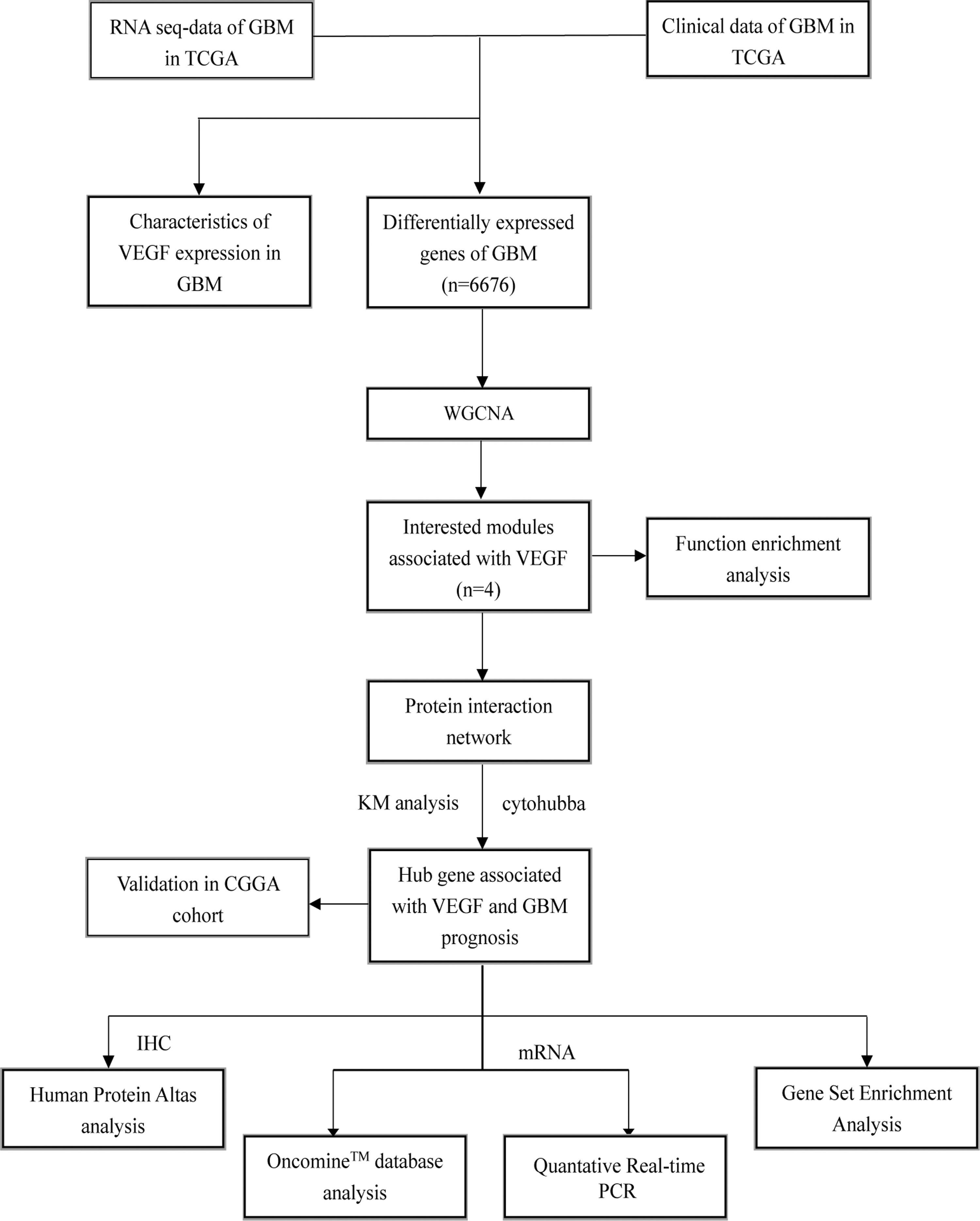
Overview of the procedures for analyzing VEGF and VEGF-associated key gene in GBM. GBM, glioblastoma multiforme; VEGF, vascular endothelial growth factors.
WGCNA for identifying clustered gene modules
Normalization was performed using the Limma R package. The edge R package was utilized to calculate the significantly differentially expressed genes (DEGs) in GBMs, and log-fold change| >1 and p-value <0.05 were set as the thresholds for differential gene screening based on GBM in TCGA dataset. A scale-free network was constructed using the “blockwiseModules” function of WGCNA R package (version 3.6.1). The soft threshold for network construction was chosen for the adjacency matrix to be the continuous value between 0 and 1 so that the constructed network can be closer to the real biological network state. Following the module partition analysis, the gene coexpression modules were identified. Then, the module eigengene, which represents the expression level for each module, was calculated. These modules were analyzed by their eigengene correlation to VEGF expression.
Functional enrichment analyses
Gene Ontology (GO) and Kyoto Encyclopedia of Genes and Genomes (KEGG) analyses were performed on the interested modules to investigate their functional role in VEGF expression.
Identification of key genes correlated with VEGF expression
Using STRING database protein-protein interactions (PPIs) between the interested module genes were assessed. In addition, CytoHubba plugin of the Cytoscape software was used to rank nodes by their network features, selecting the top genes as hub genes in the “Maximum Correlation Criteria.” Furthermore, we evaluated the impact of expression level of hub genes and their corresponding VEGF members on GBM prognosis. Together with the expression levels of VEGFs, the hub genes that correlated with GBM prognosis were regarded as key genes.
Verification of expression and prognostic value of the key gene
We assessed the expression of key gene in GBM and the corresponding normal brain tissues using Oncomine™ database. Utilizing the Human Protein Atlas (HPA) online database, we detect the expression of key gene at a translational level. Further, we performed Kaplan-Meier survival analysis to detect the prognostic value of the key gene followed by result validation using ROC curve. To investigate the biofunction of the key gene, gene Set Enrichment Analysis (GSEA) was performed in the enrichment of the MSigDB collection (c2.cp.kegg and h.all. v6.2. symbols) based on expression levels of the key gene.
Quantitative reverse transcription PCR
A total of 55 samples, including 40 tumor samples from patients with GBM and 15 peritumoral brain tissues, were subjected to quantitative reverse transcription PCR (qRT-PCR) to verify the expression of key gene. Total RNAs was extracted using TRIzol reagent (AG21102; Accurate Biotechnology, Hunan, China). cDNA was synthesized from the extracted RNA (0.5 μg) using the RT Premix for qPCR (AG11706; Accurate Biotechnology) in Reverse Transcription System (Biometra Advanced). The RT-PCR was performed at 37°C for 15 min, 85°C for 5 s, and 4°C hold. qRT-PCR was performed on cDNA, using the SYBR Green Kit (AG11701; Accurate Biotechnology) on the Bio-Rad CFX96 connect Real-Time PCR System. The mixes were predenatured at 95°C for 30 min, followed by 40 cycles of denaturation at 95°C for 15 s, and 60°C for 30 s. The 2−ΔCt method was utilized to calculate the relative expression level of COL6A1. The primers used are as follows: COL6A1 forward 5′-GGCCTGGACTGGACATGA GA-3′, reverse 5′-AAAGTCAGCAACATGGATATGGTT C-3′; GAPDH, forward 5′-GCCATCACAGCAACACAGAA-3′, reverse 5′-GCCATACCAGTAAGCTTGCC-3′.
Statistical analysis
Wilcoxon test was used to compare the difference of VEGFs and expressions of key genes between the groups. OS is presented as the Kaplan-Meier curve. Kaplan-Meier survival curve with the log-rank test was calculated and plotted using survminer R-package. p < 0.05 was regarded as statistically significant.
Ethics Approval and Informed Consent
The study was approved by the Research Ethics Committee of Guangdong Provincial People’s Hospital, Guangdong Academy of Medical Science [No. GDREC20190145H(R2)]. All subjects provided written informed consent.
Results
VEGF expression in GBMs
Expression of VEGF-A and VEGF-B was upregulated, whereas that of VEGF-D was reduced in GBM tissue compared to normal brain tissue (Supplementary Fig. S1A). Although VEGFs influenced the angiogenesis in glioma, no strong correlation between VEGF expression and OS of patients with GBM was observed in TCGA cohort (Supplementary Fig. S1B-F).
WGCNA of DEGs in GBM
Totally, 6676 DEGs (3227 downregulated and 3450 upregulated genes) were identified, screened out, and analyzed (Fig. 2). After eliminating any substandard samples, soft power was set as 4, and DEGs were clustered into 18 modules (Fig. 3 and Supplementary Fig. S2). The cyan module showed significant association with VEGF-A expression (Cor = 0.43, p = 4e-08), whereas the purple module was associated with VEGF-C expression (Cor = 0.46, p = 2e-09). The green module was significantly associated with the expression of VEGF-B (Cor = 0.32 p = 4e-05) and PIGF (Cor = 0.7, p = 2e-24), while the tan module had an intensive association with VEGF-D expression (Cor = 0.29, p = 3e-04) (Fig. 4).
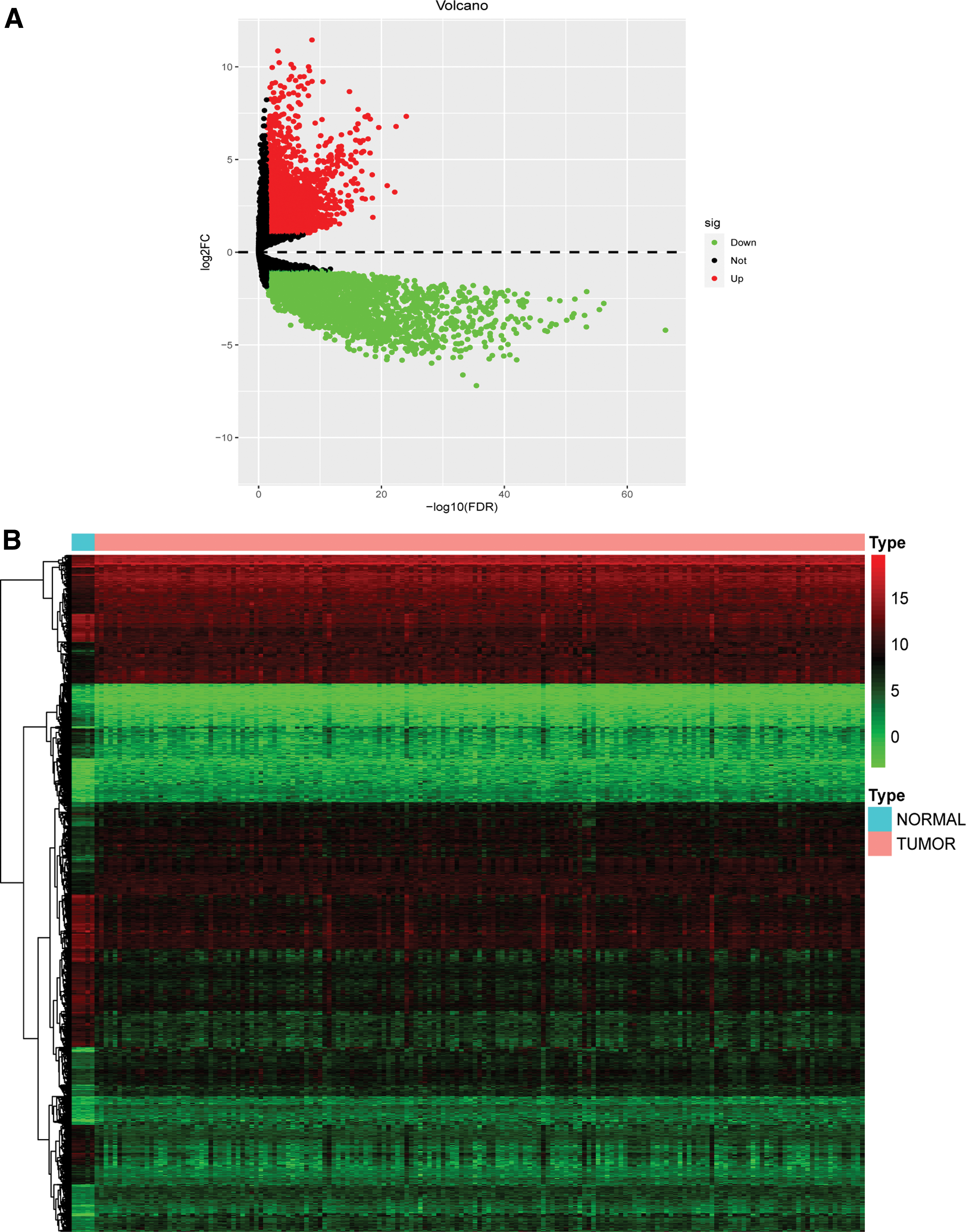
DEGs between GBM and normal brain tissues.

WGCNA analysis. Application of
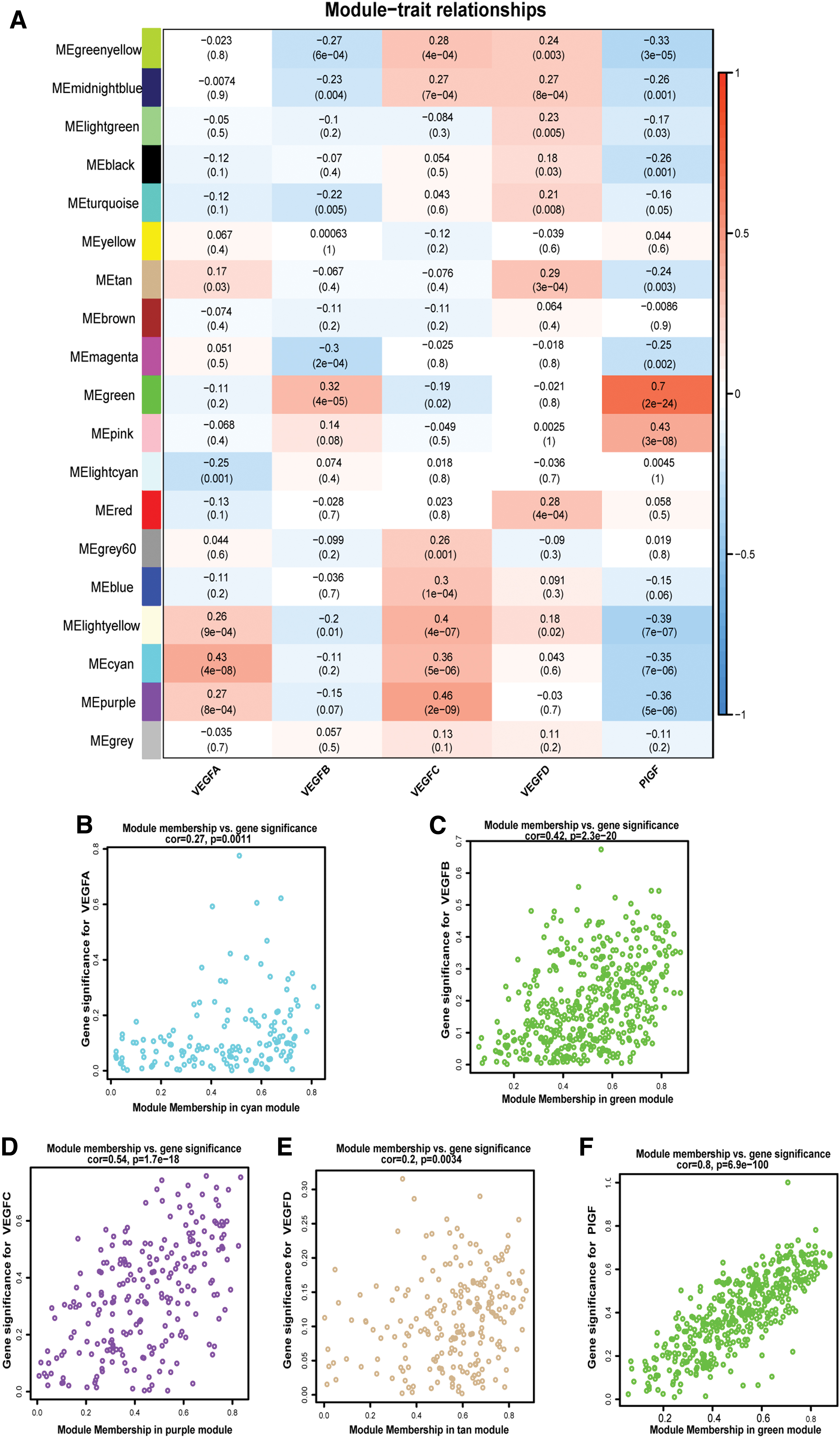
Module-trait associations.
Functional and pathway enrichment analyses
Genes in the cyan module were mainly enriched in extracellular structure organization, regulation of receptor binding, extracellular space, and ECM structural constituent (Fig. 5A), while those in the purple module were mainly enriched in the collagen metabolic process, ECM organization, and growth factor binding (Fig. 5B). Further, genes in the green module were mainly enriched in translational elongation, mitochondrial intermembrane space, glucosyltransferase activity, and protein phosphatase inhibitor activity (Fig. 5C), while those of tan module were mainly enriched in positive regulation of fatty acid beta-oxidation, glycolipid metabolic process, microtubule organizing center part, and monocarboxylic acid transmembrane transporter activity (Fig. 5D).
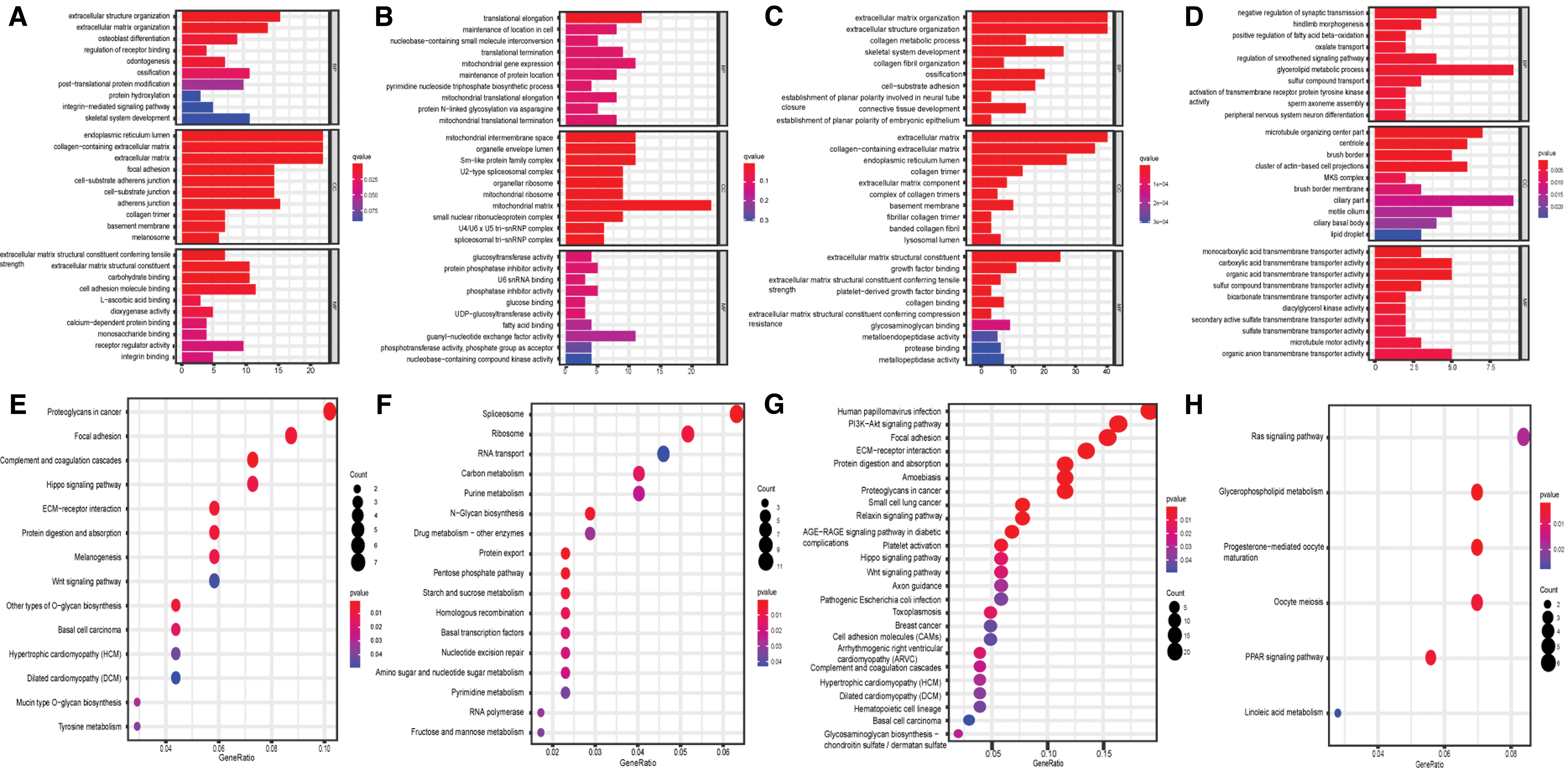
Function enrichment analyses of interested modules. GO analysis of the
KEGG analysis showed that genes in cyan module were enriched in the proteoglycans in cancer, ECM-receptor interaction, and Wnt signaling pathway (Fig. 5E). Genes in green module were enriched in ribosome, spliceosome, and carbon metabolism (Fig. 5F). Genes in purple module showed were mainly enriched in the PI3K-Akt pathway, ECM-receptor interaction, small cell lung cancer, and proteoglycan pathways in cancer (Fig. 5G). Further, genes in tan module were enriched in glycerophospholipid metabolism and PPAR signaling pathway (Fig. 5H).
COL6A1 can be regarded as key gene associated with VEGF expression in GBMs
Protein-protein interaction networks of the genes in the interested modules are shown in Figure 6. CytoHubba plugin screened COL6A1, SNRPG, COL3A1, and AHI1 as the hub genes from four modules, respectively. Of them, alterations in the expression level of COL6A1 and VEGF-A closely correlated with GBM progress (Fig. 7A, B). Therefore, COL6A1 were regarded as the key gene associated with VEGFs in GBMs. Oncomine analysis and qRT-PCR showed that the mRNA expression of COL6A1 in GBM is higher than that in the corresponding normal brain tissues (Fig. 8A, B). HPA immunohistochemistry data showed that COL6A1 was moderately upregulated in high-grade gliomas compared to normal brain tissues (Fig. 8C). Furthermore, survival analysis showed that COL6A1 was associated with poor prognosis of GBM in TCGA cohort; this finding was validated in CGGA cohort (Fig. 8D, F). The 5-year survival ROC curve indicated the expression level of COL6A1 has good performance in predicting OS (Fig. 8E, G). Further, GSEA reveal that the most significantly enriched signaling pathways were based on their NES. Genes in high-expression phenotype of the key gene were significantly enriched with ECM-receptor interaction and focal adhesion in GBM (Fig. 8H).
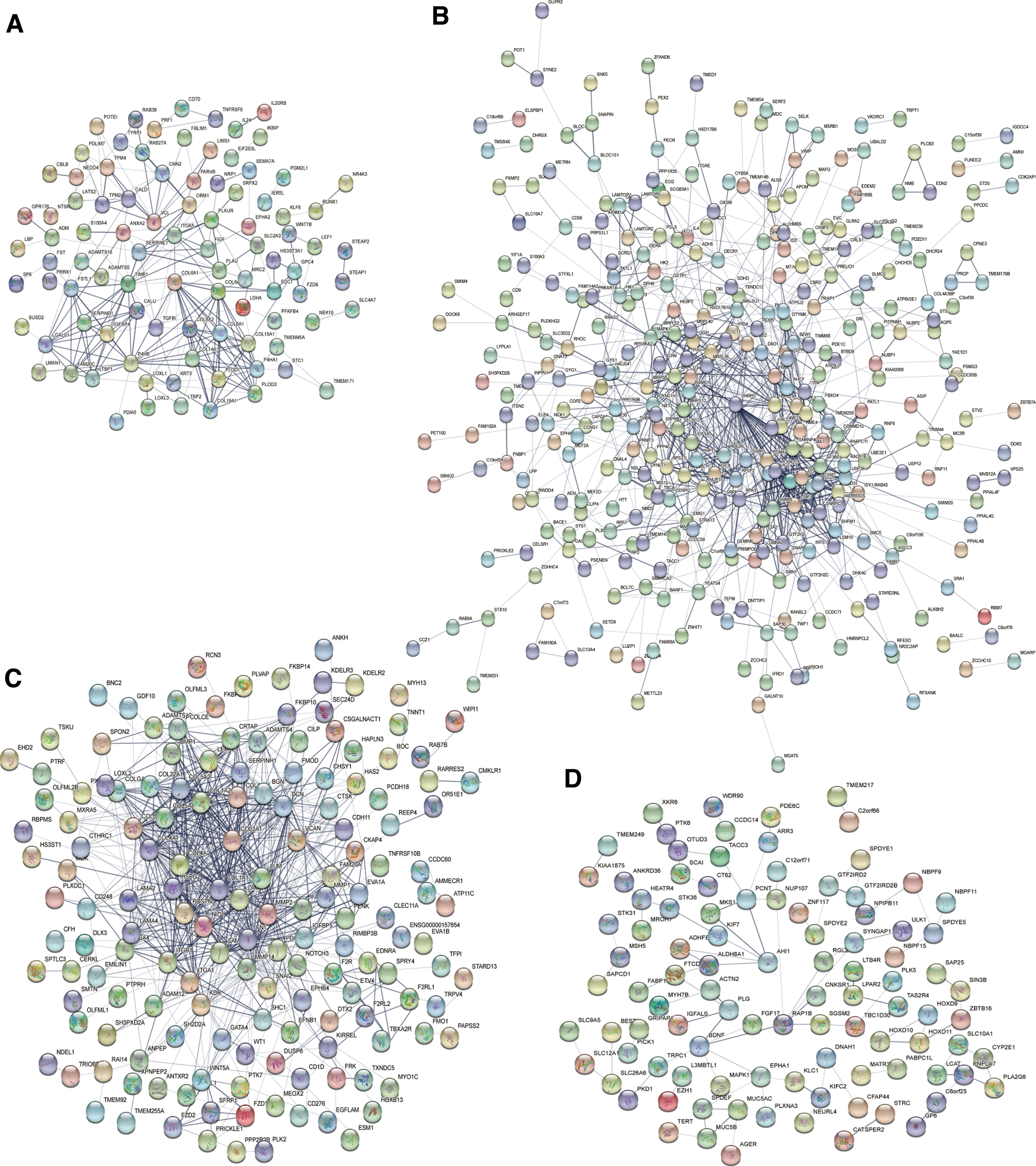
Visualization of modules. PPI network of the
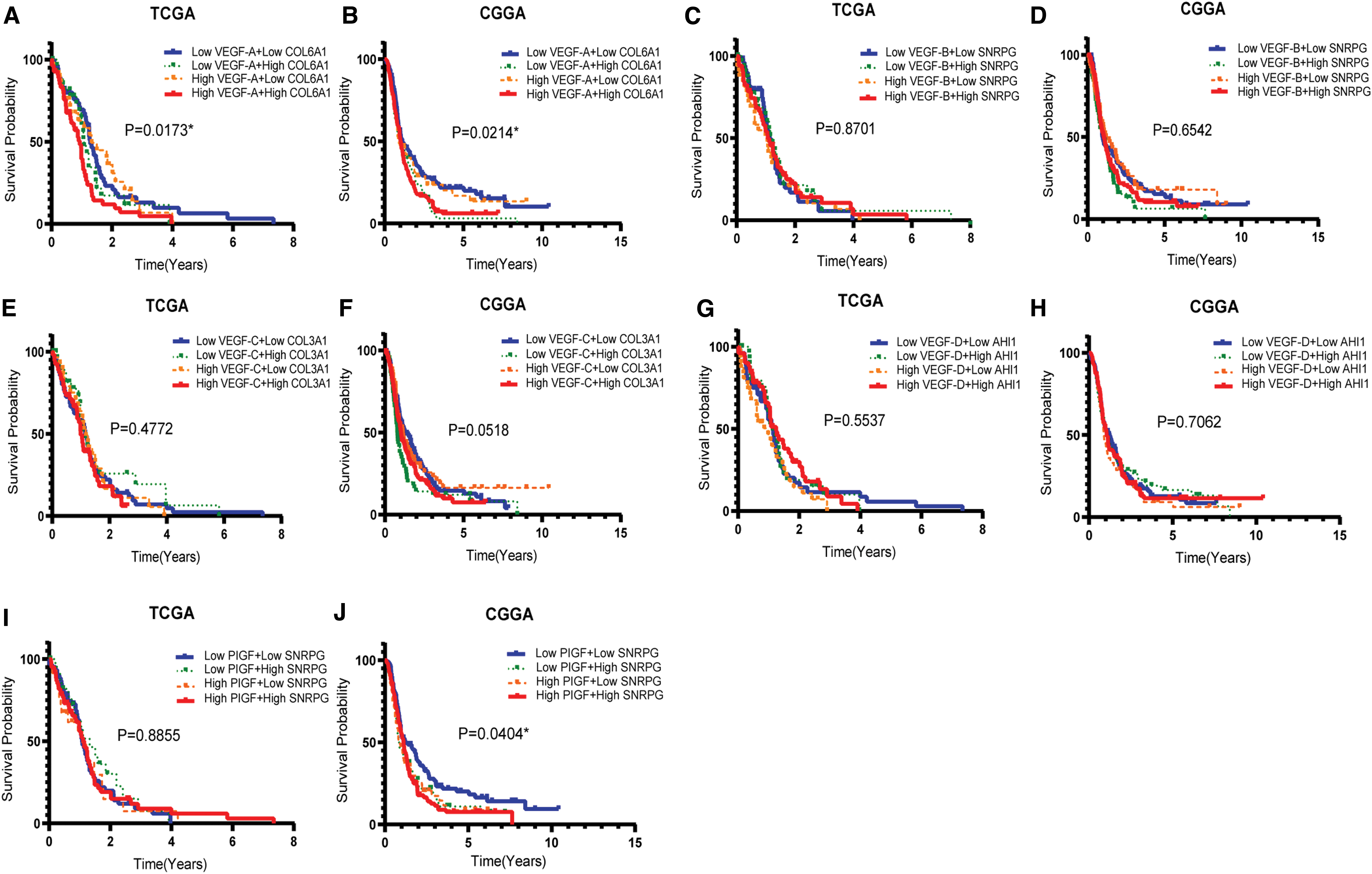
Effect of expression level of hub genes and VEGFs on the prognosis of GBM prognosis. Effect of COL6A1 and VEGF-A on GBM patients in

Key gene in module.
Discussion
Michaelsen et al. (2018) reported that VEGF-C sustains VEGFR-2 activation, thereby promoting tumor growth and that VEGF-C is involved in counteracting bevacizumab therapy in GBM. Although anti-VEGF therapy is promising in GBM management, its application has been limited. Further, escape mechanisms exhibited by GBM cells requires further exploration (Kim et al., 2018).
In the present study, we applied WGCNA to construct coexpression modules associated with VEGF expression and functional enrichment analyses to explore the biofunction of the four interested modules, namely cyan, green, purple, and tan. The biological processes or pathways that the interested module genes mainly enriched in were ECM-receptor interaction, growth factor binding, and PI3k-Akt pathway. ECM stiffness increases angiogenesis by stimulating VEGF signaling in ECs, thereby supporting growth and survival of the ECs. Besides, ECM hampers drug penetration to this niche and promotes the cancer cells proliferation (Najafi et al., 2019).
It is not surprising that the growth factor-binding pathway was an enriched process in the interested modules. Interestingly, VEGF-stimulated irregular tumor flow induces further tumor hypoxia and subsequent increases in VEGF production (Carmeliet, 2005). In addition, the PI3K-Akt pathway is pivotal for tumor progression, invasion, metastasis and angiogenesis (Martini et al., 2014). Ping et al. (2011) demonstrated that the interaction of CXCL12 with its receptor CXCR4 promotes GCS (glioma stem cells) -initiated glioma growth and angiogenesis by stimulating VEGF production via the PI3K-Akt pathway. Targeting the PI3K-Akt pathway results in the downregulation of VEGF expression and the inhibition of glioma proliferation (Zhang et al., 2012; Paul-Samojedny et al., 2015; Ramezani et al., 2017).
In our study, COL6A1 (collagen type VI α1 chain) was identified as a candidate biomarker related to VEGF expression and GBM prognosis via integrated bioinformatics analysis. COL6A1 assists in the synthesis of COL6 (collagen VI), a component of the ECM and constructs microfibrillar networks in the connective tissues of blood vessels and muscles. Previous studies have proven that COL6A1 is associated with myopathy and cardiovascular diseases (Bushby et al., 2014; Chen et al., 2019). Further, studies have also indicated that COL6A1 is significantly correlated with multifarious malignancies. Reportedly, COL6A1 expression is an independent prognostic marker of patient survival in cervical cancer (Hou et al., 2016). Owusu-Ansah et al. (2019) revealed that knockdown of COL6A1 suppresses metastasis and invasion of pancreatic cancer. These studies suggest that COL6A1 plays significant role in tumor progression.
Our studies have showed that high expression of COL6A1 is associated with worse prognosis of GBM. A previous study has revealed that infiltrative or proliferative growth of GBM results in cell compaction, thereby changing collagen and VEGF expression. Disruption of the structure and expression of collagen inhibit tumor angiogenesis, but does not promote glioma invasion and necrosis (Mammoto et al., 2013). However, fewer studies illustrated the relationship of these two genes in GBM and angiogenesis.
In the present study, we found that the high expression of COL6A1 group was significantly associated with ECM-receptor interaction as demonstrated by GSEA. An anchoring meshwork constructed by COL6 bridges cells to the surrounding environment and organizes the tissue architecture of the vasculature via directly interacting with COL6 (Marchand et al., 2019). In summary, our finding indicates that COL6A1 may regulate VEGF-associated angiogenesis and promote GBM tumorigenesis via mediating ECM-tumor interaction. More experimental support is needed to validate our result in the future.
Conclusion
In the present study, we showed that the expression of VEGFs was strongly correlated with ECM-receptor interaction, growth factor binding, and PI3K-Akt pathway. Overexpression of COL6A1 was associated with VEGF expression and poor prognosis in GBM. Therefore, we consider COL6A1 as holding significant potential to be considered as a novel strategy of anti-VEGF treatment in GBM management.
Footnotes
Authors' Contributions
H.L., C.X.H., and Y.Y. designed the study, checked the data, and prepared the article. J.T.Z. and G.Z.L. performed data collection and statistical analysis. R.M. and S.W.C. searched the literature. P.H.X. and Y.J.Z. performed the experiments. W.P. and D.Z. supervised this project.
Acknowledgments
We thank Mr. Zongtai Zheng, Mr. Zesen Chen, and Mr. Edison Zhang for the data processing.
Author Disclosure Statement
No competing financial interests exist.
Funding Information
This program was financially supported by Natural Science Foundation of China (No. 81901250), High-level Hospital Construction Project of Guangdong Province of China (No. DFJH201924), and Natural Science Foundation of Guangdong Province of China (No.2018A0303130236).

References
Supplementary Material
Please find the following supplemental material available below.
For Open Access articles published under a Creative Commons License, all supplemental material carries the same license as the article it is associated with.
For non-Open Access articles published, all supplemental material carries a non-exclusive license, and permission requests for re-use of supplemental material or any part of supplemental material shall be sent directly to the copyright owner as specified in the copyright notice associated with the article.
