




Abstract
Abstract
Introduction
In general, the prevalence of clitoral phimosis is estimated at about 22%, 6 and the rate of vulvar LS in gynecology private practice is estimated to be 1.7%. 7 The incidence or prevalence of the buried clitoris is unknown. Clitoral phimosis is defined as an incomplete foreskin retraction with cephalic force application or limited exposure of the glans clitoris. The degree of phimosis is varying, 6 and the buried clitoris is considered as an extreme form of clitoral phimosis.
In late 1980s and early 1990s, ultrapotent topical steroid (clobetasol propionate 0.05%) was introduced to treat vulvar LS.8–12 Before the clobetasol era, surgery for the buried clitoris could not be recommended due to enforcing the Koebner's phenomenon by a surgical trauma and worsening LS sequelae. In LS, the vulvar skin is already scarred. 2 In order to minimize trauma associated with surgical treatment for the buried clitoris, I introduced hydrodissection. Until now, surgical lysis of adhesions between the clitoral prepuce and the clitoris were performed with rigid, dull instruments.1,13
The aims of this study were to develop a minimally traumatic surgical technique for buried clitoris associated with LS and to evaluate the long-term clinical outcome of this technique. This study was conducted in the private gynecologic practice in the United States. Reviewing the pertinent world literature related to surgical treatment of clitoral phimosis or the buried clitoris by recognizing scientific publications, using Medical Subject Headings (MeSH), which were selected and used in a search on Medline (1980–May 2007), ACOG online database (1990–May 2007), HealthSTAR (1990–May 2007), Cochrane Library database (1995–May 2007), OviDisc database, and utilizing a manual search, indicated that hydrodissection with reverse V-plasty for buried clitoris is a new surgical technique.
Materials and Methods
Study design and setting
This study was planned and conducted as a prospective, sequential, cohort clinical trial in one treatment group. The technical term of a “sequential trial” was defined as a repeated interim analysis of clinical parameters, rather than a single data analysis at the conclusion of the study. The Department of Obstetrics and Gynecology at Howard University School of Medicine (Washington, DC) approved the study's protocol, according to the Helsinki Declaration of 1975. Written consent was obtained from each subject. All surgeries were performed by myself under local anesthetic infiltration in the office procedure room. Data collection was coordinated and monitor by myself.
Preenrollment evaluation
The initial screening included demographic data, medical history and physical examinations, (including clitoral sensory neurologic evaluation), coexisting disease(s), vulvar biopsy, pain-rating score, using the “numeric rating scale” (NRS), and functional status of the clitoris by history only.
Inclusion criteria
Subjects included those who were competent to understand the study protocol and voluntarily provide written informed consent. Symptomatic patients with buried clitoris associated with LS were fitted for the study. All subjects had to have been in an at least 3-year postmenarche period. Each subject had to complete at least 45 days of treatment with clobetasol before the operation. Subjects were willing to comply with participation in follow-up outcome measures.
Exclusion criteria
Subjects excluded were those who had undergone previous surgical intervention for clitoral phimosis, lysis of adhesions, or hoodoplasty for medical or cosmetic indications; clitoral surgery; refused local infiltration as a form of anesthesia; primenarche or fewer than 3 years postmenarche girls; negative histology for LS; pregnancy; acute or chronic infection within the last 2 weeks before surgery; subjects who were no able to be positioned in the lithotomy position; or subjects with a history of adverse reaction to anesthetic.
Subjects of the trial
Thirteen (13) subjects were diagnosed with symptomatic buried clitoris associated with LS between 1994 and 1999. Ten (10) subjects were enrolled, and surgical procedures were executed between October 1994 and December 1999. The follow-up was conducted until June 2008. Subject ages were between 15 and 85 years, and all of them underwent a reconstructive procedure of hydrodissection with reverse V-plasty.
Surgical technique
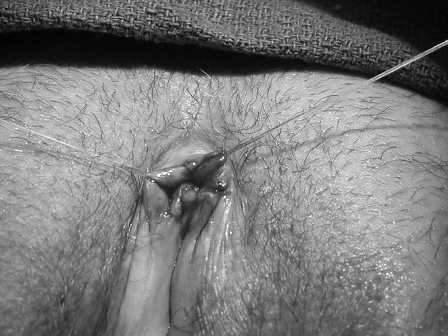
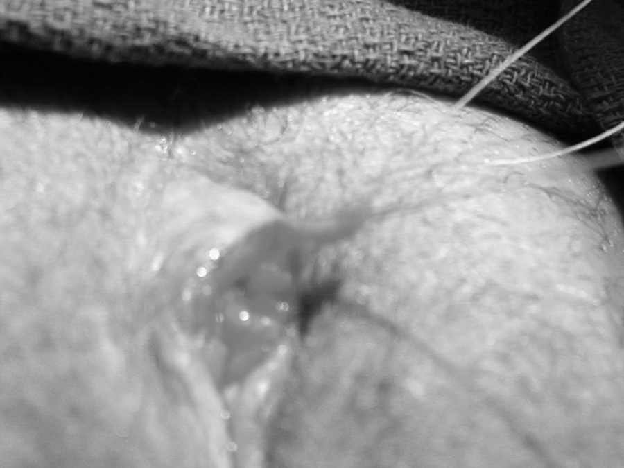
A thick layer of lidocaine-prilocaine cream (2.5%/2.5%) was applied to the preputial skin-mucosa surface and covered with gauze for 30 minutes. For the last 10 of 30 minutes, an ice pack was added to the clitoral area. After removing ice, the clitoral area was prepped with betadine solution. Marcaine (0.5% with epinephrine 1:200,000; Hospira, Inc., Lake Forest, IL) was used for local anesthetic infiltrations. Local anesthetic was injected with a tuberculin 27 G × ½ in needle and syringe (Terumo, Elkton, MD). No sedation or pudendal block were used. A small, agglutinated opening of the clitoral prepuce (i.e., small indentation) of the buried clitoral hood was identified (Fig. 1). At the lateral outer edge of the indentation of the clitoral prepuce, aside from the glans of the clitoris, two temporary-traction sterile, nonabsorbable sutures (3-0 Prolene, taper SH2 needles) were applied bilaterally through the skin-mucosa areas to the clitoral hood. The glans of the clitoris was identified under the skin by palpation and was placed between the traction sutures (Fig. 2.). Each clitoral prepuce indentation had a small, agglutinated opening which allowed for inserting sterile BD Angiocath (Infusion Therapy Systems, Sandy, UT). Before an angiocath was inserted, the needle had been removed beforehand from the angiocath set and only a flexible, plastic catheter was used. An angiocath was connected with BD 10-mL Control Syringe (Becton-Dickinson, Franklin Lakes, NJ). Different sizes of BD Angiocath (with size ranges from 24 GA, 0.75 in, 0.7 × 19 mm to 18 GA, 1.16 in, 1.3 × 30 mm) were used, depending upon the indentation size. Sterile saline was utilized for the separation of the agglutinated clitoris prepuce adhesions and removing debris. The completely obliterated space between the clitoris and the inner prepuce was opened by pressurized fluid. Debris from accumulation smegma and adhesions were flushed out. The hydrodissection was continued until after the clean saline return was free of any debridement. The mucocutaneous flap of the clitoral prepuce was developed (Fig. 3). After hydrodissection, a total preputial retraction was achieved. On average, 5 mm of incision was made into the dorsal midline of the newly developed clitoral flap. The dorsal incision was carried down at the 45-degree angle to the frenulum of the clitoris. The reverse V-shape plasty was accomplished by excising the tissues with the ophthalmology scissors bilaterally (Fig. 4.). The edges of the clitoral prepuce were approximated with a 0-6 coated Vicryl sterile, absorbable suture (Fig. 5). Also, photodocumentation was obtained from each subject before and after the operation (Fig. 6).

The arrow indicates a small, agglutinated opening (indentation) of the clitoral prepuce associated with the buried clitoris and lichen sclerosus.

Local infiltration completed and two traction sutures placed away from the glance of the clitoris.
The hydrodissection part of the operation completed (an almost bloodless procedure). The skin is overlapping in excess above the glance of the clitoris.
A reverse V-plasty in progress to remove the excessive skin of the prepuce.
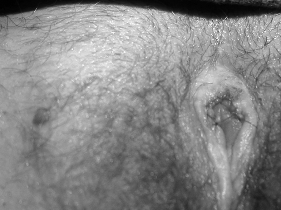
The reverse V-plasty completed, the skin is approximated with 0–6 sutures.
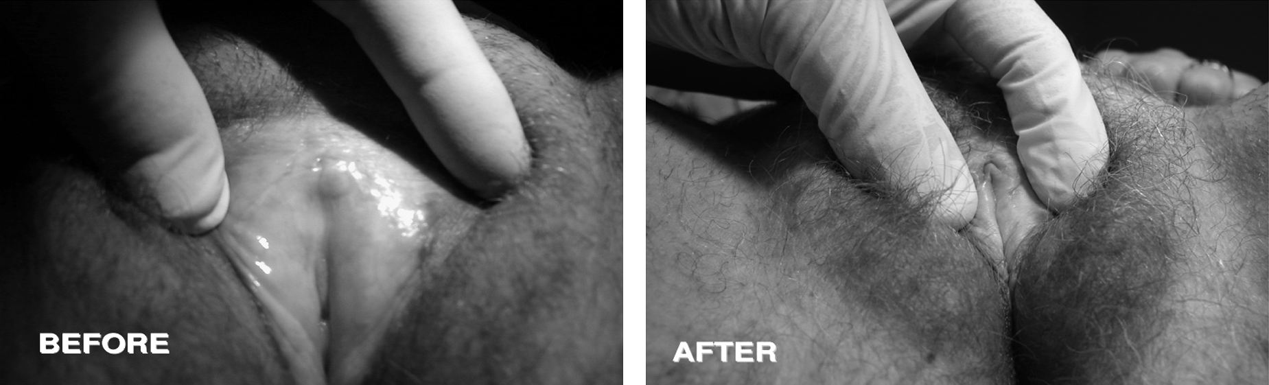
The buried clitoris associated with lichen sclerosis before and 6 weeks after the hydrodissection and reverse V-plasty operation.
Additional intervention to surgical treatment
Before surgery, all the subjects applied ultrapotent topical clobetasol (0.05%) ointment daily, until their LS symptoms and signs (i.e., pruritus and/or burning, or pain and stings of lichenification and inflammation) were in total remission. On average, it requires a 45-day therapy to observe a complete remission of LS. Postoperatively, a clobetasol (0.05%) ointment was continued daily for 2 weeks and reduced the application to twice per week.
Outcome measures
The primary-outcome measure evaluated the clinical results of surgical resolution and aesthetic outcomes; the secondary outcome measure was to apprise the reinstitution of clitoral sensation, ability to reach external (i.e., clitoral) orgasm, and symptoms responding to surgical treatment. The end-points of follow-up were at the completion of 5-year clinical observations by each subject.
Clitoral or pelvic pain before and after surgical treatment were evaluated from using a self-report numeric scale, with options ranging from 0 to 10 (0 corresponds to no pain and 10 represents the most severe pain 13 ).
Intent-to-treat analysis
Primary outcome measures involved a referring physician evaluating the individual subject's result of surgery by incorporating the following: (1) a complete surgical resolution (when the glans clitoris was exposed and clitoral foreskin was fully retractable); (2) a poor surgical resolution was defined as unattainable visualization of the clitoral glans and no retractable clitoral foreskin present; and (3) a partial surgical resolution was classified as a surgical clinical outcome between a complete and poor surgical resolution. Also, the short- and long-term postsurgical complications were evaluated by referring physicians. Overall aesthetic outcome measures were performed by physicians and patients by answering the questions (i.e., excellent outcome, very good outcome, good outcome, and dissatisfied outcome of the operation).
Secondary outcome measures involved the following: (1) symptom response to the surgical treatment was evaluated by the following criteria: (1) subsided, partial response, and no response and (2) reinstituting clitoral function, including clitoral sensation (reinstituted, improved over 50%, improve fewer than 50%, no improvement) and ability to reach external orgasm, as evaluated by subjects only (reinstituted, improved over 50%, improve fewer than 50%, no improvement).
Each patient was furnished with the questionnaire to evaluate pri- and postoperative symptoms, expectation, and overall satisfaction from surgical resolution of the symptoms, local anesthesia, and functional/aesthetic outcome from surgery.
The follow-up was conducted between 1994 and June 2008 by different referring physicians. The end-points of follow-up were at the completion of 5-year clinical observations by each individual subject. An independent research assistant had made telephone contacts with referring physicians and patients at 1-year intervals for a total 5-year of follow-up completion by each subject. Approved, confidential, and consented questionnaires, separate for physicians and patients, were administered. The results were kept in the research files.
An intent-to-treat analysis was incorporated into the study, and the same data were gathered on each follow-up visit of subjects. If subjects failed the follow-up visit, the initial or last follow-up data were used for primary or secondary measurement analysis.
Results
The ages of the subjects were between 15 and 85 years. Two (2) subjects were between 15 and 16, menarche at age 12 and 11, respectively, and had never been pregnant. The remaining women were 51 years or older and were multipara.
All subjects in this study were Caucasian. On physical examination, each and every subject had the clitoris buried beneath the agglutinated labia minora (palpable 1.5–2.00 cm firm, tender structure beneath the fused, clitoral prepuce). Also, each subject had skin changes characteristic for LS and histologically documented LS. In addition, all subjects experienced decreased clitoral sensation, decreased ability to achieve clitoral orgasm (i.e., external orgasm) and increased pain in the clitoral area with clitoral engorgement (NRS score, 10). Before surgery, all subjects reported significant cosmetic deformity of the clitoral area due to LS.
Two (2) subjects, age 15 and 16 years, had significant difficulty walking due to pain and increasing pain with fitted underwear. The older subjects experienced very little difficulty walking or increasing pain associated with fitted underwear. Before surgery, persistent clitoral pain, without clitoral engorgement, was reported by all subjects (mild pain was reported by 1 subject with an NRS of 3; moderate pain was reported by 4 subjects with average NRS of 6; and severe pain was reported by 5 women with average NRS of 9).
Time of surgery ranged between 17 and 36 minutes, and average time of the operation was 23 minutes (from the beginning of local infiltration with the anesthetic to the end of surgery). There was not any noted intraoperative, short, or long-postoperative complication related either to the surgery or local anesthesia.
Primary outcome measure results
In all subjects, complete surgical resolutions were reported by all refereeing physicians throughout the entire 5-year follow-up of the hydrodissection with reverse V-plasty operation (the glans of the clitoris was exposed and clitoral foreskin was fully retractable). Neither new scarification nor a recurrence of symptoms was observed. Pain completely subsided within the 6-week postoperative periods in all subjects. Overall aesthetic outcome measures performed by physicians were reported as excellent. Subjects reported 8 of 10 (80%) as excellent results, and 2 of 10 (20%) subjects reported as very good.
Secondary outcome measures
Clitoral and pelvic pain subsided in all subjects within the 6-week periods. Clitoral sensation was reinstituted in all the subjects shortly after the operation. Ability to reach external (i.e., clitoral) orgasm was reinstituted in all subjects within 3 months.
Discussion
Symptoms such as suprapubic (i.e., pelvic) clitoral area pain, decreases in clitoral sensation, decreases in ability to reach clitoral orgasm, and cosmetic disfiguration of the clitoral region associated with the buried clitoris was overwhelming present and required surgical intervention. However, surgical intervention should be minimally traumatic in order to decrease Koebnerization in the LS entity. Clobetasol (0.05%) ointment usually controls symptoms and signs, as well as restoring natural histology associated with LS (i.e., itching, burning, lichenification, and inflammation); therefore, it is essential to treat each patient before surgery. The reverse V-plasty was incorporated into the hydrodissection procedure, since, after separation of the clitoris from the inner prepuce surface adhesions, the excessive skin must be trimmed in order to expose the glans of the clitoris. Also, it transforms significantly in aesthetic appearance.
In clitoral phimosis, a metallic probe (a lacrimal duct probe) was used to separate adhesions between the clitoris and the inner part of the prepuce. 1 Such an approach can potentially create more traumas to the inner clitoral prepuce and the clitoris, causing more bleeding from that area than hydrodissection creates. Also, bloodless washing out of debridement and removing adhesions with hydrodissection is a bloodless process, when compared with a lacrimal duct-probe process.
A new hydrodissection technique offers a significant improvement in decreasing surgical traumas by means of separation of the adhesions at their weakest point. Also, hydrodissection is a bloodless procedure and, at the same time, removes adhesions and debridement.
Postoperatively, each woman applied daily 0.05% clobetasol to the clitoris area and the prepuce wound to minimize Koebner's phenomenon for 2 weeks and then twice-weekly thereafter. Therefore, success of this new surgical technique is a combination of surgical reconstruction and conservative medical therapy for LS.
It is imperative not to subject a pediatric female population before menarche to any form of surgery, since spontaneous resolution of clitoral phimosis or buried clitoris following puberty has been observed. 3 This new procedure can be applied in different clinical scenarios, such as any form of clitoral prepuce phimosis, clitoral prepuce adhesions, female circumcision, pseudocyst, decrease in clitoral sensation, and other causes of clitoral-prepuce hypertrophy.
Conclusions
Treatment with ultrapotent topical steroid before and after the operation is required. Hydrodissection with reverse V-plasty is a simple, reliable, reconstructive surgical procedure with good clinical outcomes and no recurrences for buried clitoris. This procedure cures the symptoms associated with buried clitoris. The operation restores clitoral function. This surgical method accomplishes a very satisfactory aesthetic transformation.
Footnotes
Disclosure Statement
No competing financial interests exist.