
Abstract
Abstract
Introduction
Case Report
A diagnosis of neurofibromatosis type 1 (NFl) was made by a specialist neurologist based on the presence of café-au-lait spots and multiple neurofibromata on the trunk, head, and neck. She reported no knowledge of family history of breast or other cancers and there was no family history of NFl.
During routine self-breast examination at age 39, she noted a retroareolar lump in her right breast. Examination revealed the presence of an 80-mm mass that was tethered to overlying skin. The prominent finding clinically was large pedunculated pigmented neurofibromas deforming the nipple (Fig. 1).
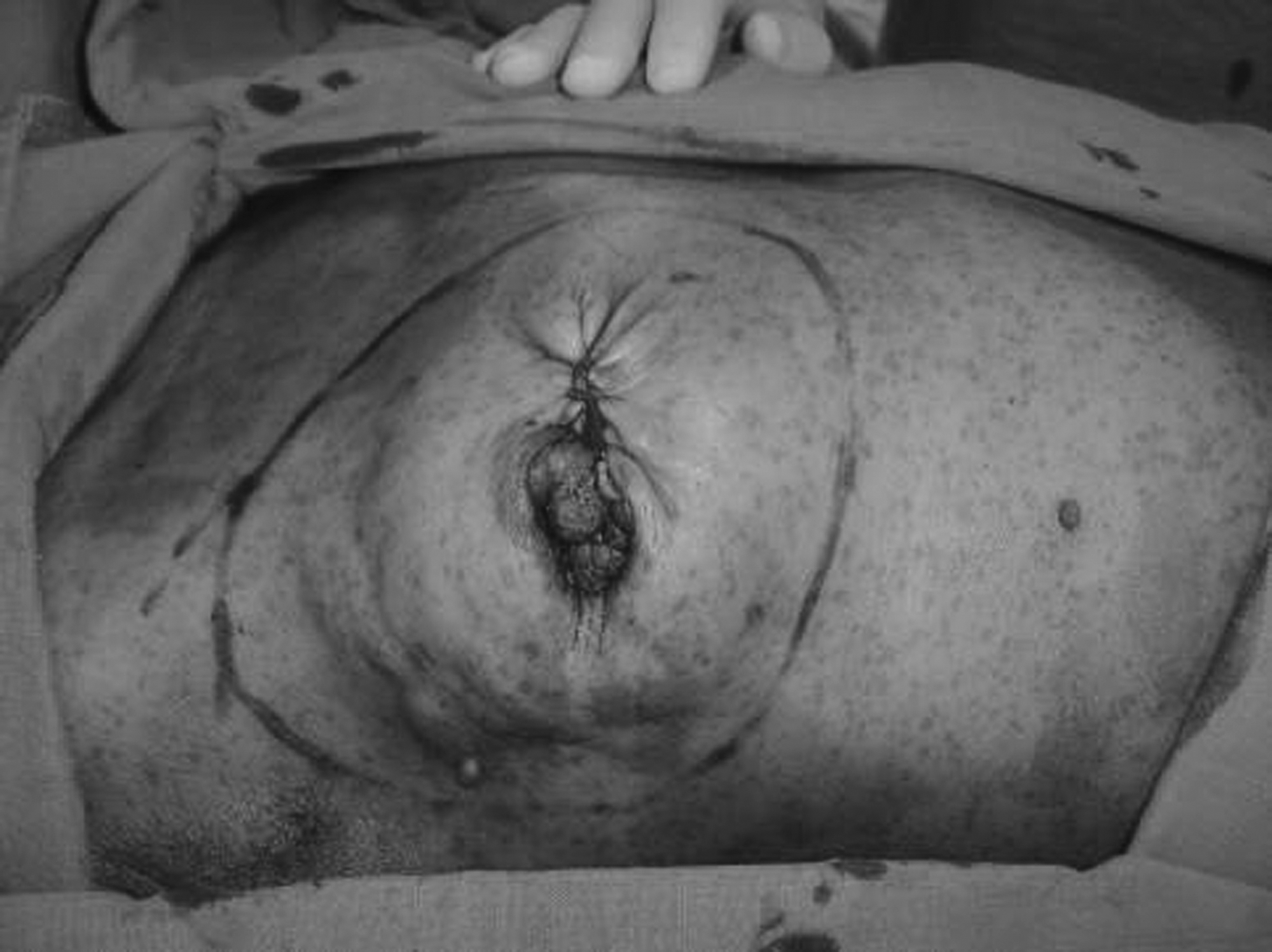
Skin retraction and neurofibromas are observed.
Mammography showed an irregular mass, and ultrasonography demonstrated an irregular, heterogeneous, and hypoechoic mass. Fine-needle aspiration cytology was indicative of a breast carcinoma. In view of the size of the lump, a right modified radical mastectomy and axillary lymph node clearance were performed, and histology revealed an 80-mm invasive ductal carcinoma. Sixteen (16) of 18 axillary lymph nodes were found to contain metastatic tumors. Following surgery, she received six cycles of chemotherapy. The patient subsequently underwent a course of postoperative radiotherapy. Twenty (20) months after her surgery, this woman remains well, with no signs of recurrence of her breast disease.
Discussion
Neurofibromatosis type I, also known as peripheral neurofibromatosis or von Recklinghausen disease, a genetic disorder almost always presenting before 10 years of age, causes many visible symptoms, which signal its presentation.
2
According to the National Institute of Neurological Disorders and Stroke, neurofibromatosis is characterized by the presence of two or more of the following symptoms
2
:
Six (6) or more light brown spots on the skin (café-au-lait spots), measuring >5 mm in diameter in children, or >15 mm across in adolescents and adults Two (2) or more neurofibromas, or one plexiform neurofibroma (a neurofibroma that involves many nerves). Freckling in the axilla or groin regions Two (2) or more Lisch nodules on the iris of the eye (known also as iris hamartomas) Optic glioma Abnormal development of the spine (scoliosis), the sphenoid bone, or the tibia A first-degree relative with NF1.
NF1 is associated with certain types of malignancy such as pheochromocytoma, neurofibrosarcoma, and childhood cancers such as myelogenous leukemia, optic glioma, malignant Schwannoma, and rhabdomyosarcoma. 1
The association of NF1 with breast cancer is rare in the literature.3,4 Murayama et al. reported 37 cases of breast cancer associated with NF1. 4 Most of the cases were diagnosed at an advanced stage with a T score greater than T2. In addition, ductal invasive carcinomas were the most commonly reported tumors. It was concluded that patients presented at a late stage, probably because the presence of numerous skin neurofibromas hindered the discovery of the breast lumps. In a previous Japanese review, Nakamura et al. found that in 18.5% of the cases of NF1, the patients with breast cancer were younger than 35 years old, compared to 6.7% reported in earlier series. 5
Sharif et al. assessed the risk of breast cancer in a cohort of 304 women with NF1 aged older than 20 years in comparison to the population risks over a period of 30 years. The results showed that women with NF1 have a fivefold risk of developing breast cancer by the age of 50 years compared with women in the general population. 6 The authors suggested that breast cancer should be considered a common association in NF1 and that all women with NF1 are at a moderately increased risk of developing breast cancer.
Concerning our case, ductal invasive breast cancer is the most commonly reported histological type in NF1 patients. In addition, our patient presented with a delay, like some patients in the published work, because she thought that the breast lump was a manifestation of her von Recklinghausen disease.
Management of breast cancer in the setting of NF1 poses a complex problem. Although lumpectomy can be performed as part of breast conservation, conventional whole breast radiation may, in theory, increase the risk of development of secondary cancer. It is well known that young adults with NF1 have a higher risk of sarcoma than the general population.7,8 Also, a known risk of radiation therapy is the increased likelihood of developing a secondary sarcoma.9,10 When both are present in a patient, these two situations, at least in theory, increase the risk of developing a secondary sarcoma.
Regarding breast reconstruction in patients who have NF1 and breast cancer and who underwent a modified radical mastectomy, we were able to find only 1 case, which was reported by Yamamoto et al. 11 A 33-year-old woman underwent a transverse rectus abdominis myocutaneous flap reconstruction followed by reconstruction of the nipple–areola complex using a star flap technique and medical tattooing. The operation, which was accomplished in the same fashion as for patients without neurofibromatosis, had a favorable cosmetic outcome. Breast reconstruction offers considerable psychologic benefits in terms of improvement in quality of life in women who have undergone a mastectomy. 11
Footnotes
Disclosure Statement
No competing financial interests exist.