
Abstract
Abstract
Introduction
Case Report
A 30-year-old woman, gravida 3, para 1, was referred to a university hospital at 23 weeks plus 2 days of gestation because of acute pain in her lower abdomen. She had experienced abdominal pain earlier and a laparoscopy had been performed on her 4 years ago. This laparoscopy revealed an asymmetric bicornuate uterus with a significantly smaller left horn. The rudimentary left horn was attached to the right side in the lower segment of the uterus. No further examination was performed. After that, she had a normal pregnancy with a vaginal delivery at week 38 and a legal medical abortion 1 year after that. Otherwise, her medical history was uneventful. The present pregnancy was a consequence of spontaneous conception.
On admission, she had focal tenderness in McBurney's point and severe lower abdominal pain. There was no history of vaginal bleeding, fever, or trauma. Her blood pressure was 123/73 mmHg and she had a pulse rate of 66 beats per minute. Her blood count was normal and her C-reactive protein (CRP) level was 14 mg/L.
Ultrasonography revealed a normal amount of amniotic fluid and normal fetal growth with a weight estimate of 772 g. According to the clinical report, the ultrasound showed a normal uterus. The uterus was enlarged and its bicornuate condition could not be visualized. The patient's symptoms suggested appendicitis, and she was admitted to a ward for observation.
After a few hours, a laparotomy was performed because she had increasing pain. During the operation, a little blood was found in the peritoneal cavity, apparently arising from numerous massive vessels of the left-sided rudimentary uterine horn. Neither active bleeding nor uterine rupture was detected (Fig. 1). The female infant, weighing 540 g, was excised from the small space of the rudimentary horn with Apgar scores of 2, 2, and 3, respectively. The left rudimentary horn had no direct communication with the uterine cavity of the unicornate right uterus and was excised together with the implanted placenta and left adnex. The right uterine horn and adnex were normal. The appendix was healthy.
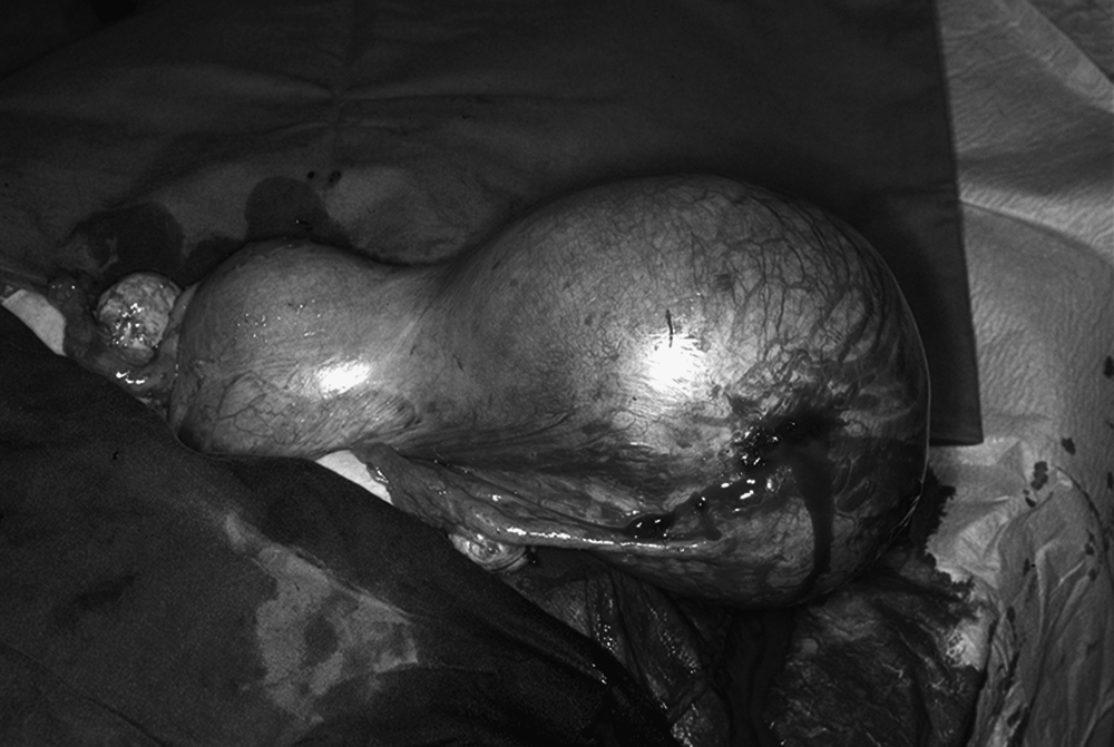
Left pregnant rudimentary horn before excision. Right uterine horn is seen in close contact. Both ovaries are macroscopically normal.
Macroscopic examination revealed an unruptured noncommunicating uterine horn weighing 670 g. The uterine wall of the rudimentary horn was filled with placental tissue, which was tightly associated with corneal structures. No abnormal placentation was found on histopathologic examination, but it confirmed the diagnosis of a noncommunicating uterine horn.
The patient had an uneventful recovery after the operation, and 2 years later, she underwent sterilization by laparoscopy. The surviving infant (a female) was extubated at 3 months' age. Today, she mainly communicates by sign language. At the age of 9 years, she has spastic triplegia, epilepsia, and is mentally retarded. She has some vision in the left eye.
Discussion
A unicornuate uterus with a rudimentary horn is a rare type of Müllerian duct malformation and results from defective fusion. Fusion defects can occur in combination with other anomalies of the genitourinary tract, such as vaginal septum or renal agenesis, which was not found in this case. The incidence of a singleton rudimentary uterine horn pregnancy is estimated to be between 1 in 76,000 and 1 in 140,000 pregnancies. 1 Neonatal survival in rudimentary horn pregnancies is poor, occurring in only 11% of the cases during the past 50 years. 1
The size of the rudimentary horn varies greatly from a small muscular lump, as in the current case, to a well-formed, almost normal-sized uterus. Given that the uterus in this case was a noncommunicating unicornuate uterus, pregnancy must have resulted from transperitoneal migration of sperm or the fertilized egg from the opposite side.
As seen here, a previous normal delivery does not rule out the possibility of a uterine horn pregnancy.
In the present report, immunohistochemical analysis revealed no abnormalities in placentation. It has been suggested that a noncommunicating rudimentary horn, which turned into a hematometra, had endometrium and that a rudimentary horn without hematometra easily results in placenta accreta. 2 In 83%–90% of cases, there is no direct communication between the horn and the functional portion of the uterus. 1
The diagnosis of pregnancy in the rudimentary horn is difficult. The sensitivity of ultrasound examination decreases as the pregnancy advances. Magnetic resonance imaging can be used to confirm the diagnosis before an invasive operation is performed.3–5
The most serious complication is rupture of the horn. Rupture of the horn is still common, but no case of maternal death has been published since 1960. 1
Conclusions
This is a unique case with long-term follow-up of the surviving child. In this case, the possibility of a rudimentary horn pregnancy was overlooked because of a previous normal pregnancy. Therefore, any pregnancy with similar complaints should be viewed with suspicion of an abnormal pregnancy.
At first, this patient's symptoms were misdiagnosed to be appendicular in origin. Control of a potentially life-threatening hemorrhage was the first priority during the laparotomy, and the pregnancy was terminated despite the small number of gestational weeks. In the authors' opinion, rudimentary horns should be removed if a patient is planning to get pregnant.
Footnotes
Disclosure Statement
No competing financial interests exist.