
Abstract
Abstract
Introduction
It is important to recognize the condition, as it is often misdiagnosed for malignancy, putting the younger patient at risk of overtreatment with the resultant loss of hormonal function and fertility. We present two cases of MOE in young females, clinically diagnosed as ovarian neoplasm followed by an oopherectomy.
Cases
Case 1
A 17-year-old female was admitted to the obstetrics and gynecology department complaining of right-sided pelvic pain and fullness in the lower abdomen for the preceding 25 days. She had reached menarche 3 years ago, her menstrual cycles were regular, and she had developed normal secondary sexual characteristics. She had an uneventful past medical history. On examination, the pain was localized in the right iliac fossa, and was constant, moderate in intensity, without radiation to other areas. A soft to firm, slightly mobile mass was palpable per rectum (as the patient was a virgin) in the right adnexa. Pelvic ultrasound revealed a solid hypoechoic mass measuring 9×7.5×5 cm with homogenous echotexture. Serum CA 125 levels were within normal limits. A clinical diagnosis of ovarian neoplasm was made, and a right salpingo-oopherectomy was performed. The specimen was sent to department of pathology. The right salpingo-oopherectomy specimen measured 9×8×3.5 cm. The external surface was grey brown and mildly congested. The cut surface was grey white, solid, soft, and homogenous in appearance (Fig. 1). Microscopically, it was reported as MOE with unremarkable fallopian tubes.

Gross specimen of right salpingo-oopherectomy revealing a markedly enlarged edematous ovary (case 1).
Case 2
A 22-year-old female undergoing investigation for primary infertility and metrorrhagia was admitted to the obstetrics and gynecology department complaining of pain in the left lower abdomen for the previous 2 months. The pain was dull, constant, and without any radiation. On examination, her abdomen was soft with mild tenderness in the left lower quadrant. Bimanual pelvic examination revealed a left adnexal mass. Ultrasonographic examination revealed a mostly solid 7×5×3 cm mass in the left ovary with the presence of cystic areas. The other pelvic organs were unremarkable. Hormonal assays were found to be normal (TSH, FSH, LH, Prolactin). Serum CA 125 levels were mildly raised (50 U/mL) and hence equivocal. The patient underwent a left oophorectomy, and the specimen was received in the department of pathology. Gross examination revealed two grey yellow to grey brown soft tissue pieces measuring together 6.5×5.5×1.5 cm. The cut surface was partly cystic, partly solid, and grey yellow to grey brown. On microscopic examination, a report of MOE was given (Fig. 2).
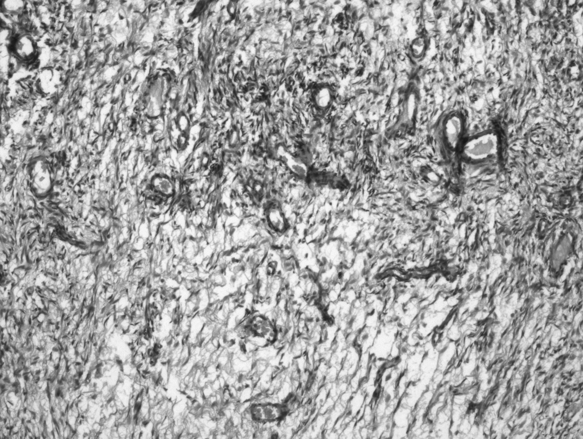
Microscopic examination showing few atretic follicles surrounded by a marked stromal edematous area (case 2: H&E staining;×100 magnification).
Discussion
Massive solid enlargement of the ovary is a tumor-like condition occurring in young women, considered to be the result of torsion of the ovary to the extent that it interferes with venous and lymphatic drainage, but is insufficient to cause necrosis. 3 It was initially described by Gustafson et al. in 1954, and characterized by Kalstone et al. in 1969. 4
The affected age group is between 6 and 33 years with a median age of 20 years. There are two types of presentation. The most common is acute or chronic abdominal pain, the other being characterized by menstrual disturbances. Endocrine disturbances like virilization and sexual precocity can occur in pediatric patients. 2 The affected ovary is usually unilateral, with a predilection for the right side. Such right-sided predominance of MOE was explained by the relatively lower tolerance of the right side to partial torsion resulting in congestion because of the higher pressure in the right ovarian vein than in the left, 5 though one of our patients had a left-sided MOE. The size of the affected ovary can be variable, and there is a case report where the ovary was massively enlarged weighing 2,400 g and attaining a size of 35×20×7 cm. 6
The incidence of MOE is unclear because the pathologic diagnosis of MOE is unfamiliar to the majority of pathologists, and the diagnosis of MOE is usually retrospective. 7 The definitive diagnosis of MOE cannot reliably be made preoperatively. Ultrasonography scanning and magnetic resonance imaging (MRI) have both been reported to aid in establishing the diagnosis. Recent reports have demonstrated that multiple ovarian follicles situated around the periphery of the cortex of the enlarged ovary are the most important indicators of MOE.3,4 Levels of tumor markers such as CA 125 have not been found helpful in definitely excluding the diagnosis of neoplasm, as both the sensitivity and specificity are not 100%.
Morphological recognition of the lesion is fairly simple. The cut surface of the specimen appears grey in color, is wet and soft, and the fluid oozes out with a bulge after being cut with a knife due to the pressure of the edema. Histopathologically, the ovarian stromal cells are widely separated by copious fluid, and atretic follicles may at times be recognized. The tunica albuginea and superficial cortical zone are characteristically uninvolved. A thin rim of compressed cortical stroma is recognized at the periphery of the mass. Necrosis and hemorrhage are unusual. Focal stromal leutinization has been noted in some of the studied cases and is thought to be a mechanical process induced by stretching the stromal cells.
The majority of cases are unilateral and the most of them are treated by unilateral salpingo-oopherectomy, as the lesions are mistaken for primary ovarian neoplasms at laparotomy. After an extensive review of the literature, Geist et al. concluded that most cases were overtreated, and this condition should be suspected in women in the fertile age range with solid enlargement of the ovary. Definite treatment should be undertaken only after confirmed pathological diagnosis.
However, when ovarian edema is suspected at surgery, the appropriate treatment is wedge resection, removing 30% or more of the ovary to exclude secondary causes. Frozen section is valuable at the time of surgery. 3 Wedge resection with fixation of the ovary to the uterus to prevent further torsion or simple ovarian suspension have also been successfully attempted as primary treatment modality.5,7–11
Neoplasia is usually the principal concern when an ovarian mass is diagnosed. MOE should be considered in the differential diagnosis particularly in prepubescent girls and in women of childbearing age. Consideration should be given to preoperative ovarian tumour markers and to frozen section biopsy to help in intraoperative management and pathological diagnosis. 4
Conclusions
In conclusion, it is observed that MOE is not a well-known condition and therefore, more extensive treatment than is necessary is often undertaken, as was the case in our patients. This condition should be considered in young women presenting with an ovarian mass. Conservative management is advised, with the preservation of fertility by retaining the contralateral ovary and biopsy with frozen section when clinically indicated.
Footnotes
Disclosure Statement
No competing financial interests exist.