
Abstract
Abstract
Introduction
Uterine perforation associated with IUDs is a rare but potentially serious complication. 2 After a patient is diagnosed as having this complication, the choice of an appropriate treatment is sometimes controversial. A single case of IUS migration in a woman who had had previous uterine surgery is reported. Concerns and treatment of the case are discussed.
Case
A 36-year-old woman, previously operated on for uterine perforation, was admitted for surgical evaluation of a retained T-shaped IUD. She had been using a levonorgestrel-releasing IUS for 5 years with no related complications. However, when it was time to remove the device, the gynecologist was unable to retrieve it, and a hysteroscopy failed to localize it inside the uterine cavity. Three months after the gynecologist's attempt, when the patient presented to surgery department for the first time, she had no symptoms and no significant history of fever and did not mention having any abdominal discomfort.
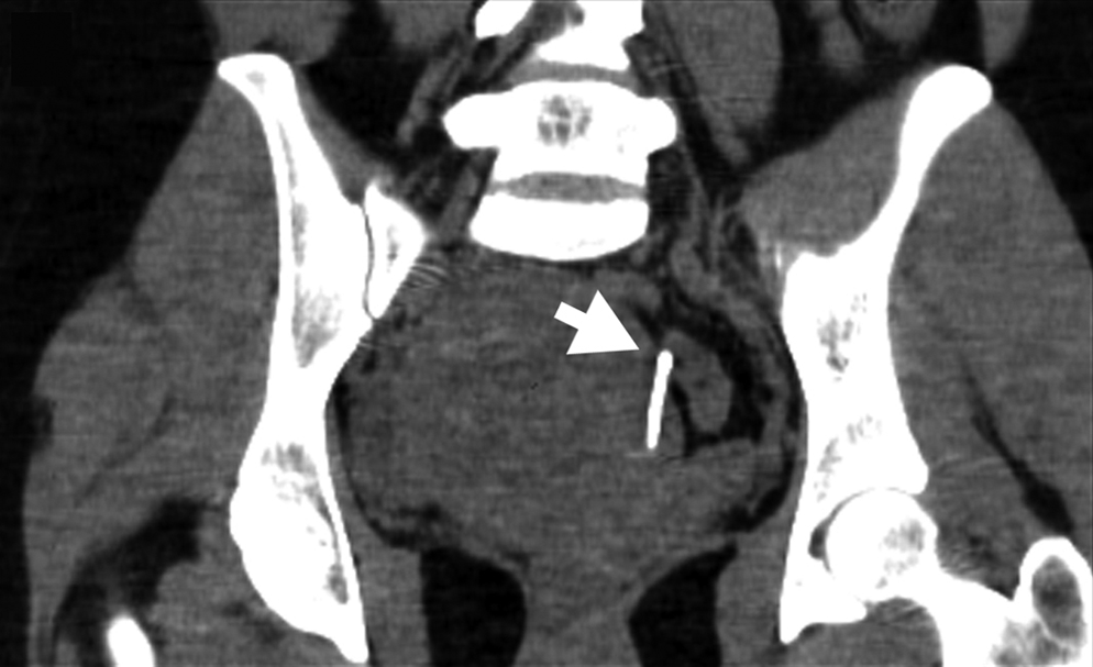
Concerns were raised about a possible intraperitoneal displacement and potential serious damage to internal organs. To localize the position of the “lost coil,” a computed tomography (CT) scan was performed. The scan showed that the device was located in the pelvis (Fig. 1, black arrow). According to the axial and sagittal sections of the scan, it seemed that the IUS lay completely inside the uterine myometrium (Figs. 2 and 3, white arrows). Although the intra-abdominal cavity was not seen to be involved in the first two planes, the coronal section showed that one of the arms of the IUS was pushing through the wall of the womb and was appearing on the surface of the uterus' serous membrane (Fig. 4, white arrow). Moreover, a large uterine fibroid was evident in all the CT sections.

Radiographic localization of the foreign body inside the pelvis.

Axial section computed tomography scan: lost intrauterine system (IUS) buried in the uterine wall.

Sagittal section computed tomography scan: the characteristic T-shape is evident inside the myometrium.
Coronal section computed tomography scan: the intrauterine system appears on the surface of the serous membrane of the body of uterus.
Considering the woman's fertile age, the absence of related symptoms, and, most importantly, the risk of a surgical procedure in a patient who had already had previous surgery, surgical removal of the IUS was not initially recommended. However, eventually, a laparotomy was planned because of the risk of visceral adherence formation.
During the operation, an extensive adhesiolysis was performed. Pelvic adhesions, which bound the uterus together with the bowel and bladder, were carefully divided. Subsequently, a myomectomy was accomplished. The T-shaped device was eventually retrieved by pulling it cautiously by the arm.
Results
There were no intraoperative or postoperative complications. Contrary to expectations, particularly as the patient had had previous uterine surgery, no significant bleeding occurred during the surgical treatment. The patient was then successfully discharged home.
Discussion
Uterine perforation is a rare but potentially life-threatening complication related to the use of IUDs. IUS-related perforation is estimated to occur in at least 2.6 per 1000 insertions, which is higher than the rate for copper-bearing IUDs, which is reported to be up to 1.3 per 1000 insertions. 2
In this case, the coil migrated slowly from its original position into the uterine wall. Even though this process would seem to be painful, it is often asymptomatic, and the perforation can remain undetected for years. In this case, the device lay almost totally embedded inside the patient's myometrium. However, it had to be considered that a small part of the body of the coil was already surfacing on the serous layer of the body of uterus, and, therefore, the possibility of a further translocation could not be excluded. The greatest risks are adhesions and fistulae formation. In the authors' previous experience, as well as in the literature,2,3 the adhesional syndrome in this case could have been massive, and the displacement of the coil could have perforated other pelvic organs, leading to fistulae formation, especially between the uterus and the bladder or the uterus and the rectum. 4 Therefore, to avoid the abovementioned complications, surgical removal of the foreign body was recommended. However, as the patient's previous uterine surgery increased the risk of intraoperative bleeding, the laparoscopic treatment option was not chosen because of the high risk of conversion to an open procedure.
Conclusions
In general, even though there is a controversy regarding which procedure to select in these cases, there seems to be a consensus for the removal of IUDs when perforation occurs, mainly because of the potential for adhesion formation. 3 According to reported experience, every single aspect of a case should be analyzed carefully. The possible benefits should always be weighed against the potential risks in order to choose the appropriate treatment for the patient.
Footnotes
Disclosure Statement
No competing financial interests exist.