




Abstract
Abstract
Introduction
These pregnancies are challenging to diagnose, and carry with them a high risk of bleeding and uterine rupture, which can require hysterectomy. A rare case of pregnancy occurring in the lower segment cesarean scar, resulting in uterine rupture in the first trimester, is reported.
Case
A 32-year-old woman, gravida 3, para 2, who was 9 weeks pregnant, presented to the emergency gynecology ward with a painful abdomen and spotting per vaginum for 2 days. Pain in the abdomen was intermittent, generalized, and severe in intensity. She gave a history of two episodes of vomiting and fainting attacks. She denied having any history of trauma. She had undergone two lower-segment cesarean deliveries because of transverse fetal positions, one delivery was 3 years prior to the current admission and the second delivery was 1.5 years prior to her admission.
On examination, her pulse rate was 110/min, blood pressure was 90/64 mm Hg, and she was afebrile and pale. Her cardiovascular system and chest examination were normal. Abdominal examination revealed a distended abdomen and a midline infraumbilical vertical scar healed by primary intention. On abdominal palpation, tenderness, guarding, and shifting dullness was present. On vaginal examination, the cervix was closed and ballooned. The exact size of the uterus could not be determined. Fullness and tenderness were present in all the fornices. An ultrasound was performed, which showed a single irregular gestational sac with fetal node, yolk sac, and a fetus of crown–rump length of 33 mm, corresponding to 10 weeks' gestation with cardiac activity, intramyometrial in the lower uterine segment. There was a massive amount of free fluid in the peritoneal cavity. The patient's hemoglobin was 5 g%, white cell count - 6800, differential leukocyte count (DLC)- 74, 22, 2, 2, platelets −1.5 lac, blood nitrogen urea −26 mg%, serum Na+ −140 mEq/L, serum K+ −3.7 mEq/L. The diagnosis of ectopic pregnancy was made in view of the history of cesarean deliveries and the location of the gestational sac on the ultrasound examination. Laparotomy was performed, as frank blood was obtained on abdominal aspiration.
A hemoperitoneum of 2 L was present; the upper part of the uterus was normal. The lower part of the uterus at the level of isthmus was distended. A rent of 2.5×2cm was present on the right side of the previous cesarean scar area. The opening was extended bluntly with a finger along the scar; the gestational sac was seen coming out through the rent (Fig. 1). The gestational sac along with placenta was removed. Active uncontrolled bleeding from the lower uterine segment was present. A decision was made to perform a hysterectomy. The bladder was adherent to the lower uterine segment scar; therefore, sharp dissection was done and bladder was reflected downward. Total abdominal hysterectomy was performed and the biopsy specimen was sent for histopathologic examination. The abdomen was closed in layers after achieving complete hemostasis. Four units of blood and two units of fresh frozen plasma were transfused. The cut section of the uterus showed a normal upper uterine segment and pregnancy in the isthmus part (Fig. 2). The histopathology report of the hysterectomy specimen revealed chorionic villi and trophoblastic tissue in the lower uterine segment, invading the myometrium.
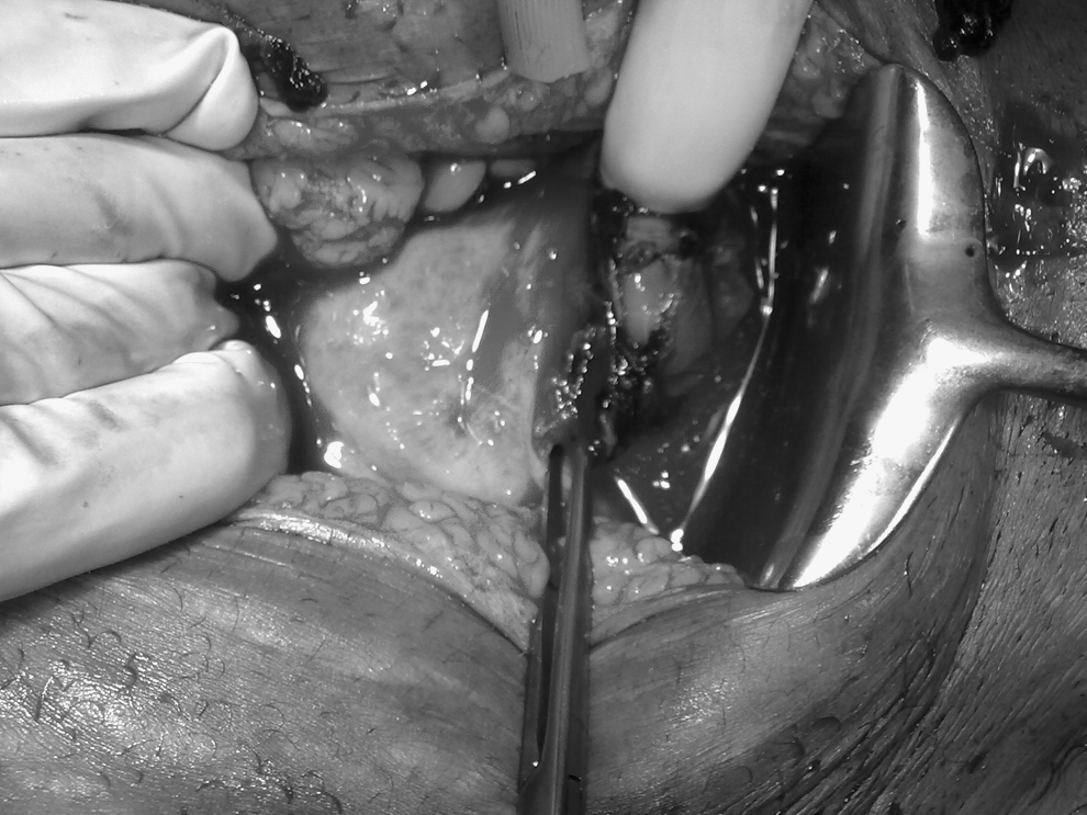
Gestational sac at the level of the uterine isthmus during laparotomy.

Hysterectomy specimen showing scar pregnancy with normal upper uterine segment and cervix.
Results
The patient's postoperative course was uneventful, and she was discharged from the hospital in a stable condition.
Discussion
A cesarean scar pregnancy is a gestation completely surrounded by the myometrium and the fibrous tissue of cesarean scar, and separated from the endometrial cavity and endocervical canal. 4 The most probable mechanism through which this can occur is invasion of the myometrium through a microscopic tract. The tract is believed to develop from trauma from previous uterine surgeries, such as dilatation and curettage, myomectomy, metroplasty, and cesarean delivery. 5
Vial et al 6 theorized that these pregnancies may be partially implanted in the uterine cavity, where they might proceed to term, or else implant deep in the scar, and that they are predisposed to early rupture in the first trimester, as in this case.
In this case, the woman had undergone two cesarean deliveries which agrees with Jurkovic et al., 2 who believed that multiple cesarean deliveries are another risk factor for scar pregnancy. The indication for previous cesarean deliveries in the present case was transverse fetal lie, in a woman whose lower uterine segment was not well-formed, which corroborates the findings of Maymon et al. 1 This may be because of inadequate healing, leading to a defect in the scar.
The diagnosis should be based on the patient's history and her clinical manifestations, such as abdominal pain and any amount of bleeding (ranging from spotting to a life-threatening hemorrhage). 1 The most important investigation is endovaginal ultrasonography and color flow Doppler imaging, 7 as in the present case. Proposed ultrasound diagnostic criteria are: 1) an empty uterine cavity and an empty cervical canal, 2) a gestational sac in the anterior part of the uterine isthmus, and 3) an absence of healthy myometrium between the bladder and sac. 2 When the pregnancy is not localized by ultrasonography, either laparoscopy or laparotomy can be used for the diagnosis.
When the gestational sac is seen in the lower part of the uterine cavity, it can be difficult to differentiate between spontaneous abortion in progress, cervicoisthmic pregnancy, and cesarean scar pregnancy. In cases of spontaneous abortion in progress, the gestational sac should be seen in the cervical canal on transvaginal scan and the sac should appear avascular, reflecting the fact that the sac has been detached from its implantation site, whereas in cesarean scar pregnancies, the gestational sac would appear well perfused and would be located in the anterior uterine wall at the isthmus. 5
Cesarean scar pregnancy is, however, considered to be more aggressive than placenta previa or accreta because of its early invasion of the myometrium, that is, in the first trimester. 8 In the present case study, a significant scar defect might have resulted in deep implantation within the myometrium, causing persistent pain and spotting followed inevitably by uterine rupture in the first trimester.
There are no universal treatment guidelines for cesarean scar pregnancy, because of its rarity. Expectant management of a viable scar pregnancy puts the mother at significant risk of needing an emergency hysterectomy if the pregnancy progresses beyond the first trimester. 2 Rarely, such a pregnancy may continue to the advanced stage of 35 weeks' gestation without endangering the patient. This pregnancy was terminated by emergency cesarean hysterectomy. 9 Therefore, all scar pregnancies should be terminated once the diagnosis has been made, except in the special situation of superficial implantation in a shallow scar defect where there is ultrasound evidence of continuity of the gestation sac with the uterine cavity.
Presentation of the patient often dictates the mode of treatment. If the early diagnosis has been made, a variety of surgical and nosurgical interventions have been proposed in order to terminate the ectopic pregnancy while preserving the uterus. 10 Several types of conservative treatment have been used: (1) dilatation, curettage, and excision of trophoblastic tissues using laparotomy or laparoscopy; (2) local and/ or systemic methotrexate (MTX) administration; (3) bilateral hypogastric artery ligation, associated with dilatation and evacuation under laparoscopic guidance; and (4) selective embolization in combination with curettage and/or MTX injections. 7 Salomon et al. 11 treated a heterotopic cesarean scar pregnancy successfully with potassium chloride. Curettage seems contraindicated because the trophoblastic tissue is outside the uterine cavity and can result, potentially, in a rupture of the uterine scar implantation and hemorrhage. Nonsurgical treatment options even when successful could be expected to leave the uterine scar defect that will accompany cesarean scar implantation. The potential for an unprepared scar dehiscence that will affect future pregnancies is left to speculation. 10
A delay in either diagnosis or treatment can lead to uterine rupture, hysterectomy, and significant maternal morbidity, 10 as in the present case.
Conclusions
If the gestational sac is found at the level of the uterine isthmus in a patient who has had a previous cesarean delivery, it is important to consider the possibility of a cesarean scar pregnancy, especially when the cervical os is closed. A high index of suspicion of cesarean scar pregnancy is, therefore, necessary to diagnose this condition before it can cause catastrophic hemorrhage, and while it can still be managed nonsurgically.
Footnotes
Disclosure Statement
No competing financial conflicts exist.