
Abstract
Abstract
Introduction
The traditional management for fibroids was a hysterectomy, but, recently, uterine artery embolization, often preceded with hormone treatment, using gonadotropin-releasing hormone (GnRH) agonists, has become more common. 2 For uterine artery embolization (UAE), the fibroid tumor is devascularized by embolization of uterine arteries, leading to involution of the fibroid tumor and a dramatic decrease in symptoms. 5 Surgery is usually required for removal of symptomatic myomas, with hysterectomies being performed on women who have no desire to have children and myomectomies for women desiring to preserve their fertility. 3 Hysterectomies are the only sure cure for fibroid tumors; however, most women with these tumors will not need this major surgery. 6
The current patient's suspected submucosal myoma proved to be cervical in origin. A minimally invasive approach to this large fibroid tumor allowed for a shorter recovery time than a hysterectomy would have done.
Case
This is a case of a gravida 3, para 3, 50-year-old, white female at Tampa General Hospital, Tampa, Florida. She was seen initially on February 2, 2010, and her last menstruation occurred ∼2 weeks prior to her visit. She complained of worsening vaginal bleeding and dysmenorrhea. During menses, she saturated both a sanitary pad and a super tampon every 1.5 hours for 14–21 days. Following menstruation, she had a watery vaginal discharge for the remainder of the month. In addition to dysmenorrhea, she complained of fatigue and stress urinary incontinence while denying the occurrence of dysparunia and postcoital bleeding. She reported being sexually active without using any contraception for years. She also denied having any history of a sexually transmitted infection or abnormal Papanicolaou smear results. Her last Papanicolaou test had been taken several years ago, and her surgical history was remarkable for a breast augmentation, a hiatal hernia, and an abdominoplasty.
On physical examination, her height was 62 inches, her weight was 144 lbs, her body mass index was 26, her blood pressure was 131/62 mmHg, and her heart rate 92 beats per minute. She had an Hgb level of 8.7 and an HCT of 27.6, with normal thyroid function. A pelvic examination revealed a 5–6-cm fibroid tumor seen protruding from the external cervical os. A Papanicolaou test could not be performed because of the obliteration of the cervix caused by the fibroid tumor. On bimanual exam, an 8-week size uterus was palpated, and there were no masses in the adnexa.
Her transvaginal ultrasound on March 1 revealed a hyperechoic 9.2-mm endometrium with a uterus measuring 117-mm long, 73-mm anterior posterior, and 60-mm transverse. A submucosal leiomyomata of 10×4×6.9 cm, with more than 50% projecting into the endometrial cavity and aborting through the dilated cervix was seen. A right ovarian cyst of 3×2.8×2.8 cm was also noted.
The patient was informed of the findings and was recommended for a hysterectomy because of her age, the size and the location of the fibroid tumor, and her anemia. The patient stated that her lack of health coverage was the main reason for not getting treatment earlier. She requested a conservative approach that would enable her to remain as an ambulatory patient, so that she could return to work immediately. Suppressive gonadotropin agonist treatment was offered, but she declined because of the cost of such treatment, and, instead, requested a vaginal myomectomy and an endometrial sampling. She also chose to receive iron supplementation in the meantime for her anemia. The patient consented to a vaginal myomectomy with endometrial sampling. A hysterectomy would be performed only if her bleeding could not be controlled.
The patient underwent surgery on the morning of May 20th. It was discovered that the origin of her large fibroid tumor was cervical rather than submucosal. Through a meticulous surgical technique, the tumor was removed in entirety vaginally (Fig. 1). The cervical os was now identifiable. The anterior lip of the cervix was eventually identified and, on following the cervix posteriorly, it was discovered that the fibroids were emanating from the posterior cervical wall internally. The endometrium was sampled with a curette after the anterior cervical lip was located. Heaney fixture sutures were implemented to control bleeding. The patient lost approximately 100 cc of blood during the procedure. The patient was subsequently discharged from the ambulatory suite later that morning.
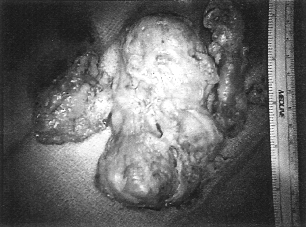
The cervical fibroid that emanated from the posterior cervical wall after removal.
Pathology analysis revealed a 12.2×11×6 cm fibroid tumor measuring 225 g. Her endometrial sampling revealed a secretory endometrium with retrieval of an endometrial polyp. Both the fibroid tumor and endometrium were negative for malignancy. The patient returned a month later for a postoperative visit and a review of the pathology report. The patient was menstruating during this follow-up visit, but stated that her bleeding was significantly less than prior to surgery. On pelvic examination, approximately 20 cc of blood was in the vault and her cervix remained dilated to approximately 1.5 cm. She subsequently underwent a Papanicolaou test 3 months later while reporting normal menstruation and was noted to have a normal-appearing cervix and uterus.
Discussion
Without adequate insurance, patients sometimes have to sacrifice their well-being for other necessities. In this case, it was demonstrated that a patient could receive treatment involving a minimally invasive ambulatory approach for a fraction of the expense of traditional therapies, while allowing her immediate return to work. Based on this patient's testimony, her menstruation returned to normal postprocedure. She inquired about options other than a hysterectomy and declined gonadotropin agonist treatments, citing the costs and her lack of health insurance. For the same reason, an MRI was not performed to determine the exact origin of the myoma prior to surgery. In 2009, there were 50.7 million people in the United States—16.7% of the population—without health insurance—an increase of 4.4 million people from the prior year. 7 As this number is expected to increase, the number of patients for whom less-invasive procedures will become attractive will also rise. Physicians must also prepare for flexibility during procedures, as proper diagnostic tests may also be out of the financial reach of many patients.
Conclusions
This minimally invasive myomectomy should be considered in instances of economic and time constraints although hysterectomy has been the standard appoach. This case demonstrated that the simpler procedure is safe and feasible for many patients who opt out of undergoing hysterectomies.
Authors' Contributions
The University of South Florida's Gynecological Ultrasound performed and interpreted the patient's imaging regarding the myoma. Tampa General Hospital Pathology department performed the histological examination of the fibroid tumor. All authors read and approved the final manuscript.
Footnotes
Acknowledgments
The authors acknowledge Sadaf Aslam, MD, Caroline Young, MSN, ARNP, William Spellacy, MD, and Mitchel Hoffman, MD for critically reviewing and editing the paper.
Disclosure Statement
No competing financial interests exist.